The knee is a hinge synovial joint between the femur (thigh bone) and
the tibia (shin bone).
The joint is protected in front by the
patella (knee cap)
.
Knee joint is a
complex compound synovial joint
(3 bone with
meniscus) with a wide range of flexion & extension and limited medial
& lateral rotation.
It is the largest joint in the body .
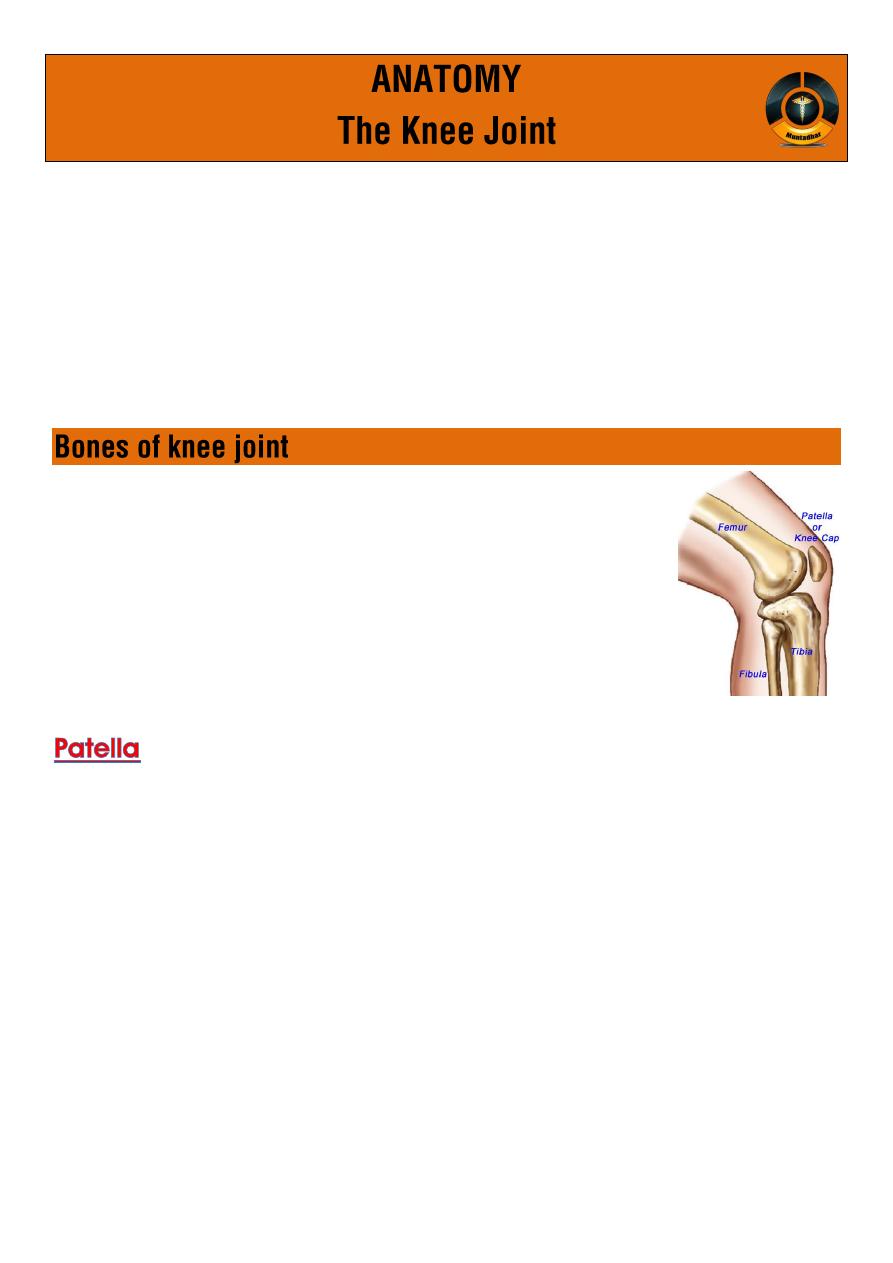
Three bones take part in forming the knee joint :
1. Lower end of the femur
2. Upper end of tibia
3. Patella (knee cup).
Also known as the
knee cap or kneepan
is a thick , circular-triangular bone which
articulates with the femur
(only !) and covers and protects the knee joint
It is the largest sesamoid bone in the human body.
It have apex (downward) and base (upward) .
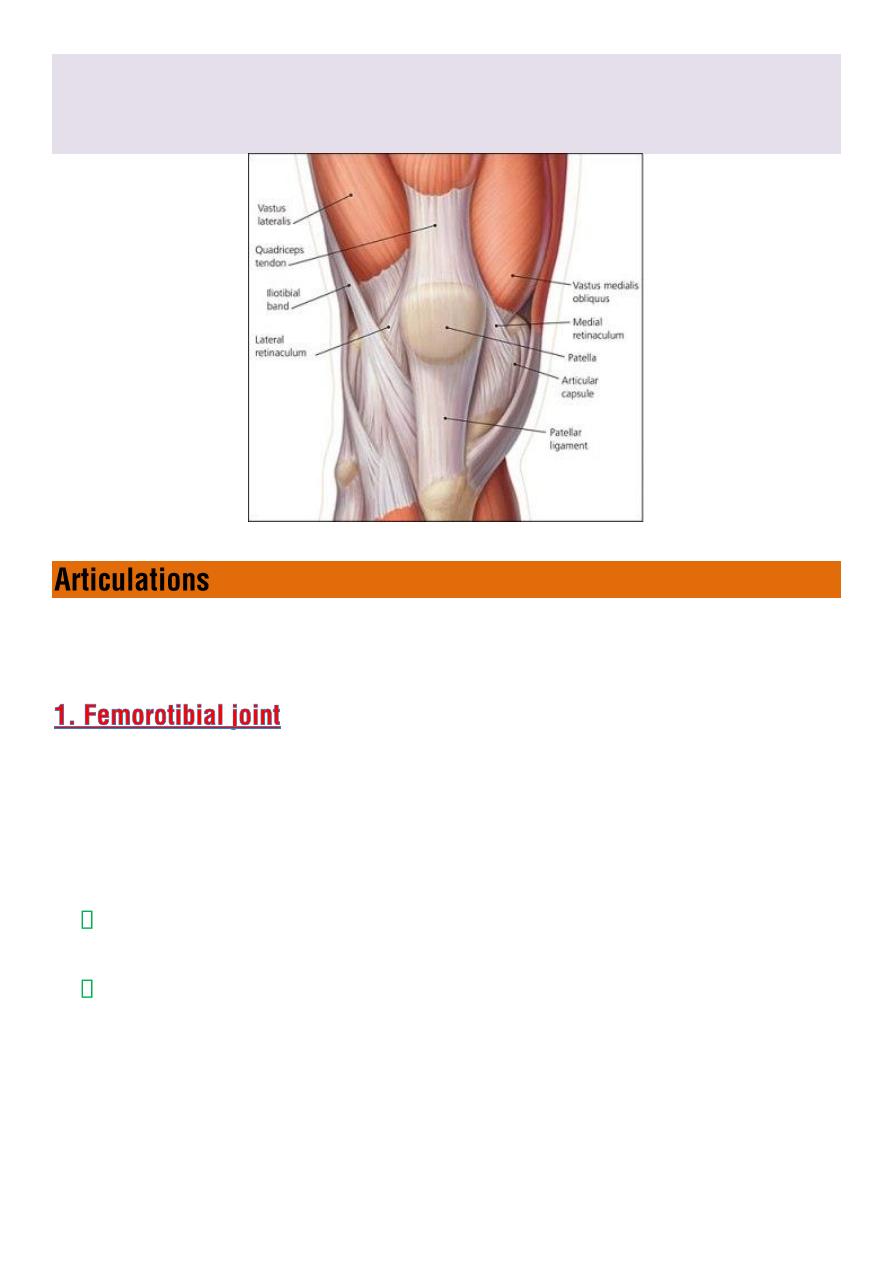
It is attached to the tendon of the
quadriceps femoris muscle
, which
contracts to extend/straighten the knee.
◊
muscle is attached to the base of patella.
◊
The
are attached to lateral and
medial borders of patella respectively.
The primary functional role of the patella is
knee extension
, The
patella increases the leverage that the tendon can exerts on the femur
by increasing the angle at which it acts .
The knee-joint is synovial hinge-joint, but is really of a much more
complicated character
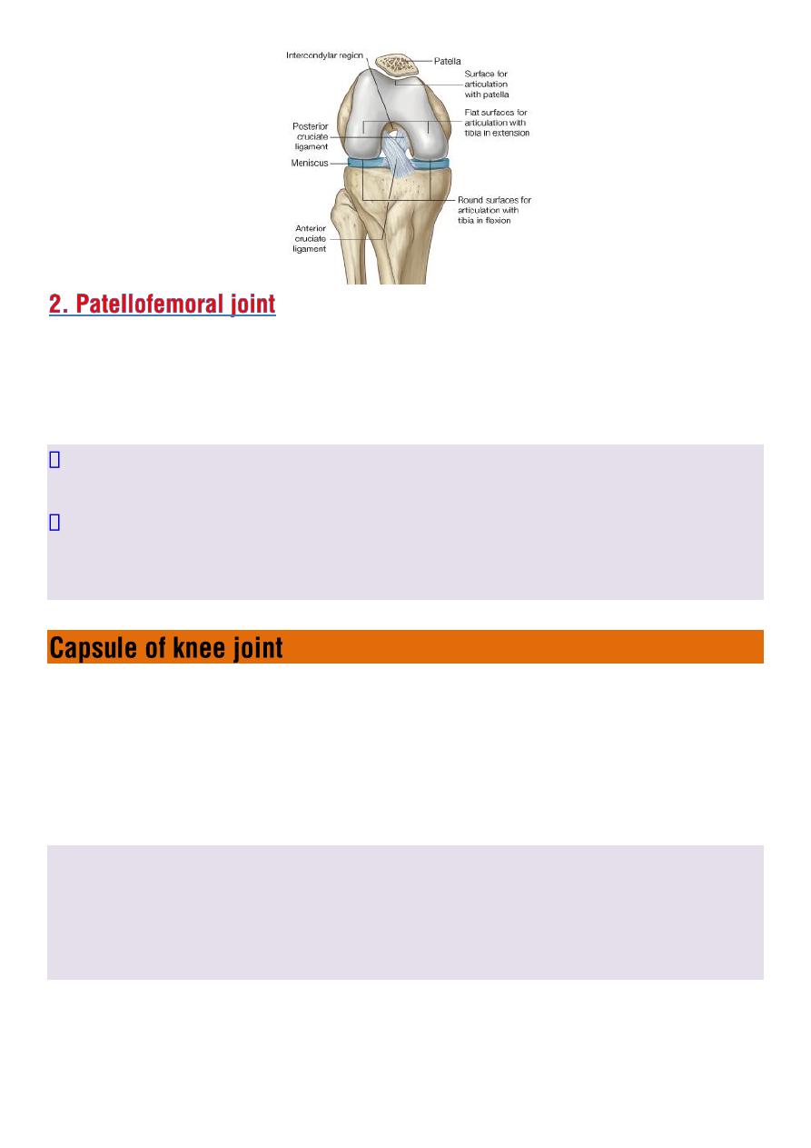
The lower end of femur has a curved articular surface, covered by
hyaline articular cartilages
;
extends backward
and called medial and
lateral condyles.
The upper end of tibia has
oval
shaped articular surfaces, medial and
lateral condyles.
The femoral and tibial condyles articulates forming femorotibial
joint.
This joint flexes and extends the knee.
Patella articulates with femur to form patellofemoral joint
is
plane gliding joint
, This joint allows the patella to glide over the
surfaces of the femur as the knee flexes and extends.
The articular surfaces of femur, tibia, and patella are
covered with
hyaline cartilage.
upper end of fibula (head)
does not adjoin
the femur .it articulates
with lateral side of tibia, just below the lateral tibial condyles .this joint
allow slight movement.
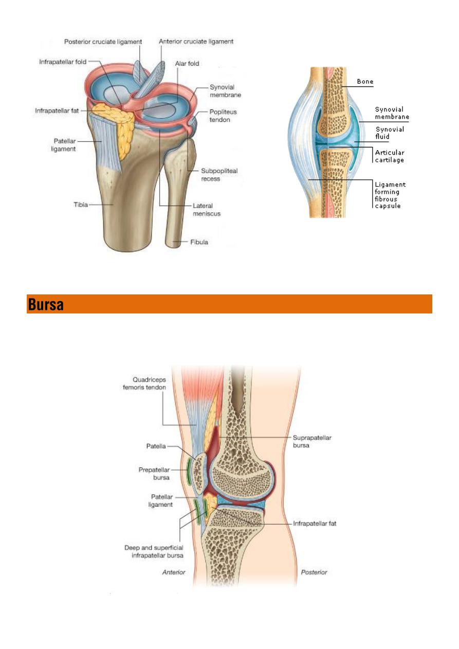
The knee joint is enclosed by a
fibrous capsule
it is
thin
anteriorly and posteriorly but is reinforced on sides by a
strong collateral ligaments.
synovial and a fibrous membrane
separated
by
fatty deposits.
Anteriorly:
the synovial membrane is attached
on the margin of the
cartilage
both on the femur and the tibia.
Behind:
the synovial membrane is attached to
the margins of the two
femoral condyles.
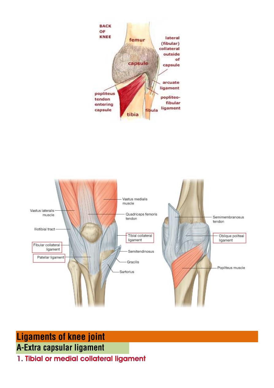
The fibrous capsule is supplemented and strengthened by
extracapsular ligaments
:
patellar ligament
,
fibular collateral ligament
,
tibial collateral ligament
,
oblique popliteal ligament
,
and arcuate
popliteal ligament.
is a broad flat, membranous band
situated medially
near to the back
than to front of the joint
Its attachments :
◊
above
to medial condyles of femur below adductor tubercle
◊
below
to the medial condyles and surface of tibial shaft.
Forced abduction of tibia on femur, result in partial tearing of this
ligament .
is cord–like fibular collateral ligament
descends from
lateral epicondyles of femur
to
styloid process and
head of fibula
separated from lateral meniscus by
popliteus muscle tendon
.
Forced adduction of tibia on femur results in partial tearing of this
ligament .
is a tendenous expansion of
semimembrenosis
muscles.
It strengthens the
posterior
aspect of the capsule.
This ligament is a broad, flat, fibrous band.
The oblique popliteal ligament forms part of the floor of the popliteal
fossa, and the popliteal artery rests upon it.
The patellar ligament connects the
patella
to the
tuberosity of the
tibia
.
It is also occasionally
called the patellar tendon
because there is no
definite separation between the quadriceps tendon (which surrounds
the patella) and the area connecting the patella to the tibia.

This very strong ligament gives the patella its mechanical leverage
and also functions as a cap for the condyles of the femur.
A triangular band in the posterior part of the knee
passes medially downward from the
lateral condyle of the femur
to
the
area between the condyles of the tibia and to the head of the
fibula
it comprised the
meniscofemoral ligament
and
anterior and posterior
cruciate ligaments
covered by synovial membrane , both are crossed each other and
serve to protect the ends of bone from rubbing on each other:
its distal attachment to
anterior intercondylar area
, passed upwards
backwards and laterally, and attached to
posterior part of medial
surface of the lateral femoral condoyle.
It's slack when the knee is flexed and taut when it is fully extended
◊
It's prevents posterior displacement of the femur on the tibia
hyperextension of the knee joint.
◊
Also prevents tibia from pulled anteriorly When the joint is flexed at
a right angle
its distal attachment to
posterior intercondylar area
, passed
upwards, forwards and medially, to attached to the
anterior part of
the lateral surface of medial femoral condoyle.
It tightens during flexion of the knee joint
◊
It's prevents anterior displacement of the femur on the tibia or
posterior displacement of the tibia.
◊
It also helps to prevent hyperflexion of the knee joint.
◊
In the weight bearing flexed knee, it is the main stabilising factor for
the femur, e.g., when walking downhill or downstairs.
Both ligaments may be cracked or torn when knee is forcefully
rotated
attaches the posterior border of lateral meniscus close to femoral
attachment of posterior cruciate ligaments.
◊ It stabilizes the meniscus during rotation of femur on tibia.
◊ It is also covered with synovial membrane.
Connects the Anterior margin of the lateral meniscus to the anterior
end of the medial meniscus .
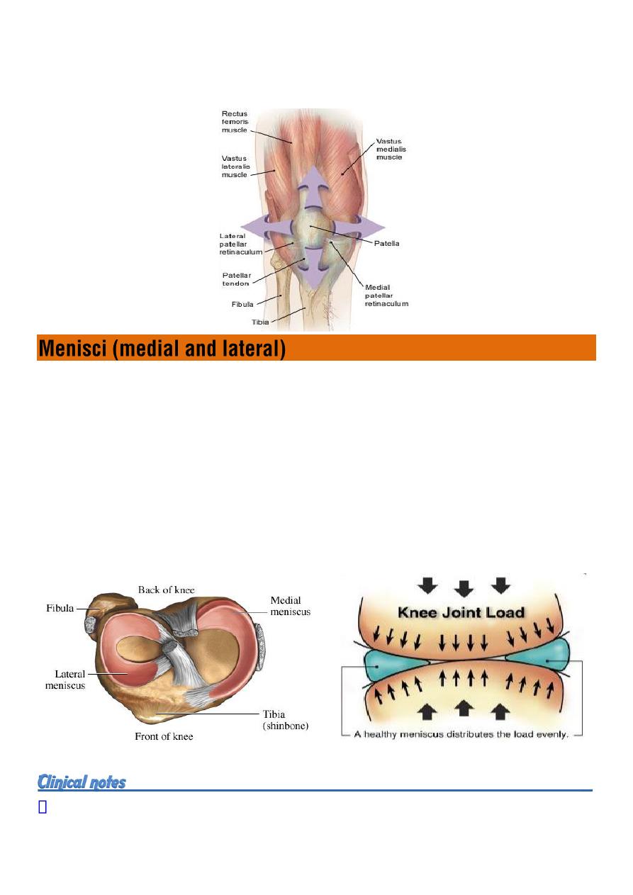
are two c-shape semi lunar pads of fibrocartilage (???)
interposed between the femoral and tibial condyles.
Menisci
deepen the articular surfaces of tibial condyle
by forming a
concave surface to receive femoral condyle , also it act
as a cushions
between the tibia and femur
.
This arrangement
distributes the weight over joint surface
, and also
helps in stabilization of the joint
by block abnormal movement between
tibia and fibula.
Injuries menisci are common.
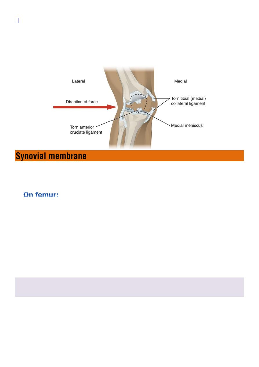
Medial menisci
is damaged more frequent than lateral
because of its
strong attachment to medial collateral ligament of the knee which
restricts its mobility
. (
Usually the foot stays fixed on the ground and
the rest of body rotates
)
The interior of knee joint capsule is lined by a synovial membrane,
which secrets
synovial fluid
to lubricate the joint
.
it is attached to the
margins of intercondylar notch
and
covers the front and sides of :
cruciate ligaments
infrapatellar pad of fat
tendon of popliteus
The synovial membrane
does not
cover the menisci.
Joint cavity:
space inside synovial membrane, both femorotibial and
patellofemoral joints are incorporated within same joint cavity.
numerous synovial sac , act as
cushions
between
tendon
and bone,
ligament
and bone, or
skin
and bone.
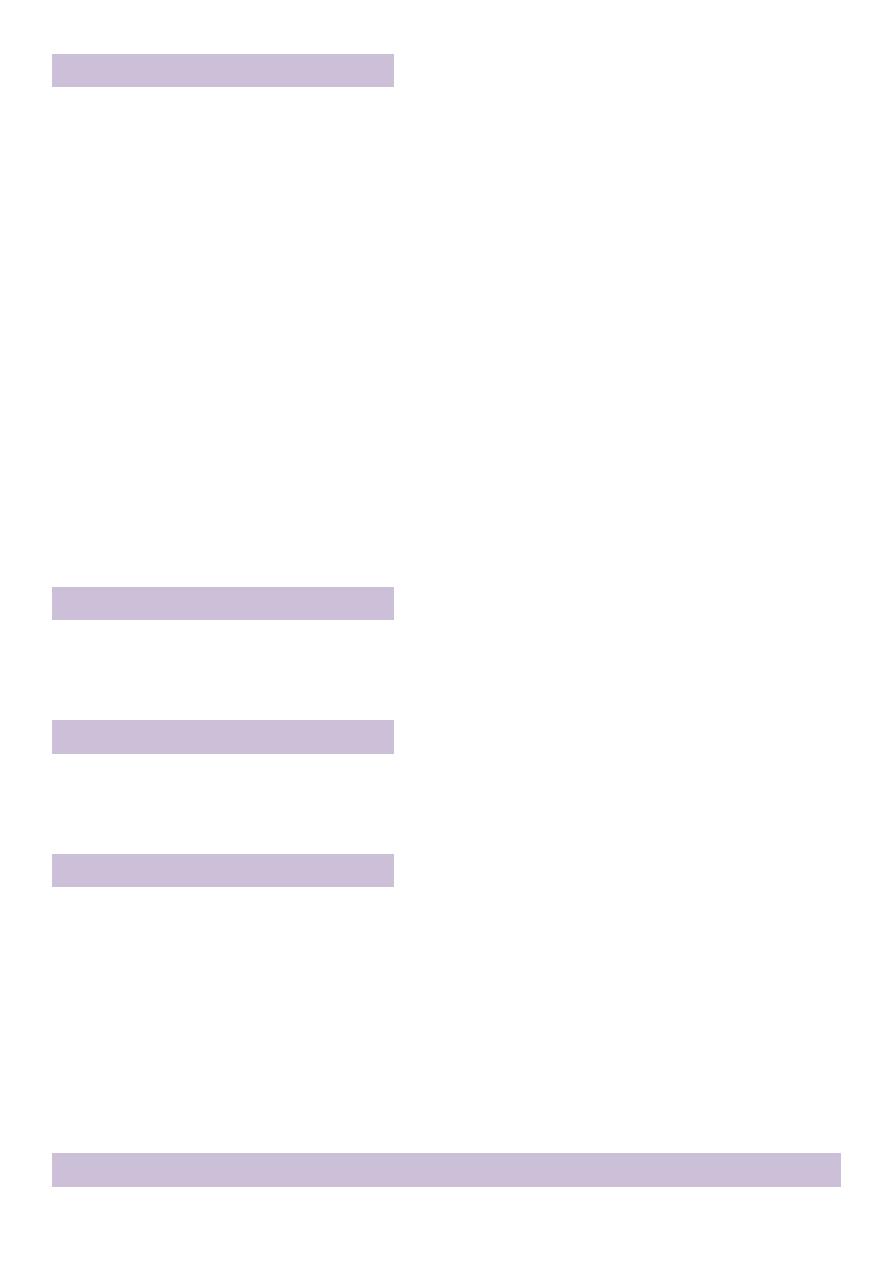
Anterior bursae
are present in front of knee
1. Suprapatellar bursa
beneath the quadriceps.
2. Prepatellar bursa
lies in the subcutaneous tissue between
skin
&
the front of the lower
half of the patella & the upper part of ligamentum patellae.
3. Superficial infrapatellar bursa
lies in the subcutaneous tissue between
the skin
and the
front of the lower part of the ligamentum patellae.
4. Deep infrapatellar bursa
lies between the
ligamentum patellae
and
the tibia
Lateral bursa
1. Between LCL & biceps femoris tendon
2. Between the LCL & the capsule ,
where it overlies popliteus tendon
Medial bursa
1. between MCL & the
tendons of sartorius, gracilis & semitendinosus
2. Between the MCL &
the tibia & joint capsule
Posterior bursae
are 6 bursae in the back of knee.
1-Popliteal bursa : Under popliteus tendon
2-Semimembranosus bursa
3-Between joint capsule & medial head of gastrocnemius
4-Between joint capsule & lateral head of gastrocnemius
Semitendnosis bursa (???????)
Inflammations of bursa is bursitis, may be due to
trauma or disease
The principle movements of the knee joint are flexion & extension
1. Flexion
Produced by
biceps femorus , semimembranosus & semitendinosus
Assisted by
Gracillus , Sartorius & popliteus
2. Extension
Produced by
quadriceps femoris & gluteus maximums
3. Medial rotation
Produced by
Sartorius , Gracillus & Semitendinosus
4. Lateral rotation
Produced by
biceps femorus
Factors responsible for knee joint stability are :
1. Muscle tone :
especially in quadriceps
2. Cruciate ligaments :
stabilize femur on tibia
preventing anteroposterior movement
3. Collateral ligaments :
assist in medial and lateral stability
4. Iliotibial tract :
stabilizes knee joint during extension
5. Oblique posterior ligament :
prevent hyperextension
Sensory fibers from femoral nerve
Genicular branches from tibial nerve and common peroneal nerve
Fiber from posterior division of obturator nerve
The knee joint receives blood supply from extensive
genicular
anastomosis
mainly from popliteal, anterior and posterior tibial artery.
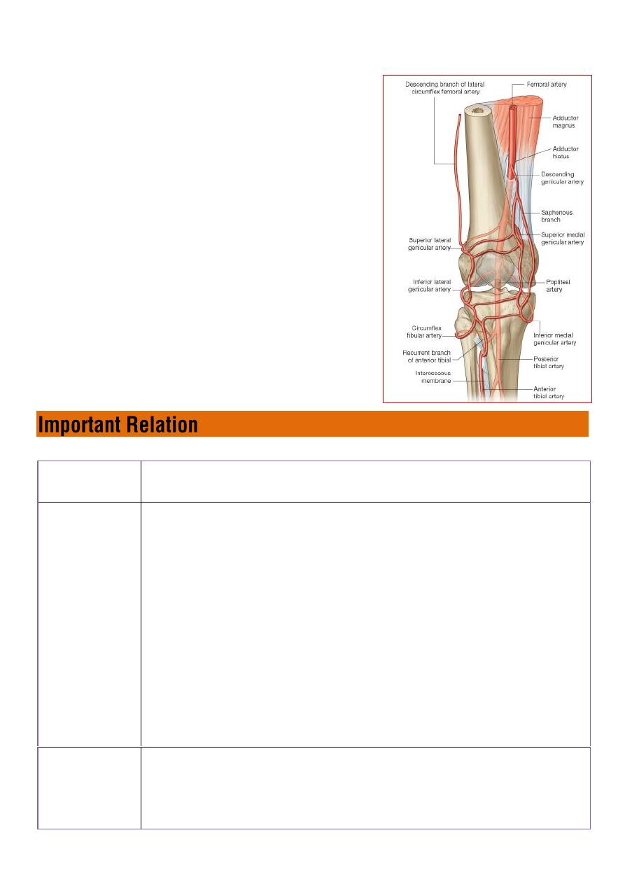
Popliteal artery
in its course through the
fossa, it gives off
medial and lateral
superior genicular arteries
to form
collateral circulation around the knee joint
Also
medial and lateral inferior genicular
arteries
encircle the leg and form
anastomosis around the knee joint.
1.
Superior medial & lateral genicular
arteries
2.
Inferior medial & lateral genicular arteries
3.
Descending genicular artery from
( femoral artery )
4.
Recurrent branch of anterior tibial artery
Anteriorly
The prepatellar bursa
Posteriorly
Popliteal vessels
tibial & common peroneal nerves
lymph nodes
the semimembranosus
semitendinosus
biceps femoris
the heads of gastrocnemius
plantaris
Medially
Sartorius
Gracilis
Semitendinosus

Laterally
Biceps femoris
common peroneal nerve