Giant cell tumor
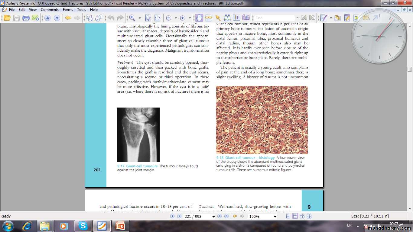
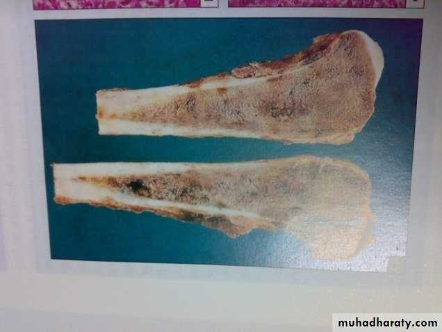
Pathology:This disease characterized by presence of giant multinucleated cells which seen in large number. The tumor cell is the stromal spindle shape cell.
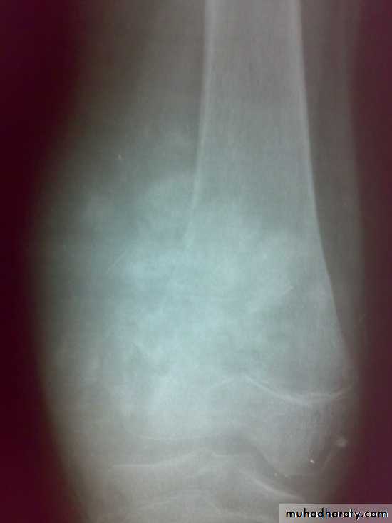
The tumor is soft and friable occupying the cavity which extend to subchondral region.
It occur in mature bone
One third of tumor is benign, one third locally invasive, and one third metastasize.
Clinical features of giant cell tumors
Patients age: usually 20-40 years.More in female.
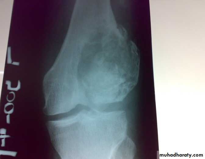
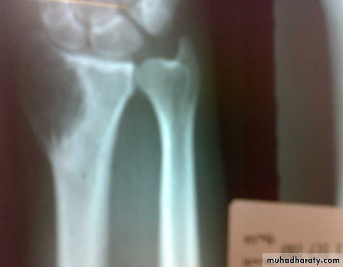
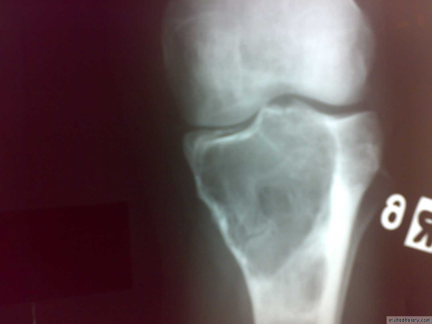
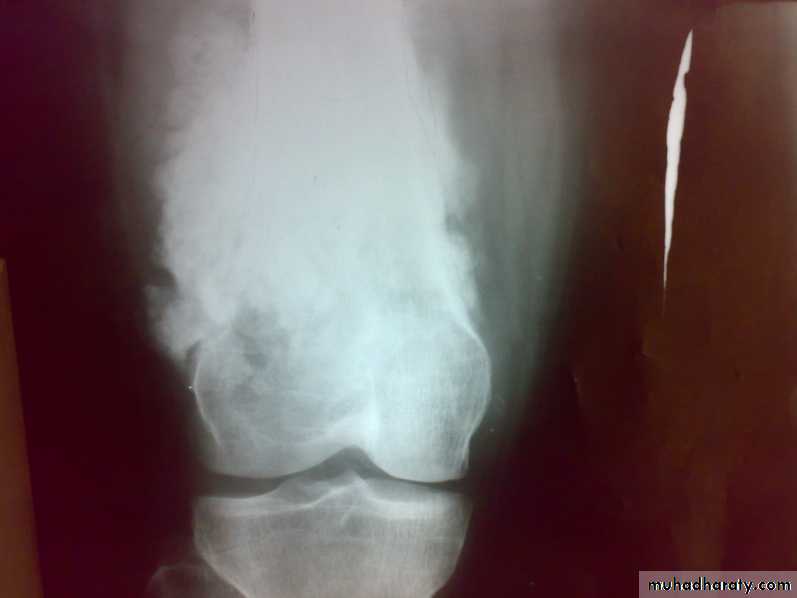
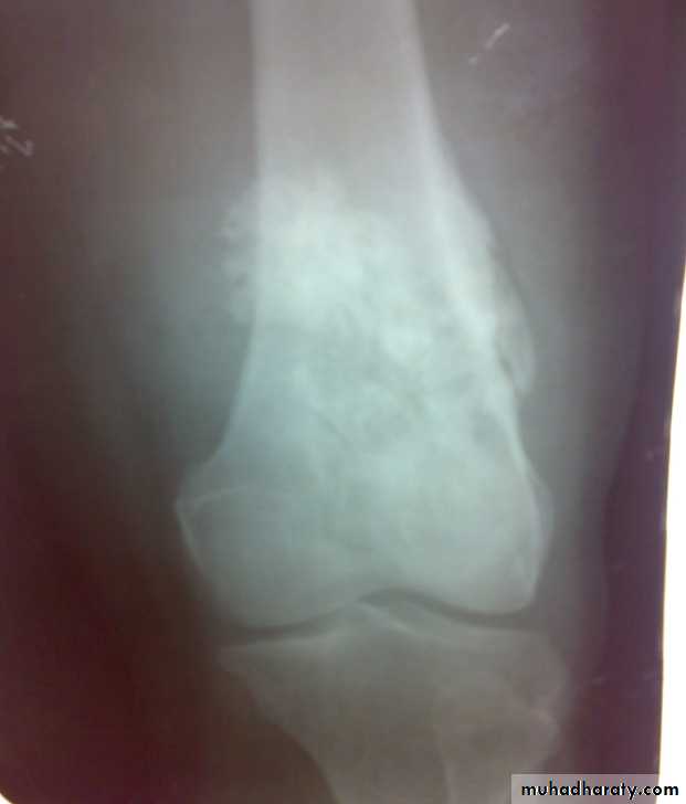
Patient presented by local pain and swelling or pathological fractures. Common site is the distal femur, proximal tibia, distal radius.
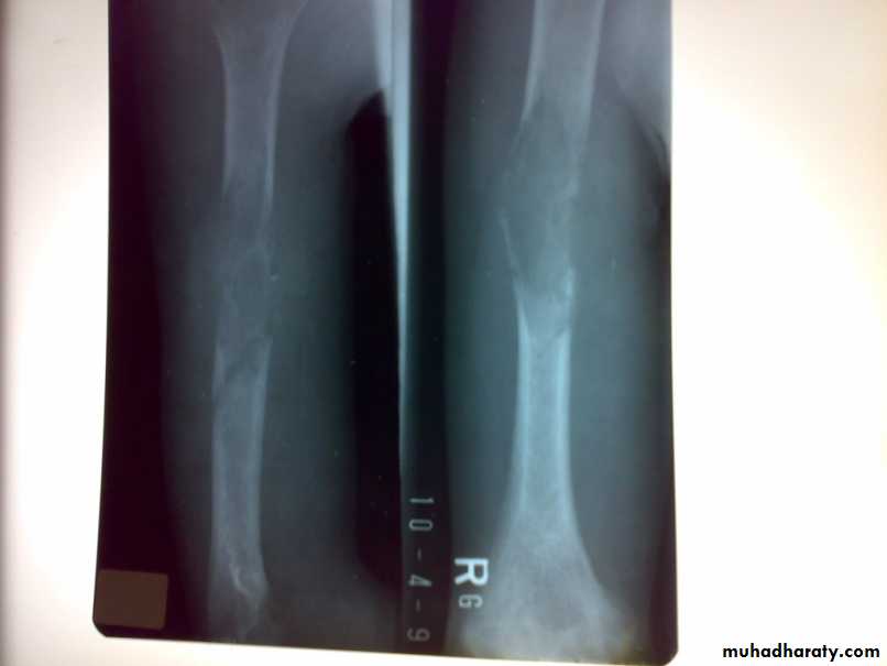
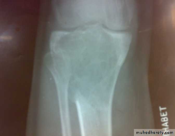
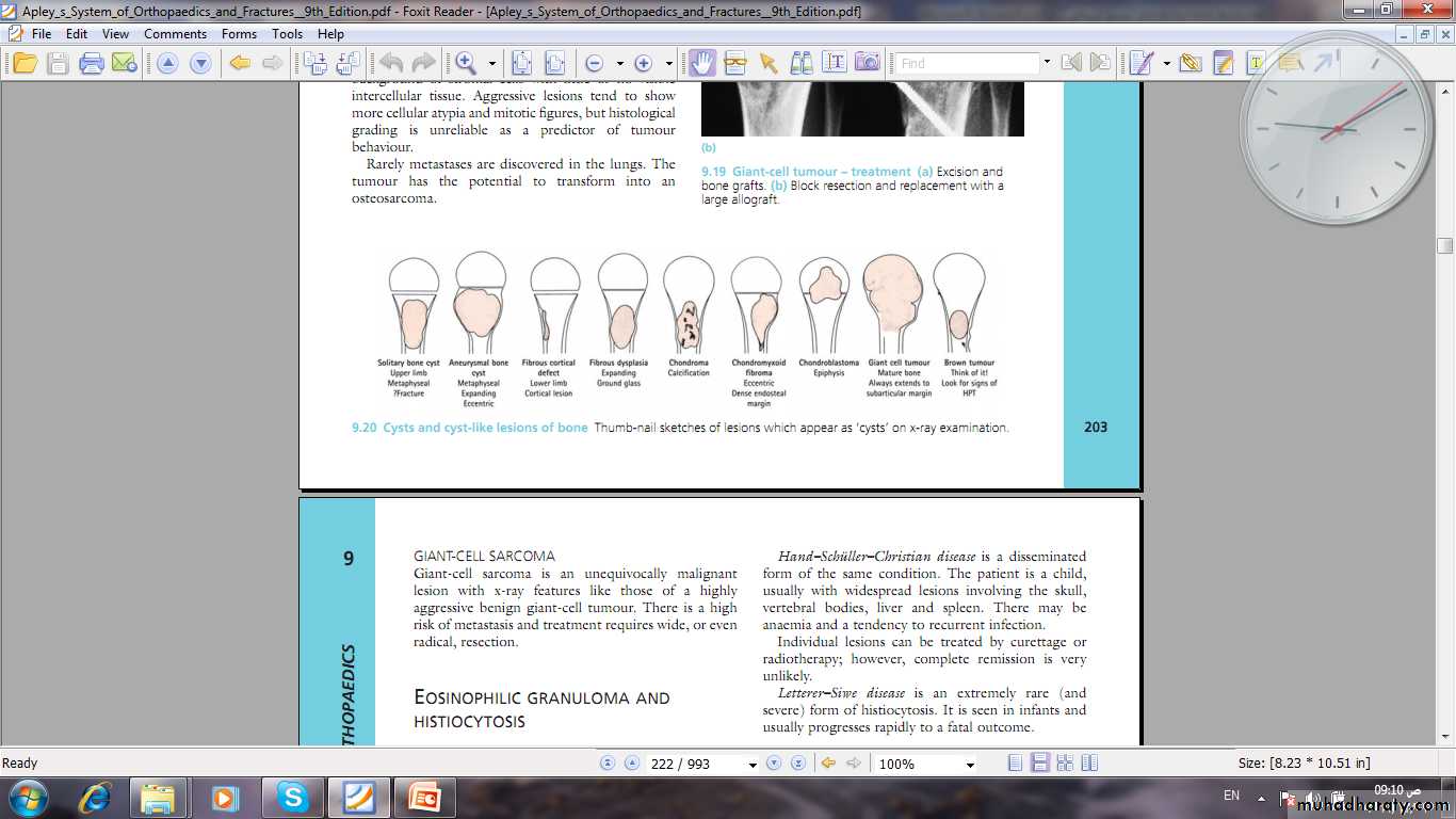
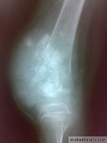
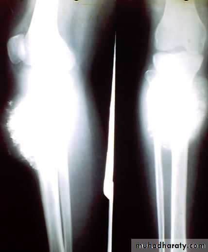
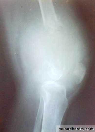
X-ray show eccentric osteolytic lesion in the end of long bone ,subchondral, trabeculated (soap bubble appearance). The cortex is thin, expanded or even perforated.
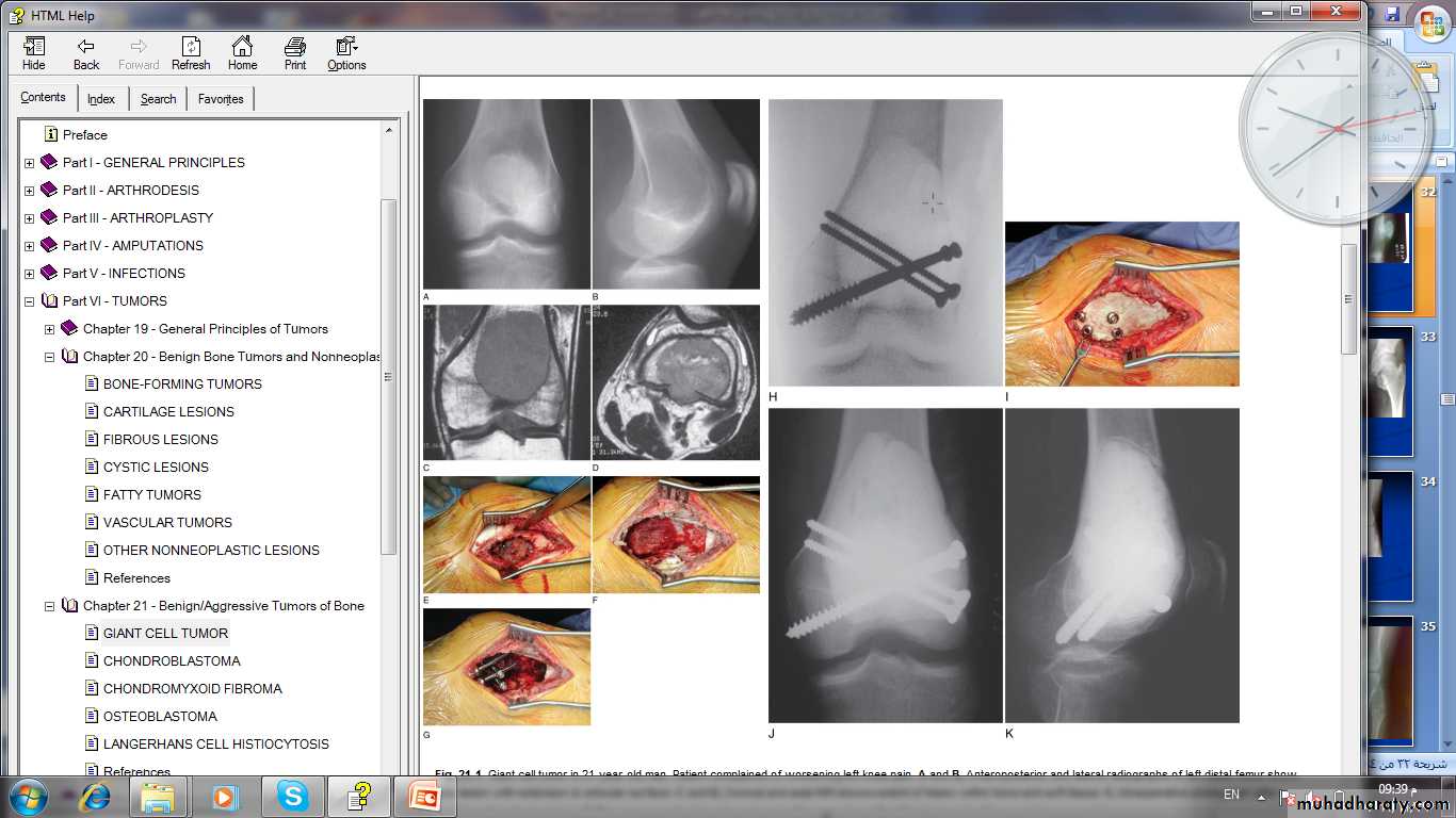
Treatment of giant cell tumors
1- curettage and bone graft.2- curettage and bone cement.
3- excision and bone grafting.
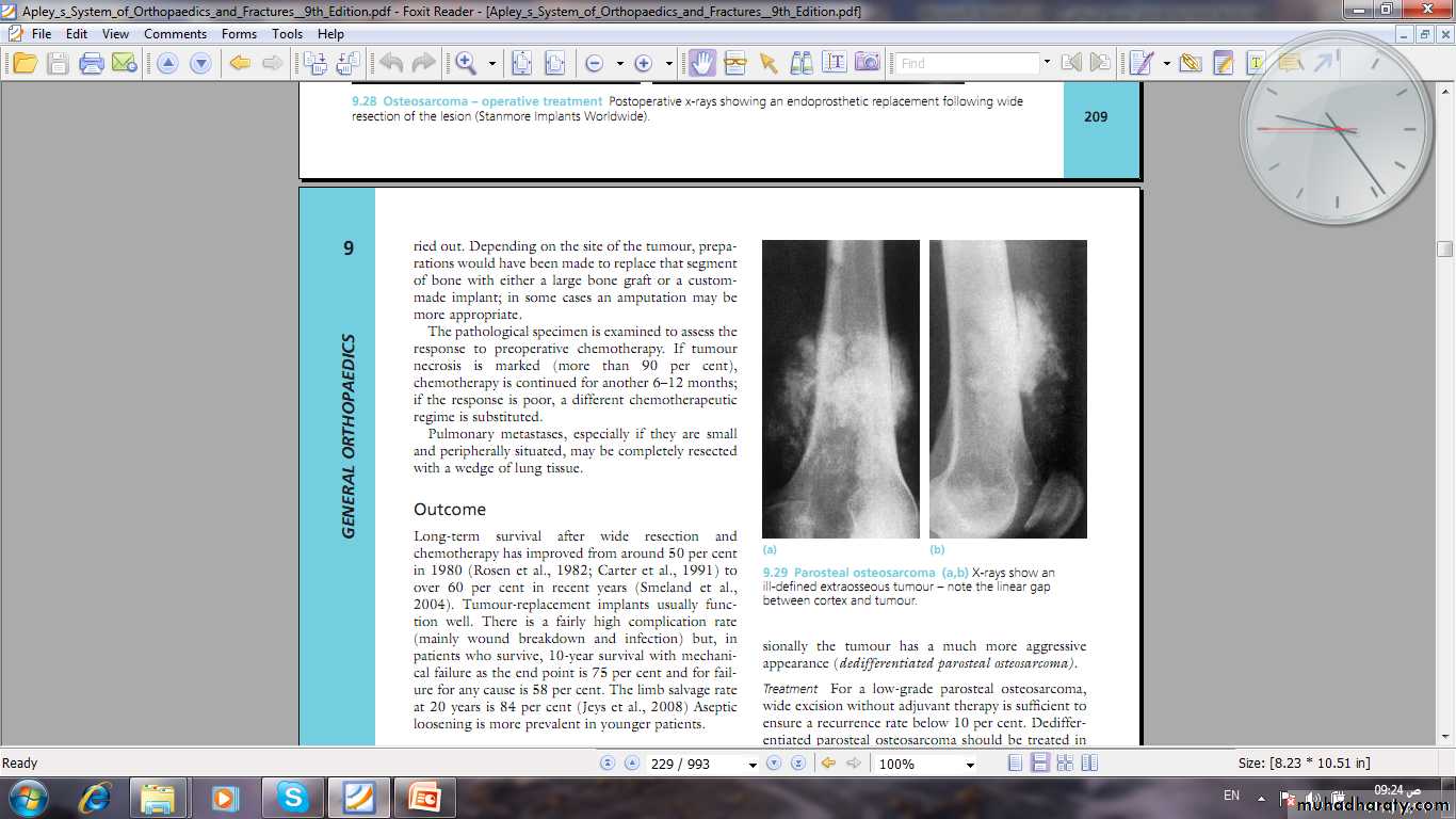
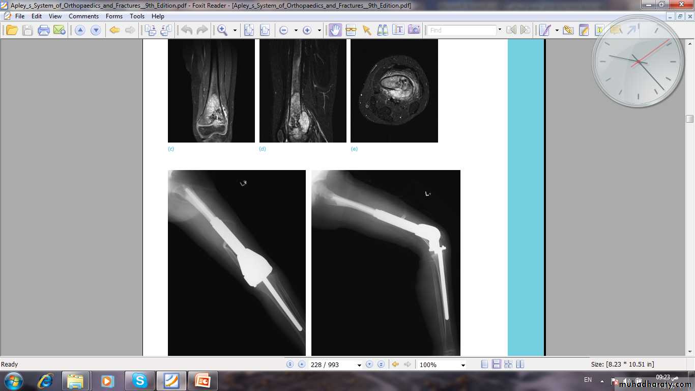
4- excision and prosthetic replacement.
5- radiotherapy for unrespectable lesion.
Osteosarcoma
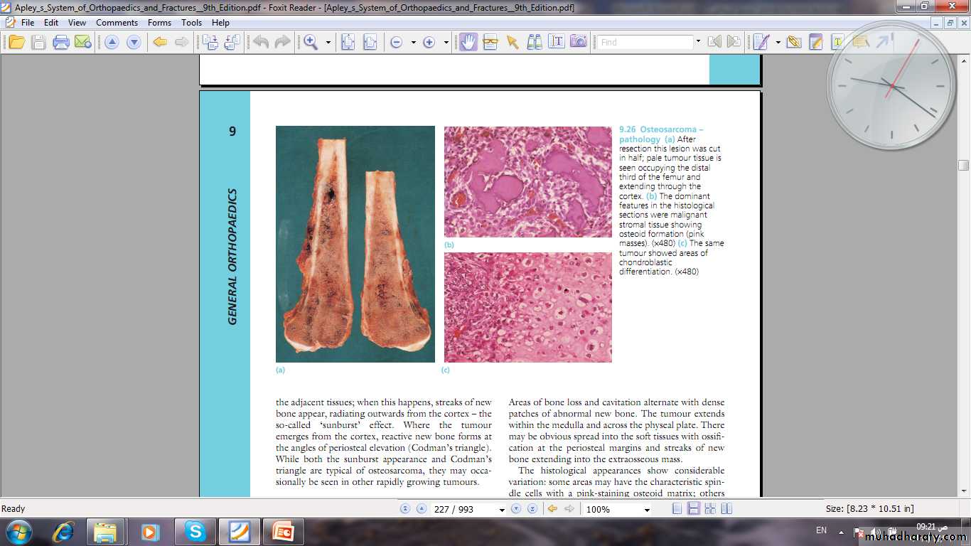
Definition:primary malignant tumor arise from bone forming cells and produce malignant osteoid tissue. In addition to malignant osteoid tissue fibroblast or cartilage tissue may be predominate. It destroys the bone and form malignant osteoid and spread to surrounding tissue and metastasized to far organs.

Clinical features
Age : 10-20 years commonly but it may occur in old following irradiation and Paget’s disease.Site: metaphysis of long bone specially around knee and proximal humerus is common sites.
History: history of trauma may present. Increasing pain, worse at night is the first symptom , swelling may be other symptom.
Examination: the affected site swollen with ill define edge , tender, and overlying skin is hot, shiny ,with dilated veins. The ESR is raised.
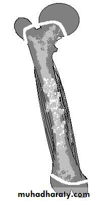
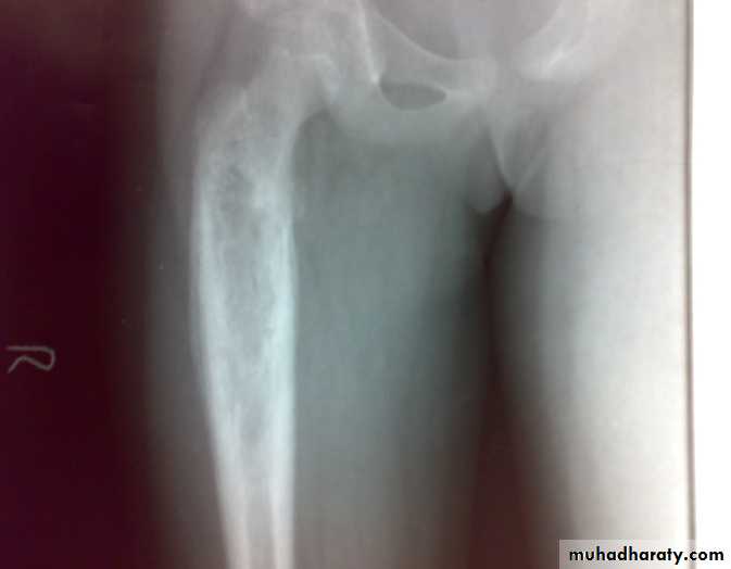
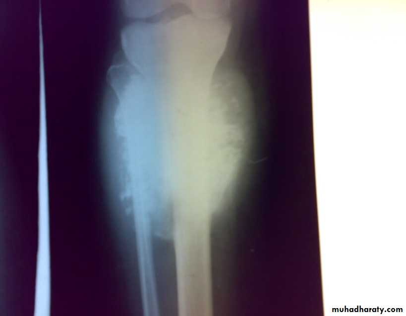
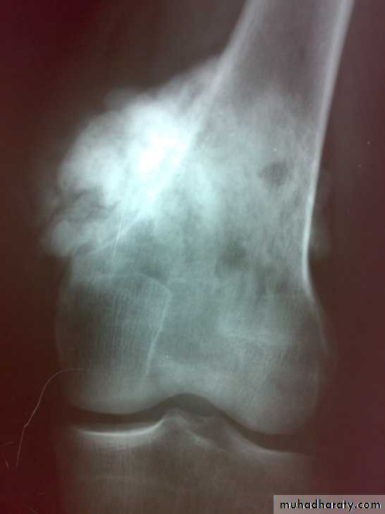
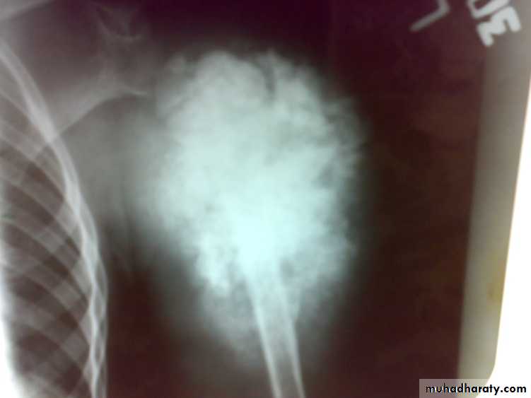
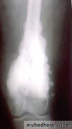
: X-ray are very variable but it show combination of bone destruction and bone formation.
The metaphysis show osteolytic and osteoblastic areas, the cortex is usually perforated and soft tissue shadow may be seen.
There is new bone formation in form of Codman's triangle at periphery of when cortex penetration cause periostium elevation and vertical streaks of calcification in the adjacent soft tissues called sunray appearance.

.
CT,MRI, RADIOISOTOPE SCANNING needed to show the extent of tumor, to stage the tumor and to plan treatment.
Chest x-ray and US to look for metastasizes.
Always biopsy is indicated to establish diagnoses.
Treatment:
in developed countries the 5 years survival more than 50% by the use of chemotherapy ,early diagnosis, proper staging and proper surgery. prognosis in our patient is bad.The most certain way of eradicating osteosarcoma is by radical amputation.
Chemotherapy used to control metastases and to limit resection to degree that limited resection and prosthetic replacement can be used (limb sparing surgery). Chemotherapy is started soon after operation and continued for at least one year.
Inaccessible tumors are treated by radiotherapy combined with chemotherapy.
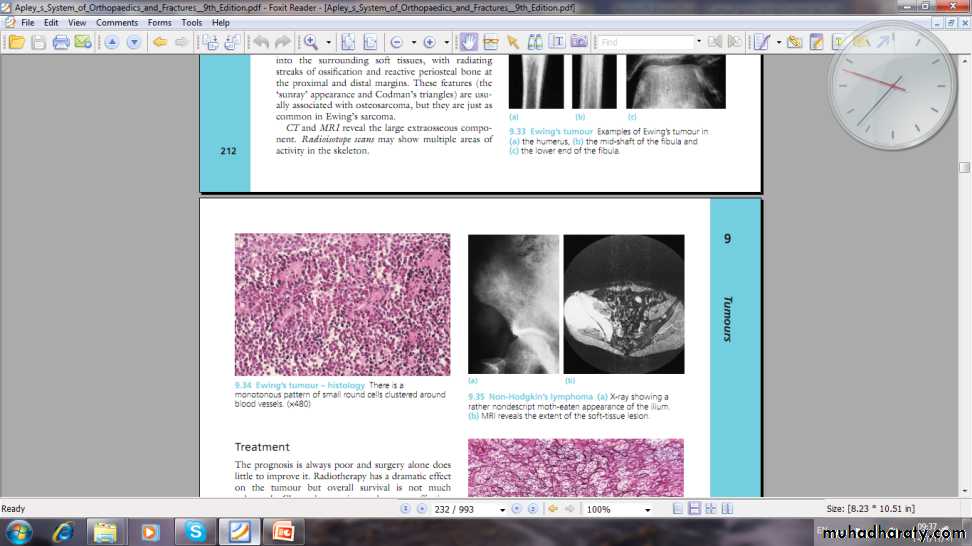
Ewing’s sarcoma
Definition: it is rare primary malignant tumor of vascular endothelial tissue of bone marrow (neuroectodermal origin).it give rise florid periosteal reaction and early metastases
Age: commonly 10-20 years.
Site: usually diaphysis of long bone.
Clinical features: pain and swelling are the chief presenting symptoms. Fever and features of osteomyelitis may seen. The swelling is ill-define, warm and tender.
ESR is elevated.