Objectives of the lecture:
1.GIT bleeding,its causes,DDx2.GERD
3.Pyloric stenosis
• Gastrointestinal Hemorrhage
• GIH• Bleeding can occur anywhere along the GIT, identification of the site may be challenging.
• Hematemesis(bloody vomitus): usually bleeding proximal to ligament of Treitz.
• Blood becomes dark & coffee-ground in appearance. Bleeding that has little or no contact with gastric acid will be bright red.
• GIH that occurs proximal to the ileocecal valve & is passed rectally will usually appear as melena(black, tarry, sticky stools) that result from the denaturing of Hb by intestinal bacteria & enzymes.
Hematochezia(red bloody stools passed rectally) usually results from distal GIH. Bd is usually mixed with stool or passed just before or just after defecation.
Occasionally, rapid bleeding from an upper GI source combined with cathartic action of bd can speed TT & cause hch.
Specific types of hch include maroon-colored stools, seen with significant bleeding usually from distal small bowel, & currant-jelly stools indicative of intestinal vascular congestion & hyperemia.
• DDx:
• red color stool is often assumed to be bd. However, many substances cause change in its color:• Foods that contain a high concentration of red pigments such as tomatoes, cranberries, beets, and red fruit juices and gelatin can cause red stools.
• Red-colored medications e.g acetaminophen & amoxicillin can be passed in stools, especially if diarrhea is present. Spinach, licorice, iron, and bismuth often lead to dark, black stools, which can be confused with true melena.
• In infants, Serratia marcescens can cause red diaper syndrome as a result of the formation of red pigment in soiled diapers stored for longer than 1 day.
• Causes of GI Bleeding:
• Newborn:
• Common causes of GI bleeding in the first 24 hours of life include:
• maternal blood swallowed during delivery and local trauma after nasogastric suctioning.
• Hemorrhagic disease of the newborn as a result of inherited deficits of coagulation factors or delay in administration of postnatal vitamin K occasionally produces GI bleeding
• Premature infants and newborns who have birth asphyxia are at increased risk for having gastric ulcerations and erosions that can bleed & Necrotising enterocolitis "NEC".
Infant
Child & AdolescentBacterial enteritis
Milk protein allergyIntussusception
Anal fissure
Bacterial enteritis
Anal fissure
Colonic polyps
Peptic ulcer/gastritis
Swallowed epistaxis
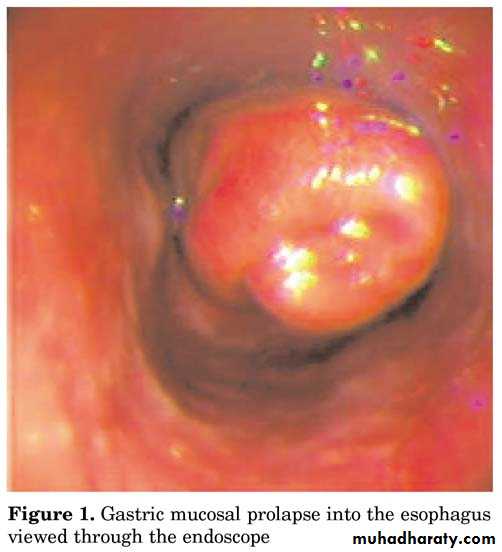
Mallory-Weiss syndrome& Prolapse gastropathy producing subepith hm
Esophagitis/Esophageal varices
Meckel diverticulum
Henoch-Schonlein purpura
Foreign body
Hemangioma, AVM
IBD
Coagulopathy
• Diagnosis:
• Upper intestinal bleeding is evaluated with OGD• Evaluation of the small intestine by capsule endoscopy. The capsule-sized imaging device is swallowed in older children or placed endoscopically in younger children.
• Lower GI bleeding is investigated with a colonoscopy
Occult blood in stool:
is usually detected by using commercially available fecal occult bd testing cards( based on a chemical reaction bw chemical guaiac & oxidizing action of a substrate (Hb), giving a blue color.It is very sensitive, but random testing can miss chronic bd loss, which can lead to IDA.
Foods that have peroxidase activity may cause false-positive results if eaten within 3 ds of testing: red meat, liver, processed meats, and raw fruits and vegetables, especially melon, turnip, radishes,& horseradish.
High vitamin C intake can cause false-negative results.
Similarly, outdated guaiac cards & prolonged storage may affect the accuracy of the test. Stool guaiac cards are not accurate for testing emesis for the presence of bd because gastric acid can affect the reaction.
• Gastroesophageal Reflux Disease
• (GERD)• GER signifies the retrograde movement of gastric contents across the LES into the esophagus & is usually phsiological in young infant.
• GERD symptoms or complications resulting from exposure of esophagus, oropharynx, or airway to gastric refluxate (acid, food, bile);
• i.e. pathological GER & it is the most common esophageal disorder in children of all ages.
Measurement of lower esophageal pH shows that in normal individuals there is acidity from reflux of stomach contents for less than 4% of a 24-hour period. Reflux occurring more frequently than this results from functional immaturity of the LES leading to episodes of inappropriate relaxation, so Transient LES relaxation (TLESR) is the primary mechanism allowing reflux to occur. A short intra-abdominal length of esophagus probably also contributes.
It is common in 1st yr of life. By 12 ms of age, nearly all symptomatic reflux will have resolved spontaneously, presumably due to a combination of:
maturation of LES, assumption of an upright posture & more solids in the diet.
A sliding hiatus hernia is present in some symptomatic infants, but many children with H.H are symptom-free.
• Clinical Manifestations :
• Infantile reflux manifests more often with regurgitation (especially postprandially), signs of esophagitis (irritability, arching, choking, gagging, feeding aversion) FTT• Older children can have regurgitation during the preschool years; complaints of abdominal & chest pain supervene in later childhood and adolescence.
• Occasional children present with neck contortions (arching, turning of head), designated Sandifer syndrome.
• The respiratory presentations:
• infants : obstructive apnea, stridor or lower airway disease in which reflux complicates primary airway disease such as laryngomalacia or BPD. OM, sinusitis, lymphoid hyperplasia, hoarseness, vocal cord nodules & laryngeal edema have all been associated with GERD.• older children commonly related to asthma or to otolaryngologic disease such as laryngitis or sinusitis.
• Infant reflux becomes evident in the 1st few ms of life, peaks at ∼4 mo, resolves in up to 88% by 12 mo & nearly all by 24 mo. Symptoms in older children tend to be chronic, waxing & waning, but completely resolving in no ˃ half, which resembles adult patterns.
• Diagnosis:
• For most of the typical GERD , thorough Hx & PE suffice initially to reach Dx .
• Contrast (usually barium) radiographic study of eso. & upper GIT is performed in pt with vomiting & dysphagia to evaluate for achalasia, esophageal strictures & stenosis, H.H, & gastric outlet or intestinal obstruction.
• It has poor sensitivity & specificity due to its limited duration & the inability to differentiate physiologic GER from GERD.
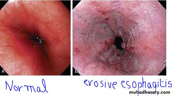
• Endoscopy allows Dx of erosive esophagitis & complications such as strictures or Barrett esophagus; esophageal bx can diagnose histological reflux esophagitis in the absence of erosions while simultaneously eliminating allergic & infectious causes.
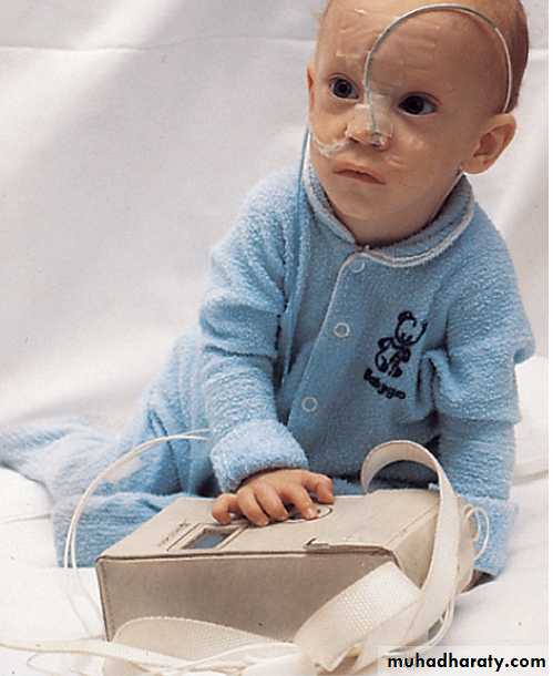
• Extended esophageal pH monitoring of the distal esophagus:
• no longer considered the sine qua non(not necessary ) of a GERD Dx, provides a quantitative & sensitive documentation of acidic reflux episodes.• The distal eso pH probe is placed at a level corresponding to 87% of the nares-LES distance, Normal values of distal eso acid exposure (pH <4) are generally established as <5-8% of the total monitored time, but these quantitative normals are insufficient to establish or disprove Dx of pathologic GERD.
• The most important indications for esophageal pH monitoring are for :
• assessing efficacy of acid suppression during Rx, evaluating apneic episodes in conjunction with a pneumogram & perhaps impedance,& evaluating atypical GERD presentations such as chronic cough, stridor & asthma.
PH study in GERD
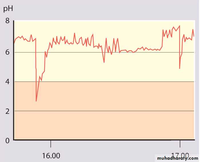
Part of a normal oesophageal pH study.
The lower oesophageal pH is above 4 for most of the time.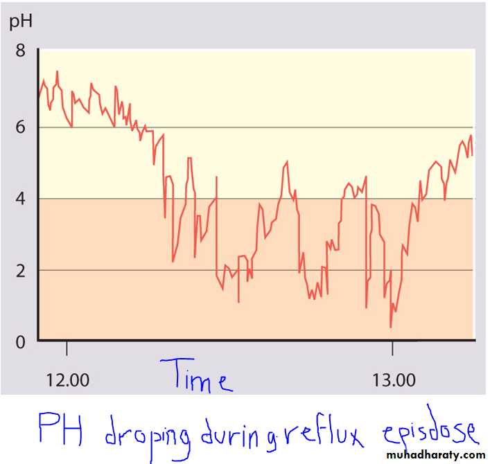
• Empirical antireflux therapy, using a time-limited trial of high-dose proton pump inhibitor (PPI), is a cost-effective strategy for Dx in adults; although not formally evaluated in older children, it has also been applied.
• Management:
• Dietary measures for infants include normalization of any abnormal feeding techniques, volumes & frequencies.
• Thickening of feeds or use of commercially prethickened formulas ↑ % of infants with no regurgitation, ↓ the frequency of daily regurgitation & emesis, & ↑ infant's wt .
• The addition of a Tbsp of rice cereal/ oz of formula results in a greater caloric density (30 kcal/oz), & reduced crying time, although it might not modify the number of nonregurgitant reflux episodes.
A short trial of a hypoallergenic diet may be used to exclude milk or soy protein allergy before pharmacotherapy.
Older children should be counseled to avoid acidic or reflux-inducing foods (tomatoes, chocolate, spicy foods , mint) and beverages (juices, carbonated & caffeinated drinks, alcohol), avoiding lying down after meals
Weight reduction for obese & elimination of smoke exposure are other crucial measures at all ages.
• Positioning measures important for infants, who cannot control their positions independently.
• Seated position worsens infant reflux should be avoided.
• Eso pH monitoring demonstrates more reflux episodes in infants in supine & side positions compared with prone position, but as supine position reduces the risk of SIDS has led AAP to recommend supine positioning during sleep. When the infant is awake & observed, prone position & upright carried position can be used to minimize reflux.
• The efficacy of positioning for older children is unclear, but some evidence suggests a benefit to left side position & head elevation during sleep.
• The head should be elevated by elevating the head of the bed, rather than using excess pillows, to avoid abdominal flexion and compression that might worsen reflux.
• Pharmacotherapy to ameliorate gastric acidity or promote the aboral movement of its content.
• Antacids: most commonly used antireflux & available .
• They provide rapid but transient relief of symptoms by acid neutralization. The long-term regular use of antacids cannot be recommended because of SE ; diarrhea (Mg antacids) & constipation (Al antacids) .
• Histamine-2 receptor antagonists (H2RAs: cimetidine, famotidine, nizatidine, & ranitidine)
• widely used, act by selective inhibition of histamine receptors on gastric parietal cells. There is a definite benefit in Rx of mild-to-moderate reflux esophagitis.
• They have been recommended as first-line therapy because of their excellent overall safety profile, but they are being superseded by PPIs in this role.
• PPIs (omeprazole, lansoprazole, pantoprazole, rabeprazole, and esomeprazole)
• provide the most potent antireflux effect by blocking the H+_K+ ATPase channels of the final common pathway in gastric acid secretion.• PPIs are superior to H2RAs in Rx of severe & erosive esophagitis. Pharmacodynamic studies have indicated that children require higher doses of PPIs than adults on a per-wt basis. rabe-&panto- are not FDA-approved in children.
• Prokinetic agents:include metoclopramide (dopamine-2 and 5-HT3 antagonist), bethanechol (cholinergic agonist), and erythromycin (motilin receptor agonist). Most of these increase LES pressure; some improve gastric emptying or esophageal clearance. None affects the frequency of TLESRs.
• Baclofen is a centrally acting γ-aminobutyric acid (GABA) agonist that has been shown to decrease reflux by decreasing TLESRs in healthy adults and in a small number of neurologically impaired children with GERD.
• Surgery, usually fundoplication( fundus of the stomach is wrapped around the intra-abdominal oesophagus) is effective therapy for intractable GERD in children, particularly those with refractory esophagitis or strictures & those at risk for significant morbidity from chronic pulmonary disease. It may be combined with a gastrostomy for feeding or venting.
• Complications of GERD:
• Reflux is often problematic:
• in children with cerebral palsy or other neuro developmental disorders
• in preterm infants who develop bronchopulmonary dysplasia (chronic lung disease of prematurity)
• following surgery for esophageal atresia or diaphragmatic hernia.
• 1.Esophageal:
• Esophagitis can manifest as irritability, arching, and feeding aversion in infants; chest or epigastric pain in older children; and, rarely, as hematemesis, anemia(iron deficiency), or Sandifer syndrome at any age.
• Prolonged & severe esophagitis strictures producing dysphagia, & requiring repeated esophageal dilations & often fundoplication.
• Long-standing esophagitis predisposes to metaplastic transformation of the normal esophageal squamous epithelium into intestinal columnar epithelium, termed Barrett esophagus, a precursor of esophageal adenocarcinoma.
• 2.Nutritional:
• Esophagitis & regurgitation may be severe enough to induce FTT bec of caloric deficits.• 3. Extraesophageal:
• Respiratory (“Atypical”): GERD can produce respiratory symptoms chronic cough, stridor, and hoarseness, aspiration pneumonia & chronic pulmonary fibrosis & may occur without esophagitis" by direct contact of the refluxed gastric contents with the respiratory tract (aspiration, laryngeal penetration, or microaspiration) or by reflexive interactions between the esophagus & respiratory tract (inducing laryngeal closure or bronchospasm).
• Often, GERD & primary respiratory disorder, such as asthma, interact & a vicious cycle between them worsens both diseases.
• Apnea & stridor had been linked in some studies. Apparent life-threatening events (ALTEs) & SIDS are controversial .
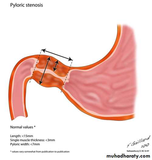
• Hypertrophic Pyloric Stenosis
• It is hypertrophy of the pylorus causing gastric outlet obstruction. It is more common in boys (4 : 1), particularly first-borns, there may be a family Hx, especially on the maternal side.• The incidence is ↑ in infants with B & O bd groups. It is occasionally associated with other congenital defects, including TEF.
• Etiology
• unknown, but many factors have been implicated. PS is usually not present at birth . An association has been found with the use of erythromycin in neonates with highest risk if the medication is given within the 1st 2 wk of life.
• Investigators have proposed that deficiency of nitric oxide, a ubiquitous mediator of smooth-muscle relaxation, may be associated with the development of PS because NO synthase is selectively depleted in pyloric m. of pt with PS.
• Clinical Manifestations:
• Nonbilious vomiting is the initial symptom of PS. The vomiting may or may not be projectile initially but is usually progressive, occurring immediately after a feeding.• Emesis might follow each feeding, or it may be intermittent. The vomiting usually starts after 3 wk of age ( irrespective of GA), but symptoms can develop as early as the 1st wk of life and as late as the 5th mo.
• About 20% have intermittent emesis from birth that then progresses to the classic picture.
• After vomiting, the infant is hungry and wants to feed again. Greater awareness of PS has led to earlier identification of pts with fewer instances of chronic malnutrition & severe dehydration & at times a subclinical self-resolving hypertrophy.
• Persistent vomiting caused by gastric outlet obstruction results in the continuous loss of gastric HCl. Dehydration causes ↑ in aldosterone production, leading to ↑ renal excretion of K+ hypochloremic, hypokalemic metabolic alkalosis.
• The depletion of Cl- in bd leads to an exchange of H+& K+ for Na in the distal tubule, resulting in a paradoxical aciduria. However, a spectrum of electrolyte abnormalities may be seen.
• Hypoglycemia may be present and may cause seizures.
Pathophysiology:
• Hyperbilirubinemia is the most common clinical association of PS, also known as icteropyloric syndrome. Unconjugated hyperbilirubinemia is more common than conjugated & usually resolves with surgical correction.
• It may be associated with ↓ level of glucuronyl transferase as seen in ∼5% of affected infants.
• Other coexistent clinical diagnoses have been described, including:
• eosinophilic gastroenteritis, hiatal hernia, peptic ulcer, congenital nephrotic syndrome, CHD, & congenital hypothyroidism.
• Diagnosis:
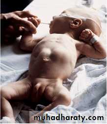
• traditionally established by palpating the pyloric mass( firm, movable, ∼2 cm in length, olive shaped, hard, best palpated from the Lt side, and located above & to the Rt of the umbilicus in the mid-epigastrium beneath the liver's edge. The mass is easiest palpated after an episode of vomiting.• After feeding, there may be a visible gastric peristaltic wave that progresses across the abdomen.
• U/S : confirms Dx in the majority of cases. Criteria include pyloric thickness 3-4 mm, an overall pyloric length 15-19 mm, and pyloric diameter of 10-14 mm. It has a sensitivity of ∼95%.
• Contrast studies demonstrate an elongated pyloric channel (string sign), a bulge of the pyloric muscle into the antrum (shoulder sign), and parallel streaks of barium seen in the narrowed channel, producing a “double tract sign”
Visible gastric peristalsis in an infant with pyloric stenosis
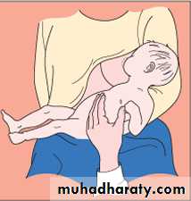
Diagram showing a test feed being performed to diagnose pyloric stenosis
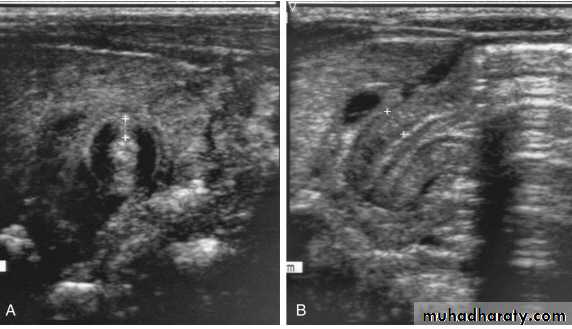
A, Transverse sonogram demonstrating a pyloric muscle wall thickness of >4 mm (distance between crosses). B, Horizontal image demonstrating a pyloric channel length >14 mm (wall thickness outlined between crosses) in an infant with pyloric stenosis.
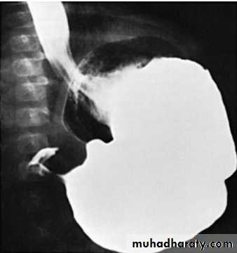
Barium in the stomach of an infant with projectile vomiting. The attenuated pyloric canal is typical of CHPS.
• DDx:
• GERD, adrenogenital syndrome, Inborn errors of metabolism, pyloric membrane or pyloric duplication, Duodenal stenosis proximal to the ampulla of Vater.• Rx:
• Preoperative Management
• The anatomic correction of PS is not a surgical emergency because, although PS is a form of intestinal obstruction, gangrene & intestinal perforation do not occur with this condition. Infants should not undergo surgery until the fluid and electrolyte deficits have been corrected.
• If infants undergo surgery with uncorrected alkalosis, then the profound effect that surgical stress has on the urinary excretion of Na may intensify the electrolyte abnormalities. Given that gastric decompression is necessary for surgery and induction of anesthesia, NG decompression should be performed during fluid and electrolyte resuscitation.
Surgical Management
Once the volume and electrolyte status is corrected, the infant is ready for surgery. Ramstedt pyloromyotomy through a right upper quadrant transverse incision has been the traditional treatment for hypertrophic PS.
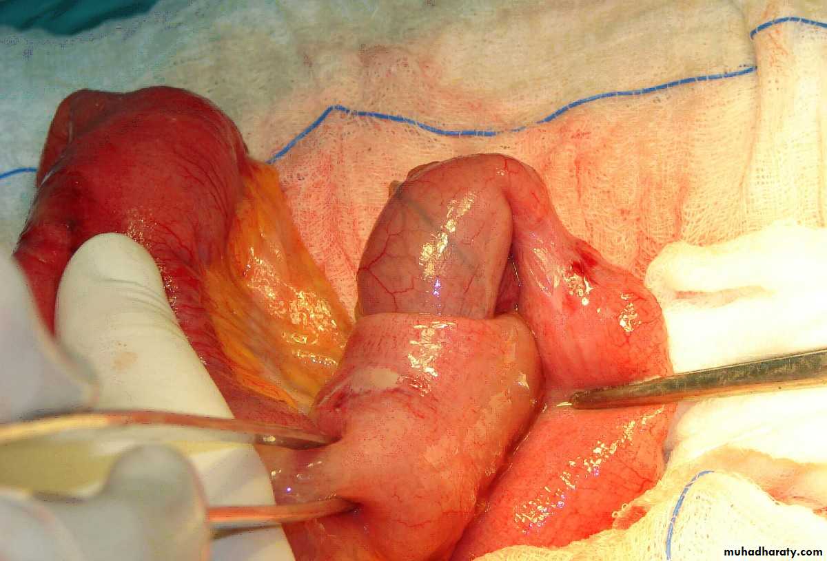
Pyloric stenosis at operation showing pale, thick pyloric muscle and pyloromyotomy incision