STUDY OF THE ACTION OF DRUGS ON THE RABBIT’S EYE
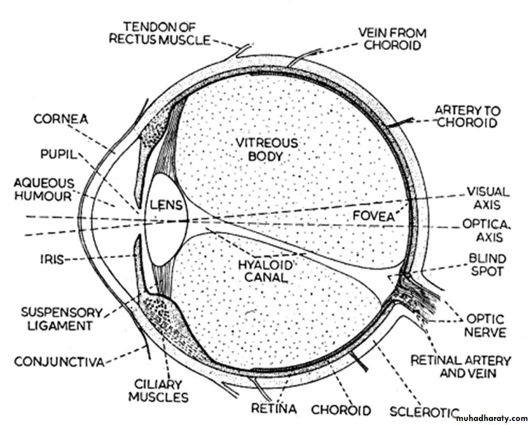
د. شذى هانيThe main compartments of the human eye (as shown in figure 1) are:
- cornea القرنية , the convex transparent membrane forming part of the of the outer coat of the eye .- iris القزحية which is the circular colored membrane in the front of the eye.
- Lens العدسة
-ciliary body and vitreous humour.
Iris:the circular coloured membrane in the front of the eye القزحية That involves: Circular muscle (Muscarinic receptors) and Radial muscle (Alpha-receptors).
Miosis: smallness of the pupils of the eyes is due to either contraction of circular muscle or relaxation of radial muscle.
Mydriasis: dilatation of the pupils of the eyes is due to either contraction of radial muscle or relaxation of circular muscle.

In Alpha-agonist (α-1 receptor agonist such as phenylephrine) → Contraction of radial muscle of Iris (Mydriasis) also ↑the out flow of aqueous humor from the eye and reduce IOP.
In Fear (Sympathetic discharge).
In Death (Lack of muscular tone due to lack of Ach.).Lens: Attached to the ciliary body by ligaments
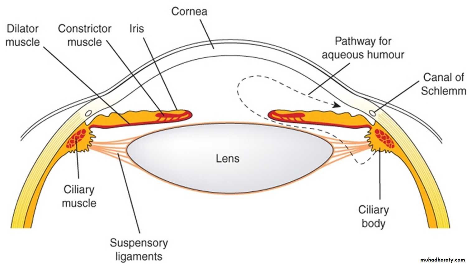
Ciliary body: that involves
- Ciliary epithelium (B2 receptors): responsible for secretion of aqueous humor.- Ciliary muscle (M receptors): responsible for near or far vision.
Ciliary Muscle (Muscarinic receptors)
M-agonist → Ciliary M. Contraction →relaxation of suspensory ligaments of Lens→ lens bulge more &↓ focal length → accommodation of the eye for near vision.M-agonist also cause → ↑ drainage of aqueous humor through canal of schlemnm→ ↓IOP.
Anti-Muscarinic (atropine ) → Ciliary M. Relaxation → Lens relaxation →accommodation for far vision.
Also This allow full visualization of retina (cycloplegia) & proper prescription of glasses by tropicamide eye drops .

Ciliary Epithelium (B-Receptors)
Responsible for secretion of aqueous humor
Ciliary muscle contraction → Increases flow → Decreases IOP.
-Ciliary muscle Relaxation → Decreases flow → Increases IOP (Glaucoma).Methods:
Place few drops of the agents in the following table into the eyes of rabbits and check for the parameters mentioned in the same table, and the results are as follows:
Parameter
Pupil SizeLight Reflex
Accommodation
IOP
Agent
Adrenaline
↔+ve
↔
↔
Pilocarpine
Miosis
+ve
Near Vision
Dec.
Atropine
Mydriasis
-ve
Far Vision
Inc.
Xylocaine
↔
+ve
↔
↔
(+ve) indicates the presence of the reflex
(-ve) indicates the absence of the reflex
(↔) indicates that there is no change