1
|
By:Fatima Ehsan AvcI
P a g e
CARDIO-VASCULAR DISEASES
SYMPTOMS
• CHEST PAIN
• SHORTNESS OF BREATH(DYSPNEA)
• PALPITATION
• SYNCOPE
PHYSICAL EXAM.(SIGNS)
• GENERAL
• PULSE
• BLOOD PRESSURE
• JUGYLAR VENOUS PRESSURE
• PRECORDIUM
• INSPECTION
• PALPATION
• AUSCULTATION-S1,S2
• ADDED
• EXTRA
•
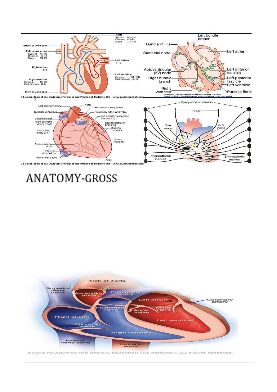
ANATOMY
GROSS-
•
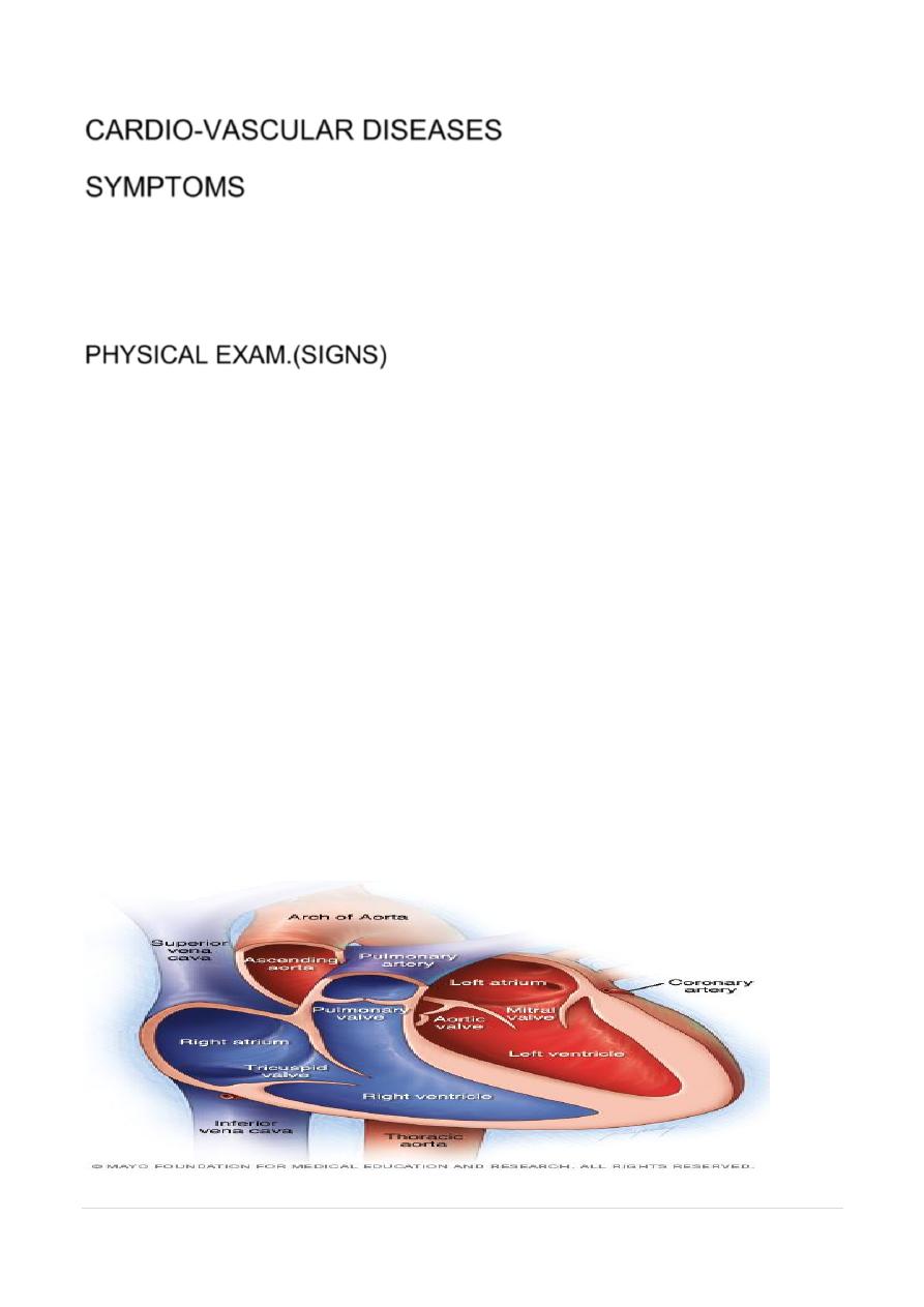
4 CHAMBERS,VALVES AND SEPTA
•
BLOOD SUPPLY
•
NERVE SUPPLY
•
MICROSCOPIC
-
•
THREE 3 LAYERS(endo,myo&pericardium)
•
CELL TYPES= MYOCARDIAL(CONTRACTILE)AND CONDUCTION SYSTEM
•
2
|
By:Fatima Ehsan AvcI
P a g e
ANATOMY-
GROSS
• ATRIA- LOW PRESSURE,STORE
• within mediastinum,
• anterior to oesophagus and descending aorta
• VENRICLES- HIGH PRESURE
• anterior to atria
• taper towards apex of heart,
• AV VALVES- STRONGE CHORDE-PAP.M
• SEMILUNAR VALVES –NO CHORDE,= FIBROUS VALVES
•
PERICARDIUM
=DOUBLE,50 ML.
•
MYOCARDIUM=
3
|
By:Fatima Ehsan AvcI
P a g e
•
heart =
two separate pumps
;-right heart
circulation to lungs .left
rest of body.
•
right atrium (RA)
- deoxygenated blood - superior and inferior
VC
right ventricle (RV),
pulmonary artery.
•
left atrium (LA)
- oxygenated blood -lungs ( four pulmonary veins)
(LV),
aorta.
•
ventricular contraction
(
systol
e)=tricuspid valve and mitral valve --
close,
pulmonary and aortic valves-- open.
•
diastole
,=pulmonary and aortic valves close, and two
atrioventricular valves open.
•
systolic pressure in LV -four times greater than right, (wall of the LV is
usually at least 1 cm thick compared with 2-3 mm for RV
.)
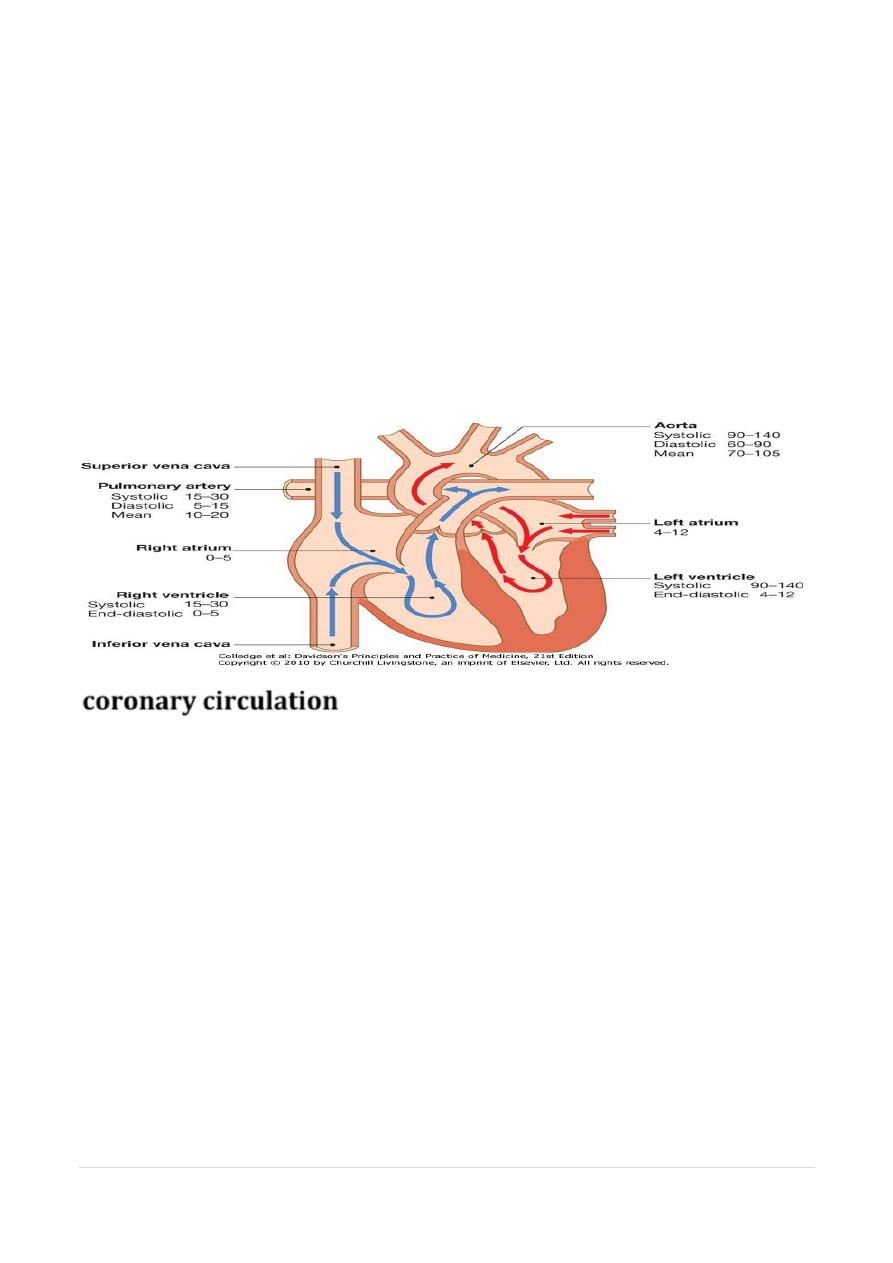
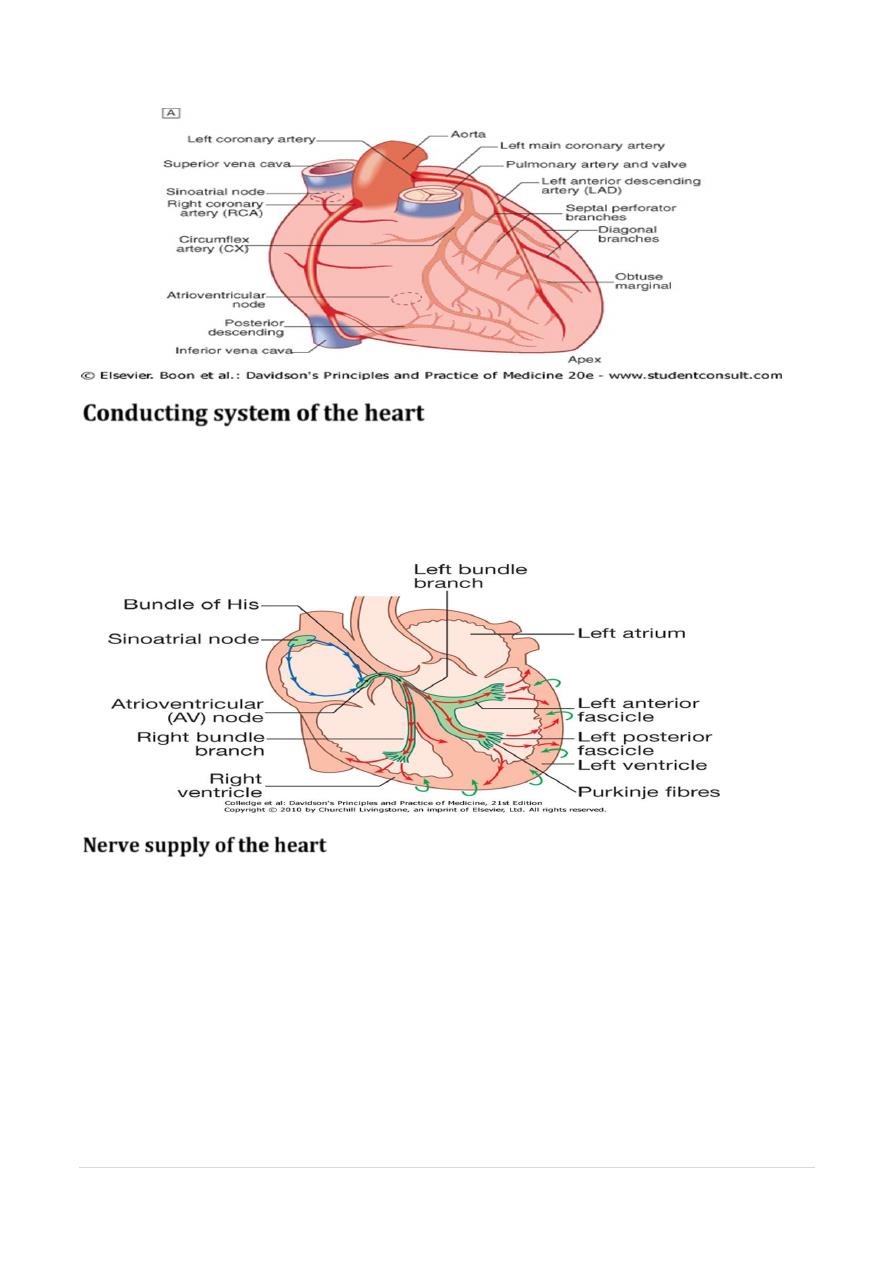
coronary circulation
left main and right coronary arteries - from left and right coronary sinuses,
(distal to
aortic valve)
.
• left main coronary artery
(Within 2.5 cm of its origin )
• 1- left anterior descending artery (LAD)-anterior interventricular groove,
• 2- left circumflex artery (CX) - posteriorly in atrioventricular groove.
•
• LAD
ant.septum (septal perforators) + ant. wall & apex of LV.(diagonal)
• CX
(obtuse marginal branches = lateral, post. and inf, segments of LV.
• Right coronary artery (RCA) –
• right AV groove,
• RA, RV and infero-post. LV.
• posterior descending artery –
• posterior IV groove + inf. IV septum.
• branch of RCA 90% (dominant Rt.system)
• supplied by CX in remainder
• (dominant left system).(5% codominat)
• RCA -sinoatrial (SA) node in about 60%
• atrioventricular (AV) node in about 90%.
4
|
By:Fatima Ehsan AvcI
P a g e
Conducting system of the heart
• The SA node –
• The annulus fibrosus =conduction barrier AV node.
• The AV node
• The His-Purkinje system
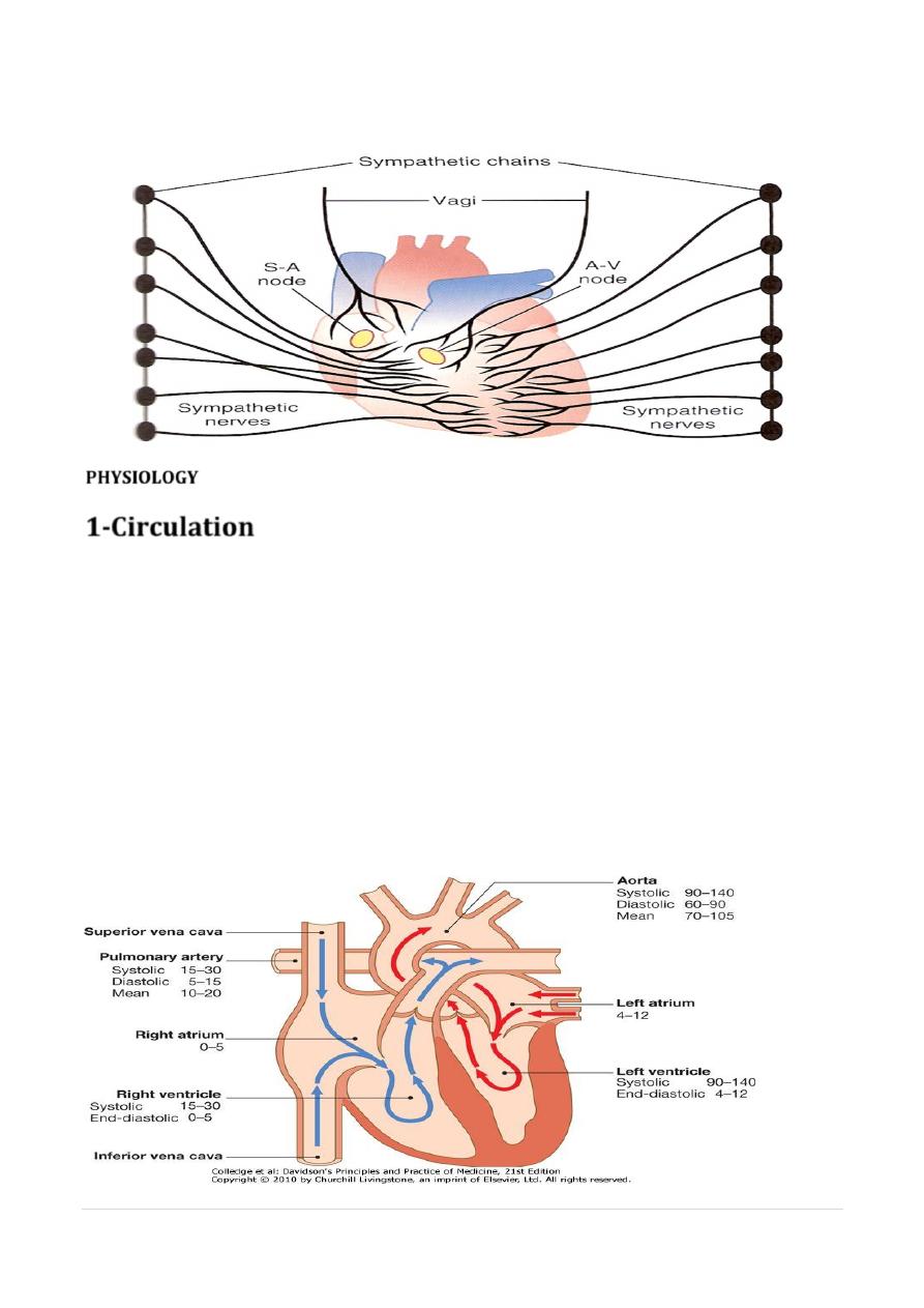
Nerve supply of the heart
innervated by Autonomic nerves- sympathetic and parasympathetic
fibres.
• 1-Adrenergic nerves -cervical sympathetic chain - muscle fibres atria
and ventricles and electrical conducting system.
• β1-adrenoceptors=Positive inotropic and chronotropic effects
• β2-adrenoceptors =vascular smooth muscle =vasodilatation.
• 2- Parasympathetic pre-ganglionic fibres and sensory fibres = vagus
nerves. Cholinergic nerves -AV and SA nodes via muscarinic (M2)
recept.
• resting conditions, vagal inhibitory activity predominates -heart rate
is slow
.
5
|
By:Fatima Ehsan AvcI
P a g e
•
PHYSIOLOGY
1-Circulation
•
heart =
two separate pumps
; right heart generates circulation to
lungs and left heart feeds rest of body.
•
right atrium (RA)
- deoxygenated blood - t superior and inferior
venae cavae
right ventricle (RV),
pulmonary artery.
•
left atrium (LA)
- oxygenated blood from the lungs ( four pulmonary
veins)
left ventricle (LV),
the aorta.
•
systole
=(ventricular contraction)=tricuspid valve and mitral valve
close, and pulmonary and aortic valves open.
•
Diastole
,=pulmonary and aortic valves close, and two
atrioventricular valves open. systolic pressure in LV normally at least
four times greater than that in right, and wall of the LV is usually at
least 1 cm thick compared with 2-3 mm for the RV
.
6
|
By:Fatima Ehsan AvcI
P a g e
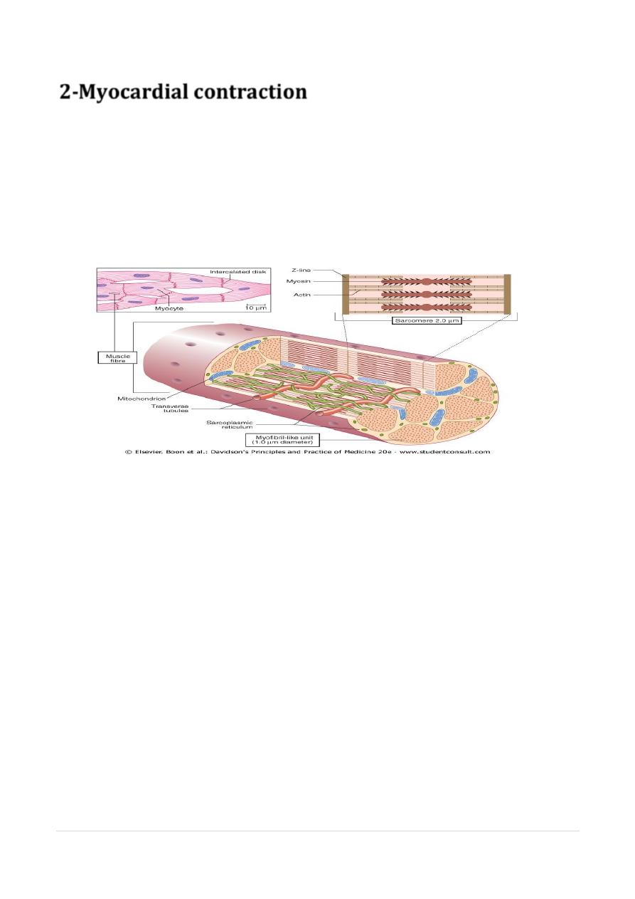
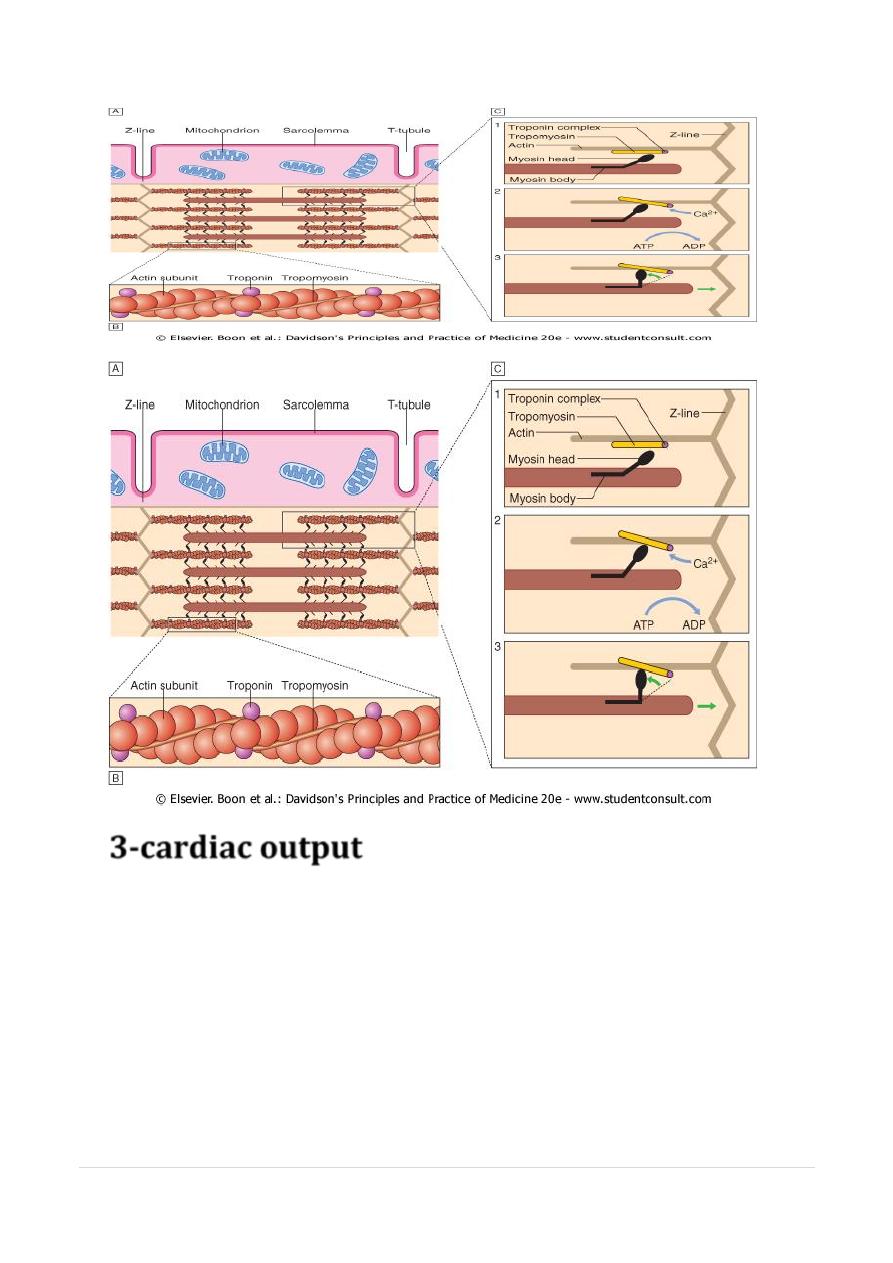
2-
Myocardial contraction
• Myocardial cells (myocytes) =50-100 μm branches and interdigitates
• intercalated disc = electrical (via gap junctions) and mechanical
conduction (via fascia adherens) to adjacent cells.
• basic unit of contraction =sarcomere (2 μm) striated -Z-lines .
• Actin filaments () = attached at right angles to Z-lines and
interdigitate with thicker parallel myosin filaments
• cross-links contain myofibrillar ATPase--breaks adenosine
triphosphate
• Two chains of actin - helical structure, with, tropomyosin, + molecule
complex= troponin.
• contraction= shortening of sarcomere ( interdigitation of actin and
myosin)
• Contraction= calcium ( plateau phase of AP) calcium ions entering
cell and being mobilised from sarcoplasmic reticulum. =
concentration rises, calcium binds to troponin, precipitating
contraction.
•
• force of contraction, or inotropic state= influx of calcium ions
• extent = sarcomere shorten = stroke volume= inotropic drugs or
severe exercise.
• depolarization ca influx release of i.c. ca.binding to
troponinactin-myosic contrction
7
|
By:Fatima Ehsan AvcI
P a g e
3-cardiac output
• Cardiac output =stroke volume x heart rate.
• Stroke volume = volume of blood ejected each cardiac
cycle
• 1- end-diastolic volume and pressure (preload),
• 2- myocardial contractility
• 3-systolic aortic pressure (afterload).
• 1-Stretch of cardiac muscle. = Starling's Law of the heart .
• 2- contractile state of myocardium = neuro-endocrine factors
(epinephrine)+ inotropic drugs and their antagonists.
• 3-Afterload
8
|
By:Fatima Ehsan AvcI
P a g e
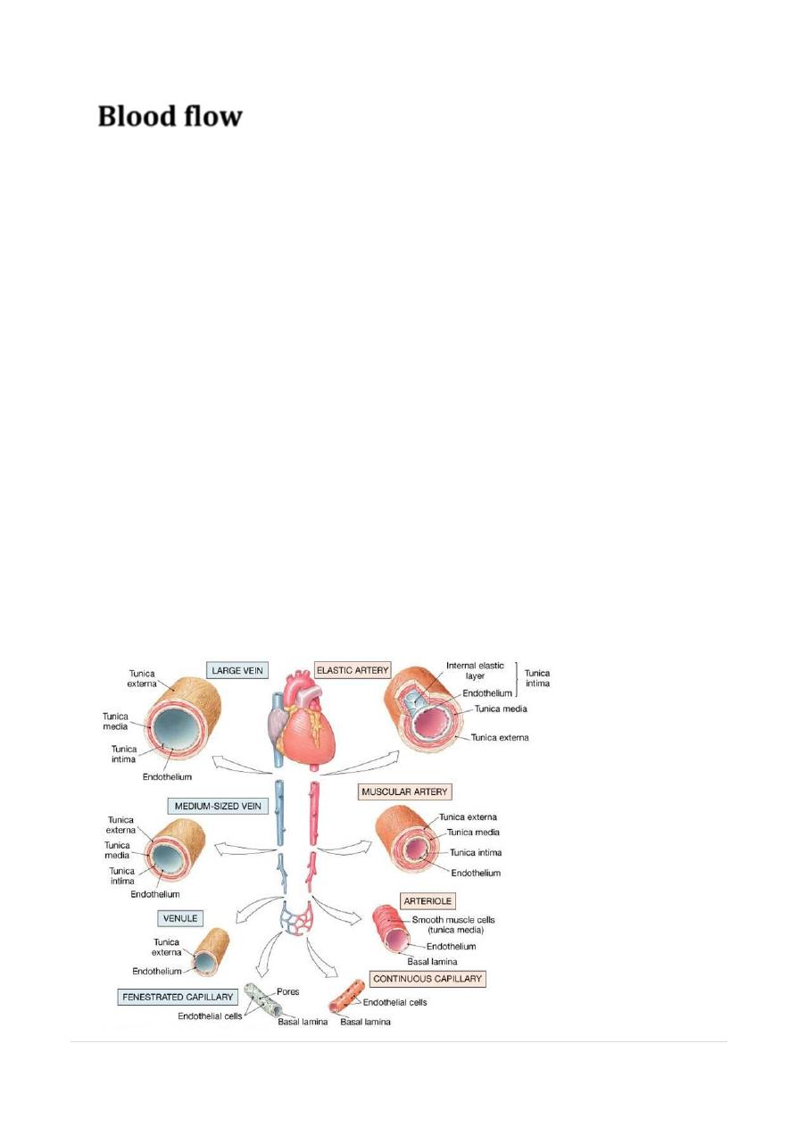
Blood flow
• SYSTEMIC
• Heart
large central Elastic A
Muscular
resistance
capilaries
• Central-compliant-expand-recoil=diastolic BP,dec.afterload
• Resistance BV=Peripheral resistance
Tone=hormone+neurogenic
(Neurogenic constriction via α-adrenoceptors, and
dilatation via muscarinic and β2-adrenoceptors.
vasoactive substances vasoconstrictors=
noradrenaline (, angiotensin II and endothelin-1.
Vasodilators=adenosine, bradykinin,
prostaglandins and nitric oxide
• CORONARY
• SYMPATH.STIM
• α-adrenoceptors
vasoconstriction;
• β2-adrenoceptors
vasodilatation;
• predominant effect of
• sympathetic stimulation =vasodilatation.
• PARA STIM=
modest dilatation of normal coronary arteries.
9
|
By:Fatima Ehsan AvcI
P a g e
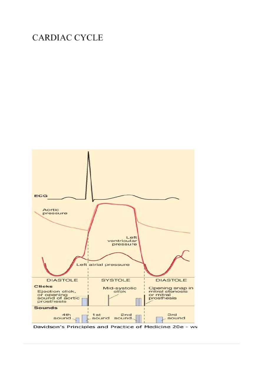
CARDIAC CYCLE
•
DIASTOLE
AND
SYSTOLE
•
1-ATRIA=80% FLOW,20% CONTRACTION
•
2-VENTRICLE
•
FILLING=
DIASTOLE-
•
EARLY
1/3RAPID FILLING
•
MIDDLE
1/3SMALL
LATE
1/3DIASTOLE-AT.CON
•
EMPYING=
SYSTOLE
-
•
ISOMETRIC
CONTRACTION
•
EJECTION
(RAPID & SLOW
•
ISOMETRIC
RELAXATION
•
11
|
By:Fatima Ehsan AvcI
P a g e
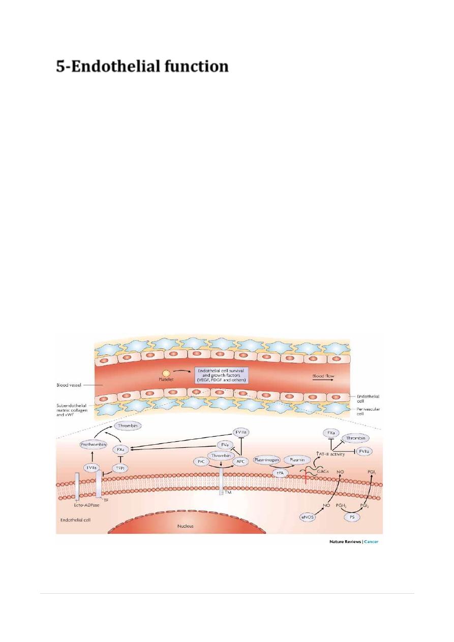
5-Endothelial function
• control of vascular homeostasis.
• A- synthesises and releases = vasoactive
mediators
• 1- vasodilatation= nitric oxide, prostacyclin and
endothelium-derived hyperpolarising factor
• 2- vasoconstriction= endothelin-1 and angiotensin
II.
• balance - maintenance and regulation of vascular
tone and BP. Damage
• B- recruitment of inflammatory cells
• C- formation and dissolution of thrombus.
• stores and releases multimeric glycoprotein, von
Willebrand factor, (thrombus formation ).
• tissue plasminogen activator = fibrinolysis and
thrombus dissolution..
11
|
By:Fatima Ehsan AvcI
P a g e