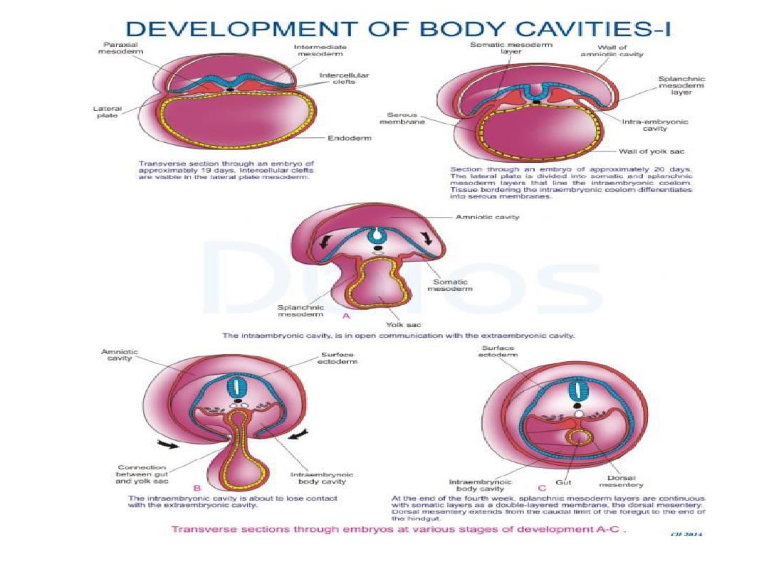
Body cavities

Formation of body cavities
• At ^ end of ^ 3
rd
week: ^ lateral plate mesoderm which is
involved in ^ formation of body cavity, it will differentiate into
2 layers ( a) ^ parietal (somatic) layer adjacent to ^ surface
ectoderm & continuous with extra-embryonic parietal
mesoderm layer covering ^ amnion. (b) ^ visceral
(splanchnic) layer adjacent to ^ endoderm & continuous
with^ visceral layer of extra-embryonic mesoderm covering ^
yolk sac.

• During ^ 4
th
week, ^ sides of ^ embryo begin to grow ventrally
forming 2 lateral body wall folds. These folds consist of ^
parietal layer of lateral plate mesoderm. ^ endoderm also
folds ventrally & closes to form ^ gut tube.
• By ^ end of 4
th
week, ^ lateral body wall folds meet in ^
midline & fuse to close ^ ventral body wall. This closure is
aided by head & tail folds that cause ^ embryo to curve into ^
fetal position.
• Closure of ^ ventral body wall is completed except in ^
connecting stalk, similarly closure of ^ gut tube is completed
except for vitelline duct.

Serous membranes
• Cells of ^ parietal layer of lateral plate mesoderm lining ^
intra-embryonic cavity become mesothelial & form the
parietal layer of ^ serous membranes lining ^ outside of ^
peritoneal, pleural, & pericardial cavities.
• Similarly, cells of ^ visceral layer of lateral plate mesoderm
form the visceral layer of ^ serous membranes covering ^
abdominal organs, lungs, & heart.
• Visceral & parietal layers are continuous with each other at
the
dorsal mesentery .
Which suspends ^ gut tube rom ^
posterior body wall into ^ peritoneal cavity.

• Dorsal mesentery extends from ^ caudal limit of ^ foregut to ^
end of hindgut.
• Ventral mesentery exists only from ^ caudal foregut to ^
upper portion of ^ duodenum & results from thinning of
mesoderm of septum transversum.
• These mesenteries are double layers of peritoneum that
provide a pathway for blood vessels, nerves,& lymphatics to ^
organs.
• ^ septum transversum is a thick plate of mesoderm occupying
^ space between ^ thoracic cavity & ^ stalk of yolk sac.


Diaphragm & thoracic cavity
• Diaphragm divides ^ body cavity into ^ thoracic & peritoneal
cavities.
• It develops from 4 components:(a) septum transversum
(central tendon) (b) pleuroperitoneal membranes. (c) dorsal
mesentery of ^ esophagus, & (d) muscular components from
somites at cervical level s three to five (C3-5) of ^ body wall.
• Since ^ septum transversum is located initially opposite
cervical segments three-five & since muscle cells for ^
diaphragm originate from somites at these segments, ^
phrenic nerve also arises from these segments of ^ ^ spinal
cord (C3,4,&5) keep ^ diaphragm alive.

• Congenital diaphragmatic hernias involving a defect of ^
pleuroperitoneal membrane on ^ left side occur frequently.
• ^ thoracic cavity is divided into ^ pericardial cavity & two
pleural cavities for ^ lungs by ^ pleural cavities for ^ lungs by
^ pleuropericardial membranes.
• Initially, ^ gut tube from ^ caudal end of ^ foregut to ^ end of
^ hindgut is suspended from ^ dorsal body wall by dorsal
mesentery.
