
By
Dr. Suhair Majeed

Urinary system
The urinary system consists of the
kidneys,
ureters, urinary bladder, and urethra. The
kidneys constitute the glandular component; the
remainder of the urinary system forms the
excretory passages.The ureters conduct urine
from the kidneys to the bladder, where it is stored
temporarily. In turn, the bladder is drained by the
urethra, through which the urine is voided from
the body.

Kidneys :
kidneys are bean-shaped organs that lie in a
retroperitoneal position against the posterior
abdominal wall, one on either side of the upper
lumbar vertebrae. Superior to each kidney is the
adrenal gland embedded in renal fat and
connective tissue. Each kidney is contained
within a thin but strong connective tissue
capsule .

Cont.
The renal artery and nerves enter the kidney
on the medial border at the
hilum, a concavity
that also serves as the point of exit for the renal
vein, lymphatics, and ureter. The hilum is
continuous with the
renal sinus, a large central
cavity surrounded by the parenchyma of the
kidney and filled with loose areolar connective
tissue that normally contains much fat. Nerves,
lymphatics, and branches of the renal artery and
vein run through the sinus.

Cont.
The
renal pelvis is a funnel shaped expansion
of the ureter where it joins the kidney; it also
passes through the sinus, dividing into two or
three short tubular structures called the
major
calyces. These in turn divide into eight to twelve
smaller units called
minor calyces. Each minor
calyx forms a cylindrical attachment around a
conical projection of renal tissue called a
renal
papilla.

Cont
.
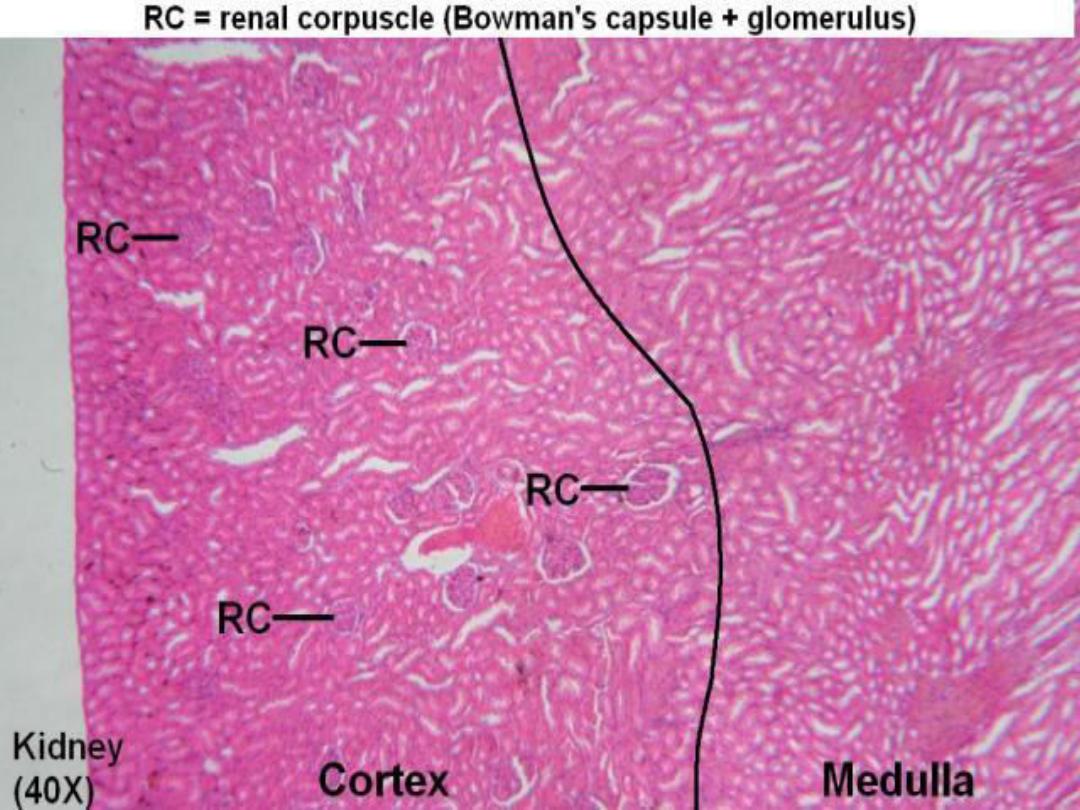
When the kidney is examined macroscopically,
two distinct regions can be seen. The darker,
granular outer region is the
cortex, a layer
beneath the capsule. The inner region, or
medulla, is paler and smoother in texture and
consists of
8 to 20 cone-shaped structures called
medullary pyramids, which are separated from
each other by inward extensions of cortical tissue.

Cont.
The cortex that separates adjacent medullary
pyramids makes up a
renal column. The bases of
the pyramids are directed toward the overlying
cortex, while their apices are oriented toward the
renal sinus and form the
renal papillae. From
the bases of the pyramids
, groups of tubules
extend into the cortex, giving it a striated
appearance. These striations represent a
continuation of medullary tissue into the cortex
and constitute the
medullary rays.

Uriniferous Tubules and Nephrons
of the Kidney :
The functional unit of each kidney is the
microscopic
uriniferous tubule. It consists of a
nephron and a collecting duct into which empty
the filtered contents of the nephron.Millions of
nephrons are present in each kidney cortex.
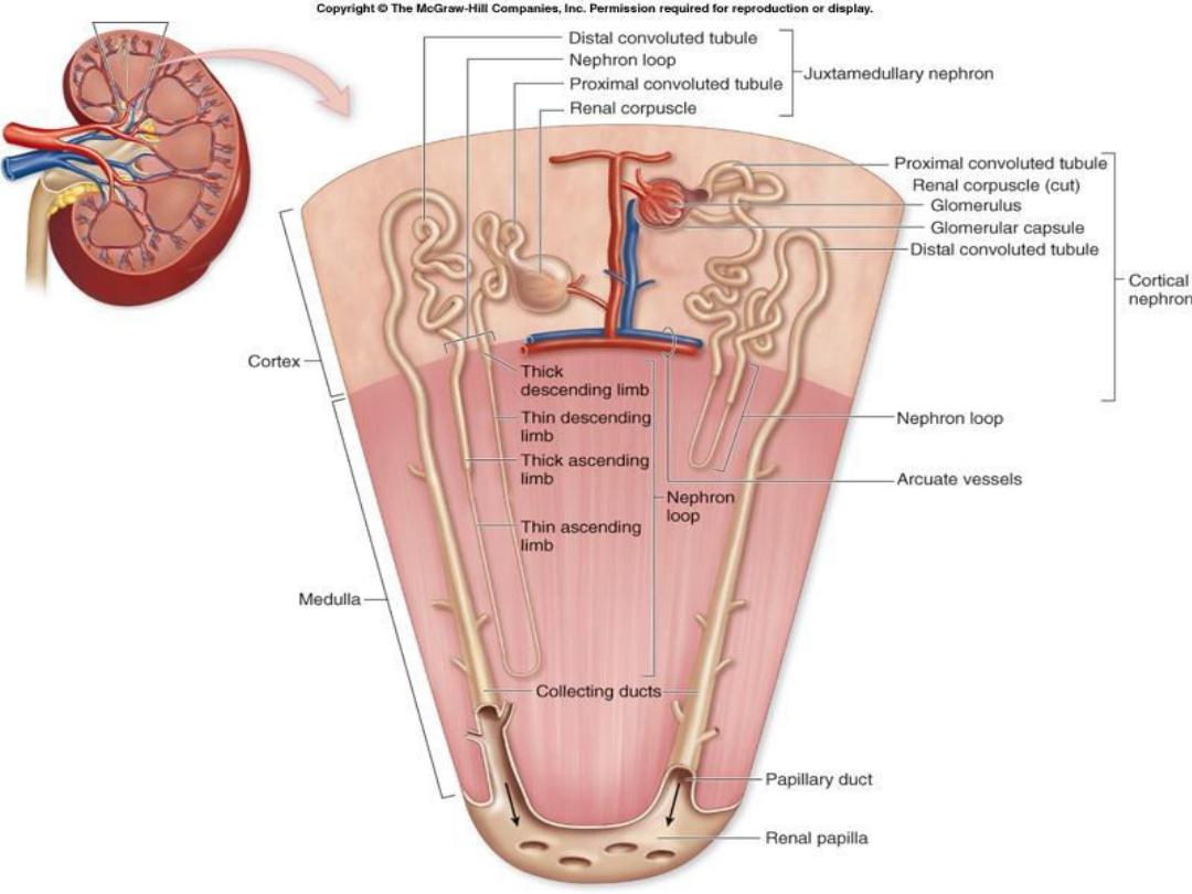
The nephron, is subdivided into two components,
1-
renal corpuscle
2-
renal tubules.

Cont.
There are two types of nephrons.
Cortical nephrons are located in the cortex of
kidney
.
juxtamedullary nephrons are situated near
the junction of the cortex and medulla of the
kidney.
Although all nephrons participate in urine
formation, juxtamedullary nephrons produce a
hypertonic environment in the interstitium of the
kidney medulla that results in the production of
concentrated (hypertonic) urine.

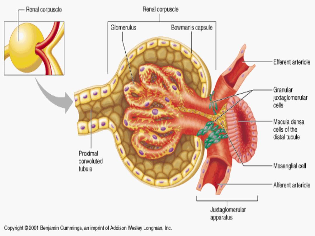
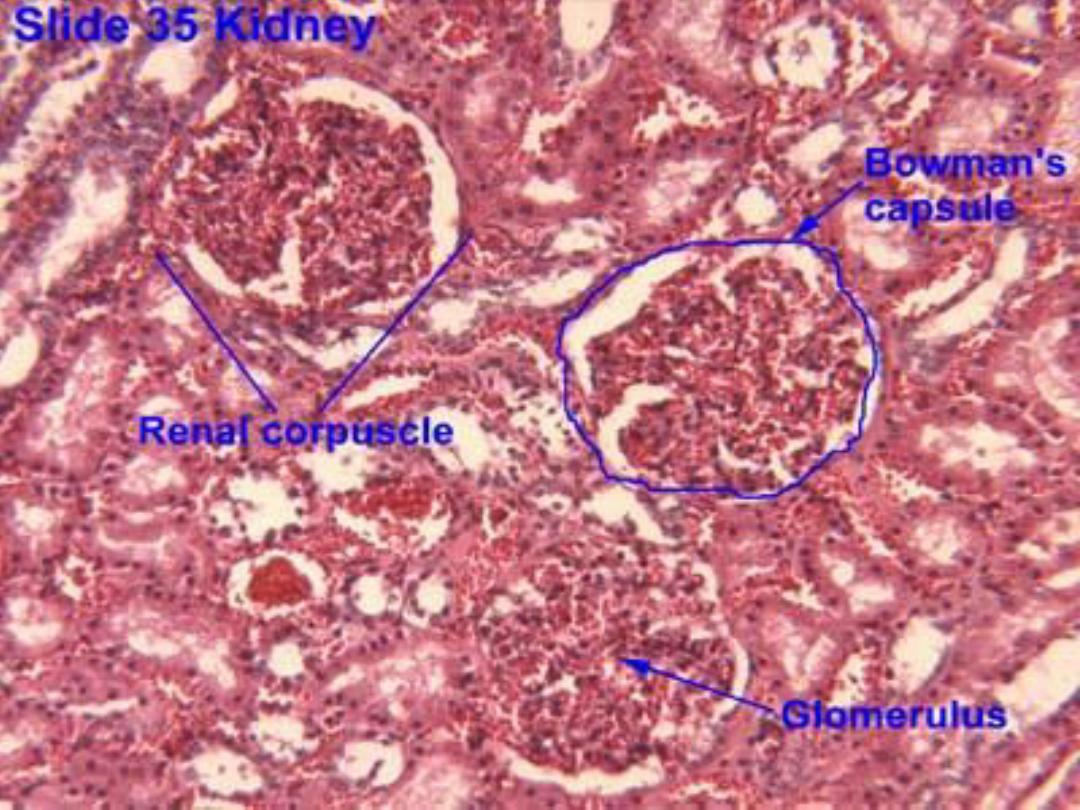
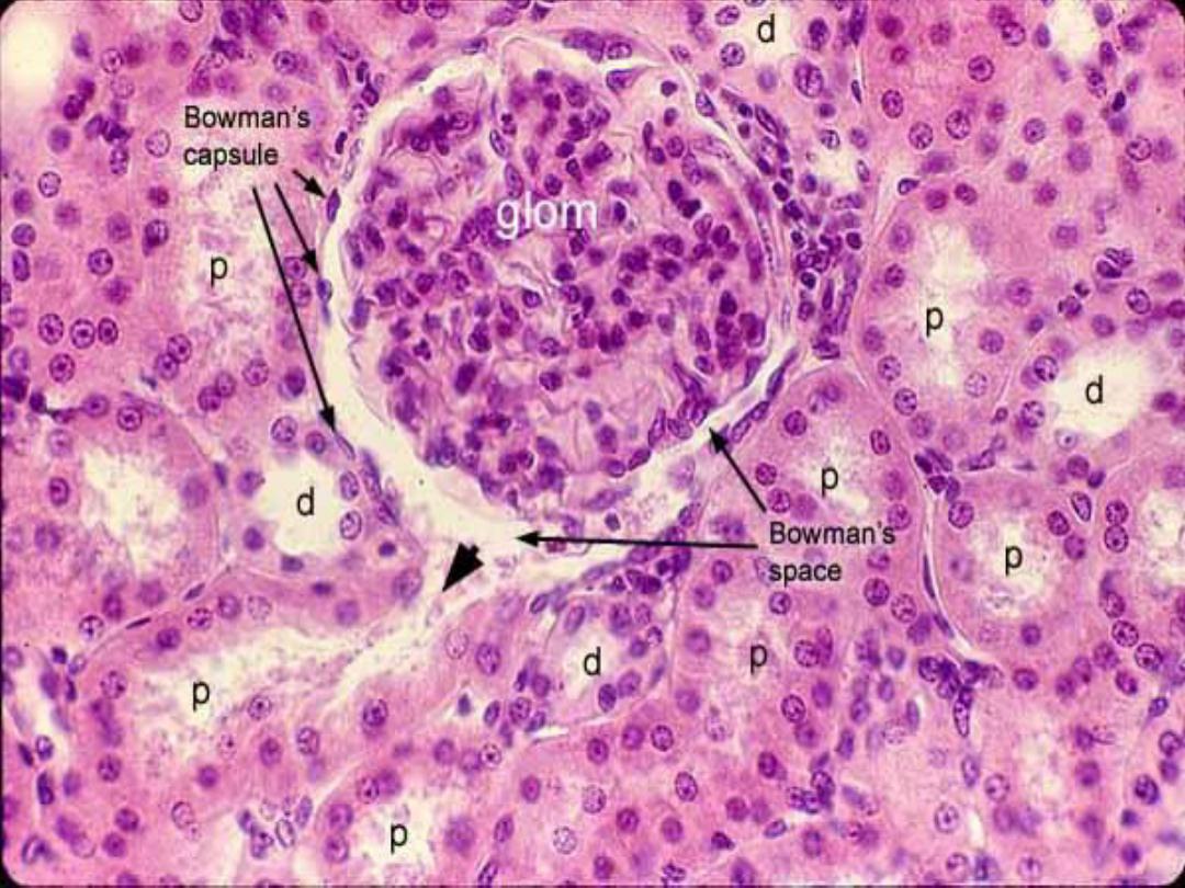
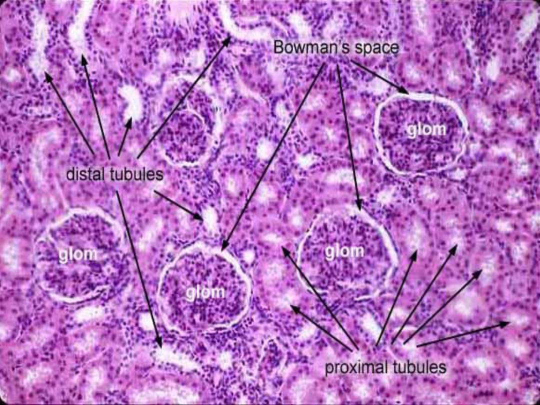
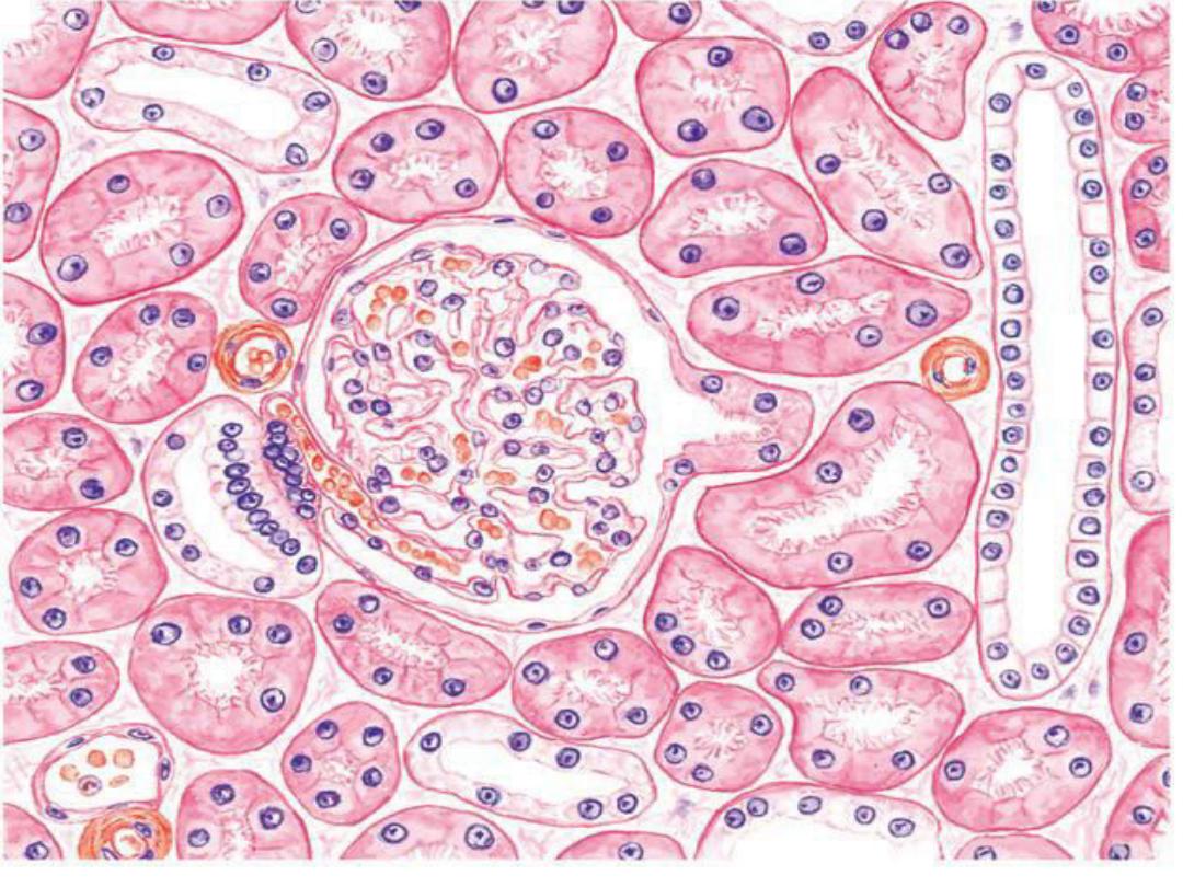
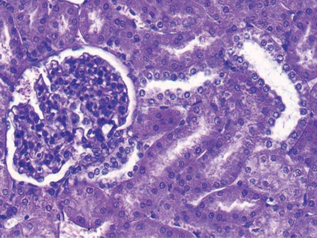
1- Renal Corpuscle :
The renal corpuscle consists of a tuft of
capillaries, called the
glomerulus, surrounded
by a double layer of epithelial cells, called the
glomerular Bowman’s capsule. The inner or
visceral layer of the capsule consists of unique
and highly modified branching epithelial cells,
called
podocytes. The podocytes are adjacent to
and completely invest the glomerular capillaries.
The outer layer of the glomerular capsule
consists of
simple squamous epithelium.

Cont.
The renal corpuscle is the initial segment of
each nephron. Blood is filtered in renal
corpuscles through the capillaries of the
glomerulus, and the filtrate enters the
capsular
(urinary) space located between the parietal
and visceral cell layers of the glomerular capsule.
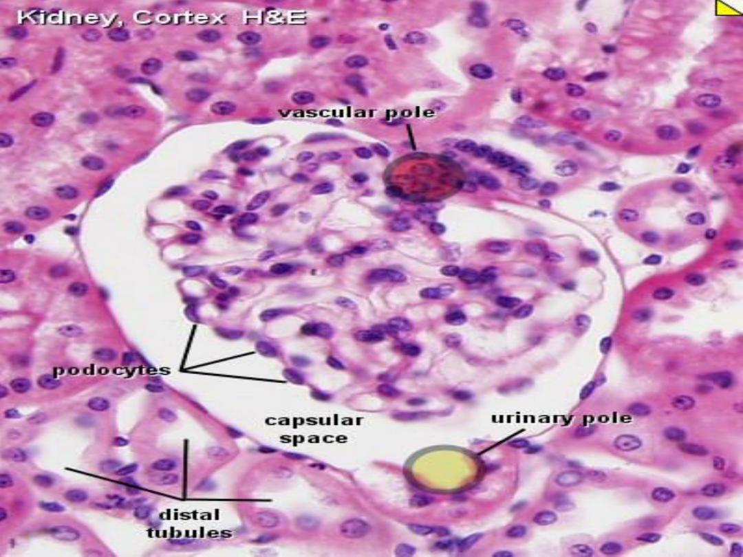
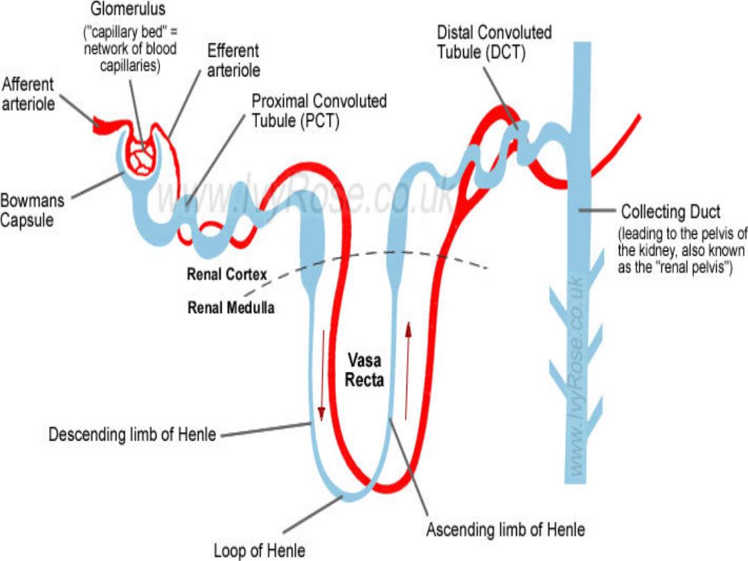
Each renal corpuscle has a
vascular pole, where
the afferent arteriole enters and the efferent
arteriole leaves the corpuscle
.

Cont.
On the opposite end of the renal corpuscle is
the
urinary pole. Filtrate produced by the
glomerulus that enters the capsular space leaves
each renal corpuscle at the urinary pole, where
the proximal convoluted tubule starts. Filtration
of blood in renal corpuscles is facilitated by
glomerular endothelium.

Cont.
The endothelium in glomerular capillaries is
porous (fenestrated) and highly permeable to
many substances in the blood, except to the
formed blood elements or plasma proteins. Thus,
glomerular filtrate that enters the capsular space
is not urine. Instead, it is an ultrafiltrate that is
similar to plasma, except for the absence of
proteins.


2- Renal Tubules :
As the glomerular filtrate leaves the renal
corpuscle at the urinary pole, it flows through
different parts of the nephron before reaching the
renal tubules called
the collecting tubules and
collecting ducts. The glomerular filtrate first
enters the renal tubule
, which extends from the
glomerular capsule to the collecting tubule. This
renal tubule has several distinct histologic and
functional regions.

Cont.
The portion of the renal tubule that begins at
the renal corpuscle is highly twisted or tortuous
and is therefore called the
proximal convoluted
tubule. Initially, this tubule is located in the
cortex but then descends into the medulla to
become continuous with the
loop of Henle.
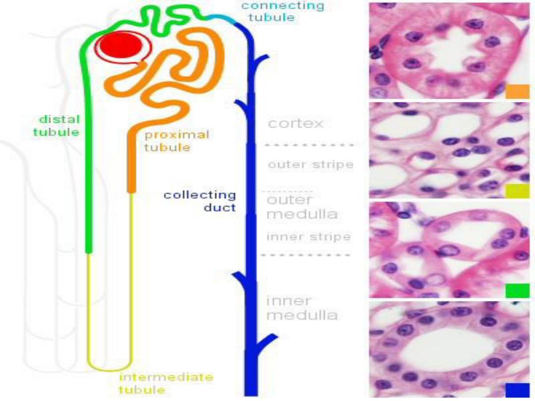

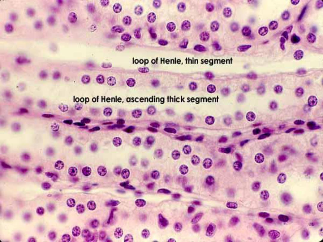
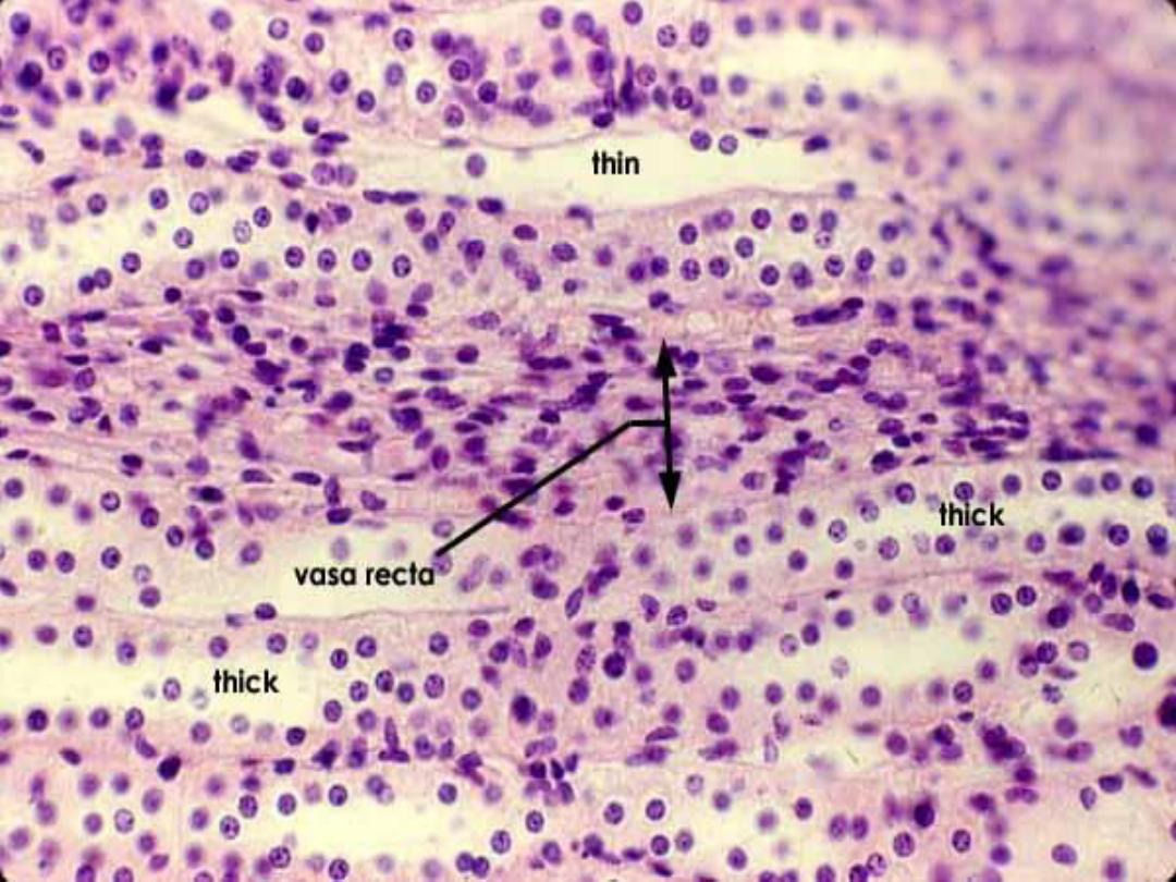
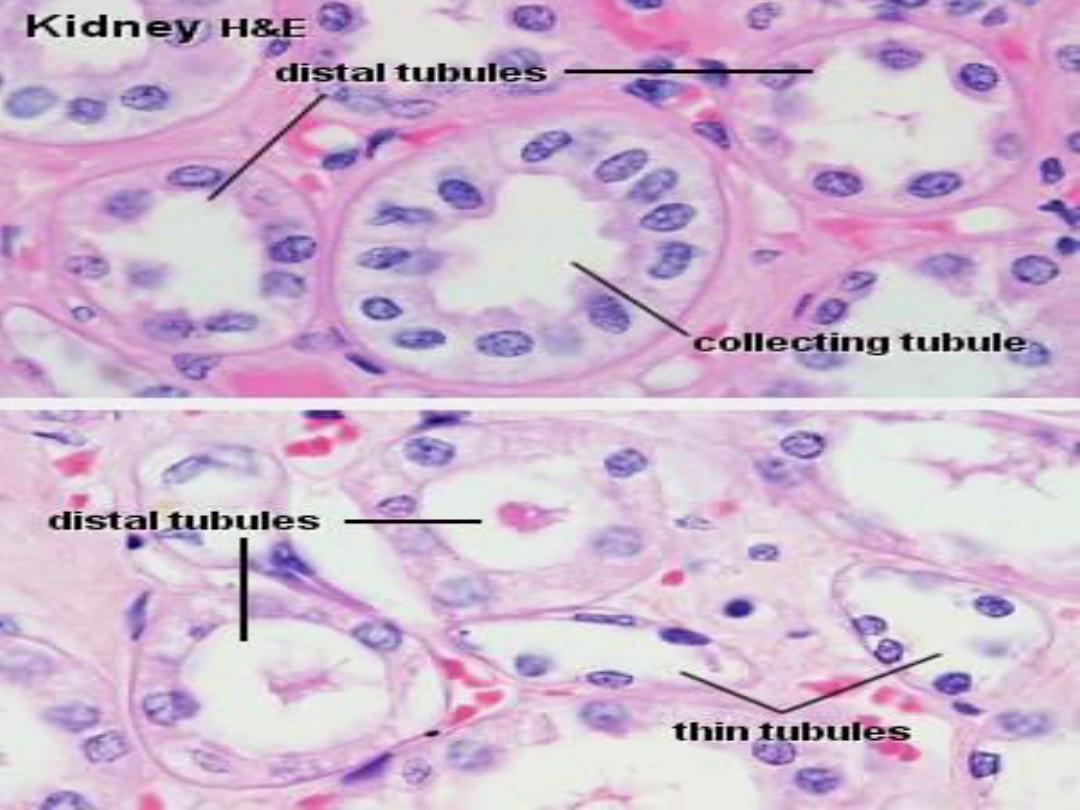
Loop of Henle :
The
loop of Henle consists of several parts:
- a thick, descending portion of the proximal
convoluted tubule;
- a thin descending and
- a thick, ascending portion called the
distal
convoluted tubule. The distal convoluted
tubule is shorter and less convoluted than the
proximal convoluted tubule, and it ascends into
the kidney cortex.

Cont.
Because the proximal convoluted tubule is
longer than the distal convoluted tubule, it is
more frequently observed near the renal
corpuscles and in the renal cortex.

Cont.
Glomerular filtrate then flows from the distal
convoluted tubule to the
collecting tubule.
In juxtamedullary nephrons, the loop of
Henle is very long; it descends from the kidney
cortex deep into the medulla and then loops back
to ascend into the cortex . The collecting tubule is
not part of the nephron . A number of short
collecting tubules join to form several larger
collecting ducts.

Cont.
As the collecting ducts become larger and
descend toward the papillae of the medulla, they
are called
papillary ducts. Smaller collecting
ducts are lined by light-staining cuboidal
epithelium. Deeper in the medulla, the
epithelium in these ducts changes to columnar.
At the tip of each papilla, the papillary ducts
empty their contents into the minor calyx. The
area on the papilla that exhibits openings of the
papillary ducts is called the
area cribrosa

Cont.
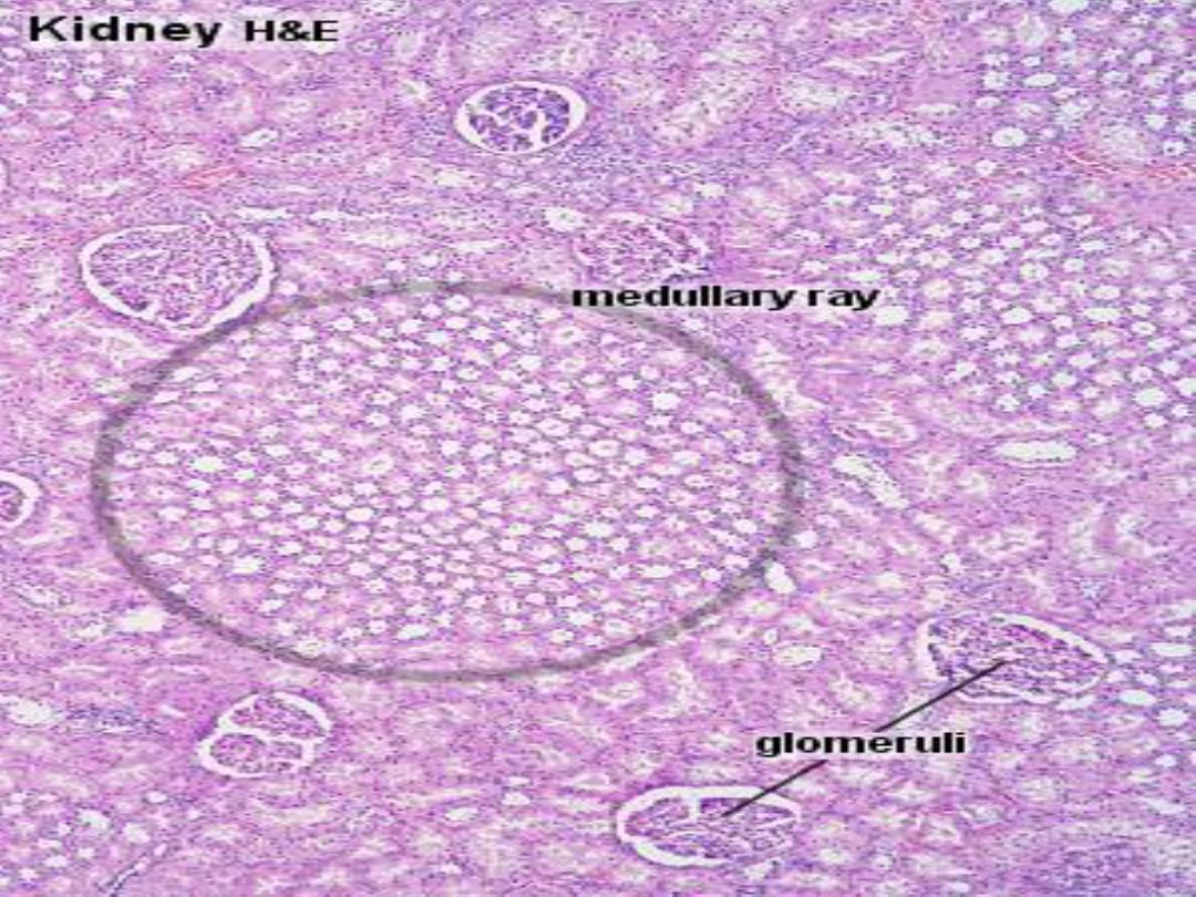
The kidney cortex also exhibits numerous,
lighter-staining
medullary rays that extend
vertically from the bases of the pyramids into
the cortex.Medullary rays consist primarily of
collecting ducts, blood vessels, and straight
portions of a number of nephrons that penetrate
the cortex from the base of the pyramids
.

Kidney Cells and Tubules :
1-
Mesangial Cells :
In addition to podocytes that surround the
capillaries, there are other specialized cells in the
glomerulus, called
mesangial cells, that are also
attached to the capillaries.
Mesangial cells synthesize the extracellular
matrix and provide structural support for the
glomerular capillaries.

Cont.
As blood is filtered, numerous proteinaceous
macromolecules are trapped in the basal lamina
of the glomerulus. Mesangial cells function as
macrophages in the intraglomerular regions
and phagocytose material that accumulates on
the glomerular filter, thus preventing its closure
with debris. These cells also appear to be
contractile and can regulate glomerular blood
flow as a result of the presence of receptors for
vasoactive substances.

Cont.
Some of the mesangial cells are also located
outside of the renal corpuscle in the vascular pole
region. Here, they are called
the extraglomerular mesangial cells that
form part of the juxtaglomerular apparatus
.

2-Proximal Convoluted Tubules:
All nephrons participate in urine formation.
The cells of the
proximal convoluted tubules
show numerous deep infoldings of the basal cell
membrane.
These features characterize cells that are
involved in active transport of molecules and
electrolytes from the filtrate across the cell
membrane into the interstitium.


Cont.
Reabsorption of most of the substances from the
glomerular filtrate takes place in the proximal
convoluted tubules. As the glomerular filtrate
enters the proximal convoluted tubules
, all
glucose, proteins, and amino acids, almost all
carbohydrates, and about 75 to 85% of water
and
sodium and chloride ions are absorbed
from the glomerular filtrate into the surrounding
peritubular capillaries
. The presence of microvilli
(brush border
) on proximal convoluted tubule
cells greatly increases the surface area and
facilitates absorption of filtered material.

Cont.
The metabolic waste products urea and uric
acid remain in the proximal convoluted tubules
and are eliminated from the body in the urine.
The proximal convoluted tubule is longer than
the distal convoluted tubule. As a result, the
sections of this tubule are more frequently seen
in the cortex near the renal corpuscles that those
of distal convoluted tubules.

3-Loops of Henle :
The loops of Henle of the
juxtaglomerular
nephrons produce the hypertonic urine by
creating an osmotic gradient in the interstitium
from the cortex of the kidney to the tips of the
renal papillae .
In the juxtamedullary nephrons, the loops of
Henle are very long, extend deep into the
medulla, and assist in maintaining the high
osmotic gradient necessary for removing water
from the filtrate into the interstitium.

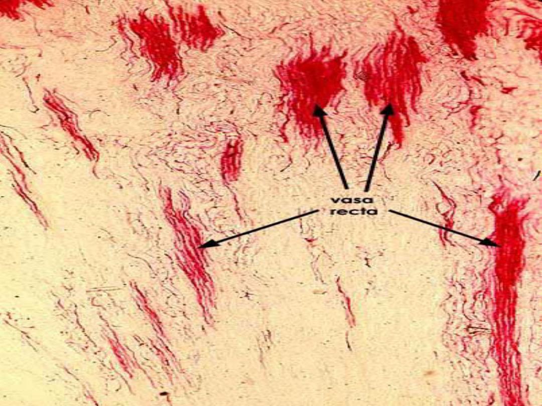
Cont.
The hypertonicity (high osmotic pressure) of
extracellular fluid in the medulla removes water
from the glomerular filtrate as it flows through
these tubules, with the
vasa recta helping to
maintain the osmotic concentration gradient in
the medulla
.

Vasa recta
Efferent arterioles from juxtamedullary
nephrons, form several long, straight vessels, the
vasa recta, that descend into the medullary
pyramid and form hairpin loops. Like the loop
of Henle, the loops of the vasa recta are scattered
throughout the medulla.
The walls of the vasa recta are thin, and the
endothelium of the ascending (venous) limb is
fenestrated.

Cont.
The vasa recta form a
vascular
countercurrent exchange system that
removes excess water and ions
. These capillary
loops are permeable to water and take up the
water from the medullary interstitium to return
it to systemic circulation
.

4- Distal Convoluted Tubules
The distal convoluted tubules are
shorter
and less convoluted than the proximal tubules.
Therefore, these tubules are less frequently
observed in the cortex and near the renal
corpuscles. In comparison with the proximal
convoluted tubules, the distal convoluted tubules
do not exhibit brush borders, the cells are
smaller, and more nuclei are seen per tubule.

Cont.
The main function of the distal convoluted
tubules is to actively reabsorb sodium ions from
the tubular filtrate. This activity is directly
linked with excretion of hydrogen and potassium
ions into the tubular fluid. Sodium reabsorption
in the distal convoluted tubules is controlled by
the hormone
aldosterone, which is secreted by
the adrenal cortex
.

Cont.
These functions of distal convoluted tubules
are vital for maintaining the acidbase balance of
body fluids and blood.

Juxtaglomerular Apparatus
Adjacent to the renal corpuscles and distal
convoluted tubules lies a special group of cells
called
juxtaglomerular apparatus. This
apparatus consists of two components,
-
the juxtaglomerular cells
-
the macula densa.
Juxtaglomerular cells are a group of modified
smooth muscle cells located in the wall of
the afferent arteriole just before it enters the
glomerular capsule to form the glomerulus.


Cont.
The cytoplasm of these cells contains
membrane-bound secretory granules of the
enzyme
renin.
The
macula densa is a group of modified
distal convoluted tubule cells. The macula densa
cells and juxtaglomerular cells are separated by a
thin basement membrane. The proximity of
juxtaglomerular cells to the macula densa allows
for integration of their functions.

Cont.
The main function of the juxtaglomerular
apparatus is to maintain the necessary blood
pressure in the kidney for glomerular filtration.
The cells of this apparatus act as both the
baroreceptors and chemoreceptors.
The juxtaglomerular cells monitor changes in
the systemic blood pressure by responding to
stretching in the walls of the afferent arterioles.
The cells in the macula densa are sensitive to
changes in sodium chloride concentration.

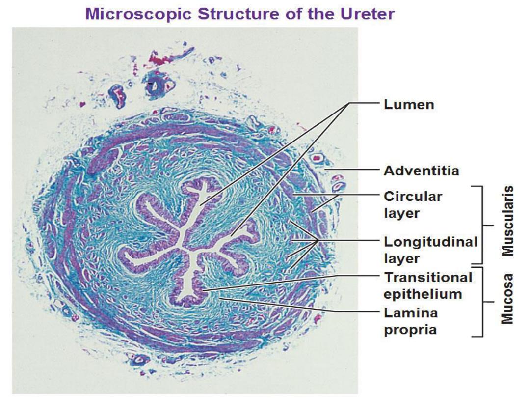
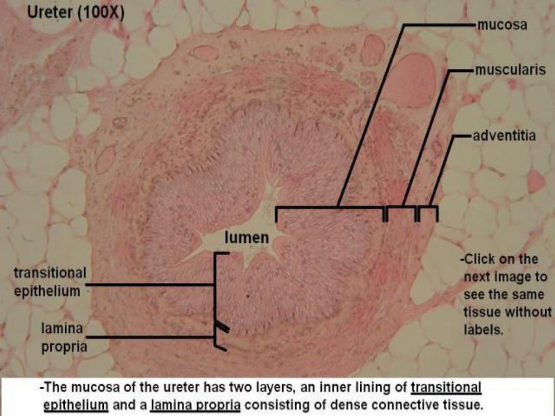
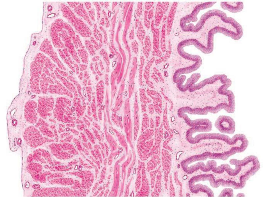
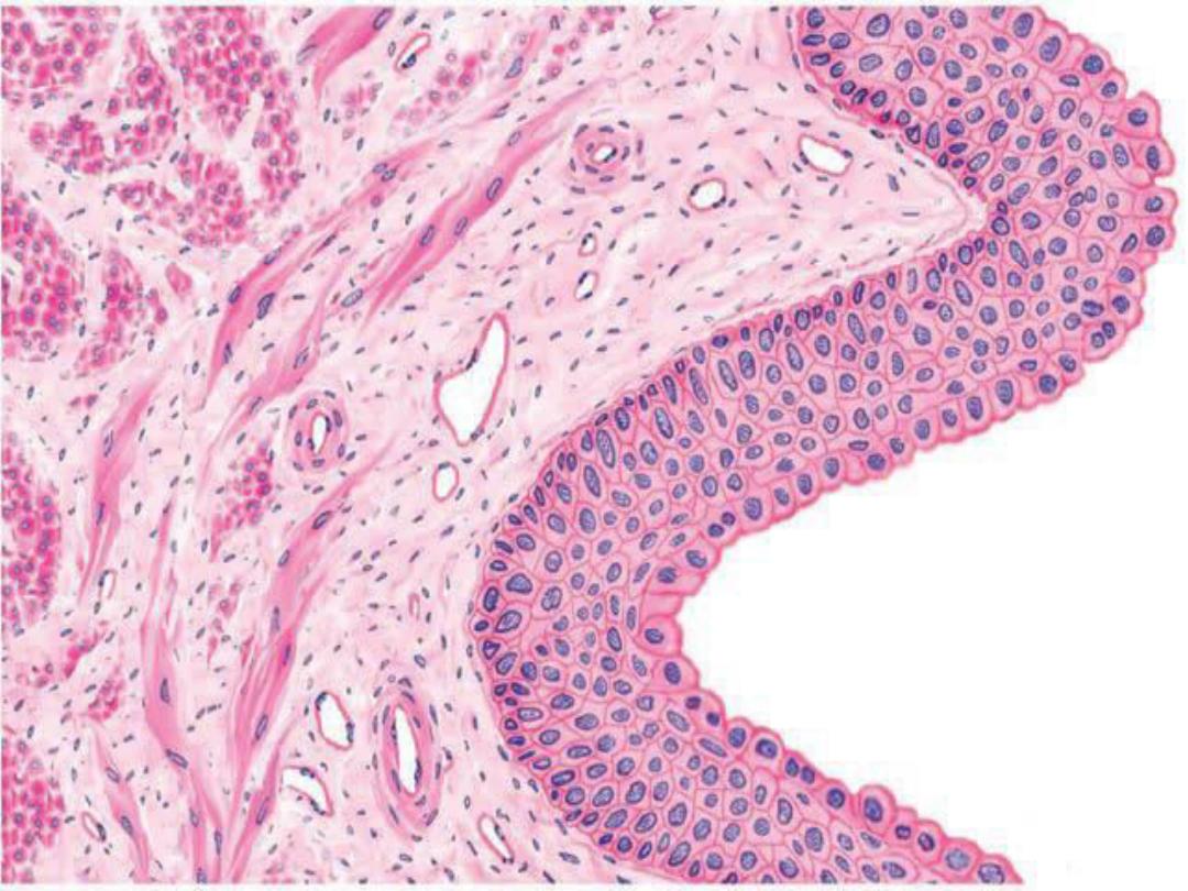
Ureter
The ureter is a muscular tube that conveys
urine from the kidneys to the bladder by the
contractions of the thick, smooth muscle layers
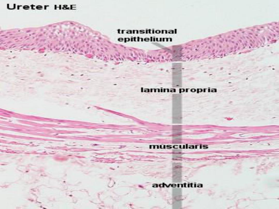
found in its wall. The mucosa of the ureter is
highly folded and lined by a thick
transitional
epithelium . Below the transitional epithelium is
the connective tissue
lamina propria .

Cont.
The muscularis of the ureter contains smooth
muscle layers, an
-inner longitudinal layer
- a middle circular muscle layer .
- A third outer longitudinal layer
A connective tissue
adventitia , with blood
vessels and adipose tissue
, surrounds the ureter
.

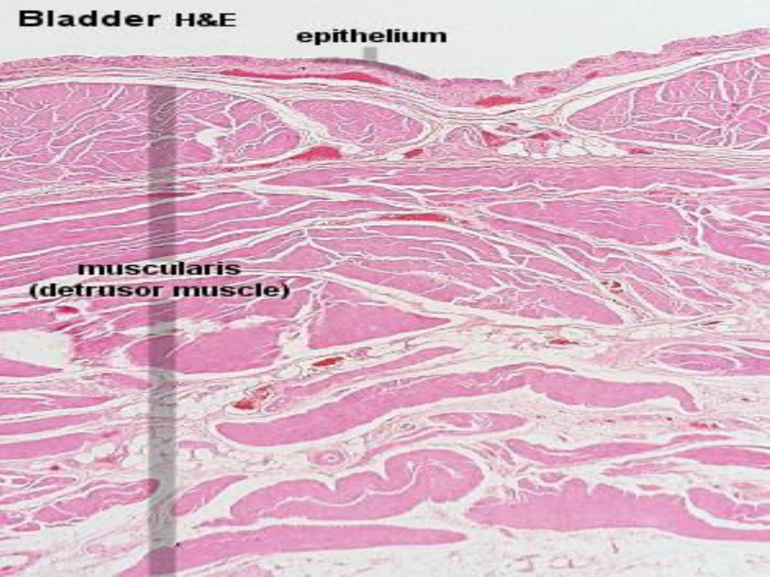
Urinary Bladder:
The bladder has a thick muscular wall. The
wall is similar to that of the lower third of the
ureter, except for its thickness. In the wall are
found three loosely arranged layers of smooth
muscle,
-the inner longitudinal,
-middle circular, and
- outer longitudinal layers.

Cont.
The three layers are arranged in
anastomosing
smooth muscle bundles
between which is found the interstitial
connective tissue
. The interstitial
connective tissue merges with the connective
tissue of the
serosa .Mesothelium covers
the connective tissue of serosa and is the
outermost layer.

Cont.
Serosa lines the superior surface of the
bladder,whereas its inferior surface is covered by
the connective tissue adventitia, which merges
with the connective tissue of adjacent structures.

Cont.
The mucosa of an empty bladder exhibits
numerous
mucosal folds that disappear during
bladder distension. The transitional epithelium
is
thicker than in the ureter and consists of about
six layers of cells , the superficial cells of the
transitional epithelium are low cuboidal or
columnar and appear dome-shaped. The deeper
cells in the epithelium are round and the basal
cells more columnar
The lamina propria ,
inferior to the epithelium, is wider than in the
ureters.
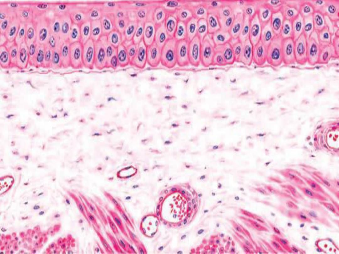

Cont.
The subepithelial
lamina propria contains
fine
connective tissue fibers, numerous
fibroblasts, and blood vessels .
Numerous blood vessels
of various sizes are
found in the
serosa , between the smooth muscle
bundles and in the lamina propria
.

Thank you