
Histology
lecture -1-

Histology
lecture -1-
Introduction to
histology and epithelial
tissue

Introduction
Histology(microscopic anatomy) is the study of tissue structure, extending
from the level of the individual cell, through organs to organ systems.
Histology is obviously related to Cell Biology (Cytology) and to Anatomy; it also
forms the structural basis for understanding function (Physiology) and is the
preparation for the study of abnormal structure and function (Pathology).
Whole body contains only 200 different types of cells

SPECIFIC OBJECTIVES Topics 1
•
Demonstrate knowledge of the structural and functional characteristics that define
a tissue.
•
Demonstrate knowledge of the mechanisms of cell differentiation, aggregation,
intercellular recognition and communication that lead to the formation of tissues.
•
Describe the constituent elements of tissues.
•
• Demonstrate knowledge of the different criteria for the classification of tissues.

Topics 2
•
Demonstrate knowledge of the structural and functional characteristics of
epithelial tissues that distinguish them from basic tissues.
•
Demonstrate knowledge of the different types of epithelial tissue and give examples
of the parts of the body in which these can be found.
•
Demonstrate knowledge of the different functions of each type of epithelial tissue
and relate them to the tissue structure.
•
Demonstrate knowledge of the specialized functions of different types of epithelial
cells and give examples of the different parts of the body in which they can be
found.

6
§ Embryonic Tissues
1.
Embryo begins as a single cell
•
divides into many cells that form layers (strata)
2.
Three primary germ layers
A.
ectoderm
(outer) gives rise to: epidermis + nervous system
B.
endoderm
(inner): mucous membranes: GI tract and
respiratory linings; digestive glands.
C.
mesoderm
(middle) forms mesenchyme (gelatinuous tissue)
and then give rise to muscle, bone, and blood

7
§ Tissue Sectioning
1.
Preparation of histological specimens
•
fixation
•
sections
•
mounted on slides & stained
2. Sectioning (slicing) an organ or tissue reduces a 3-dimensional structure
to a 2-dimensional slice (see the next 3 slides)
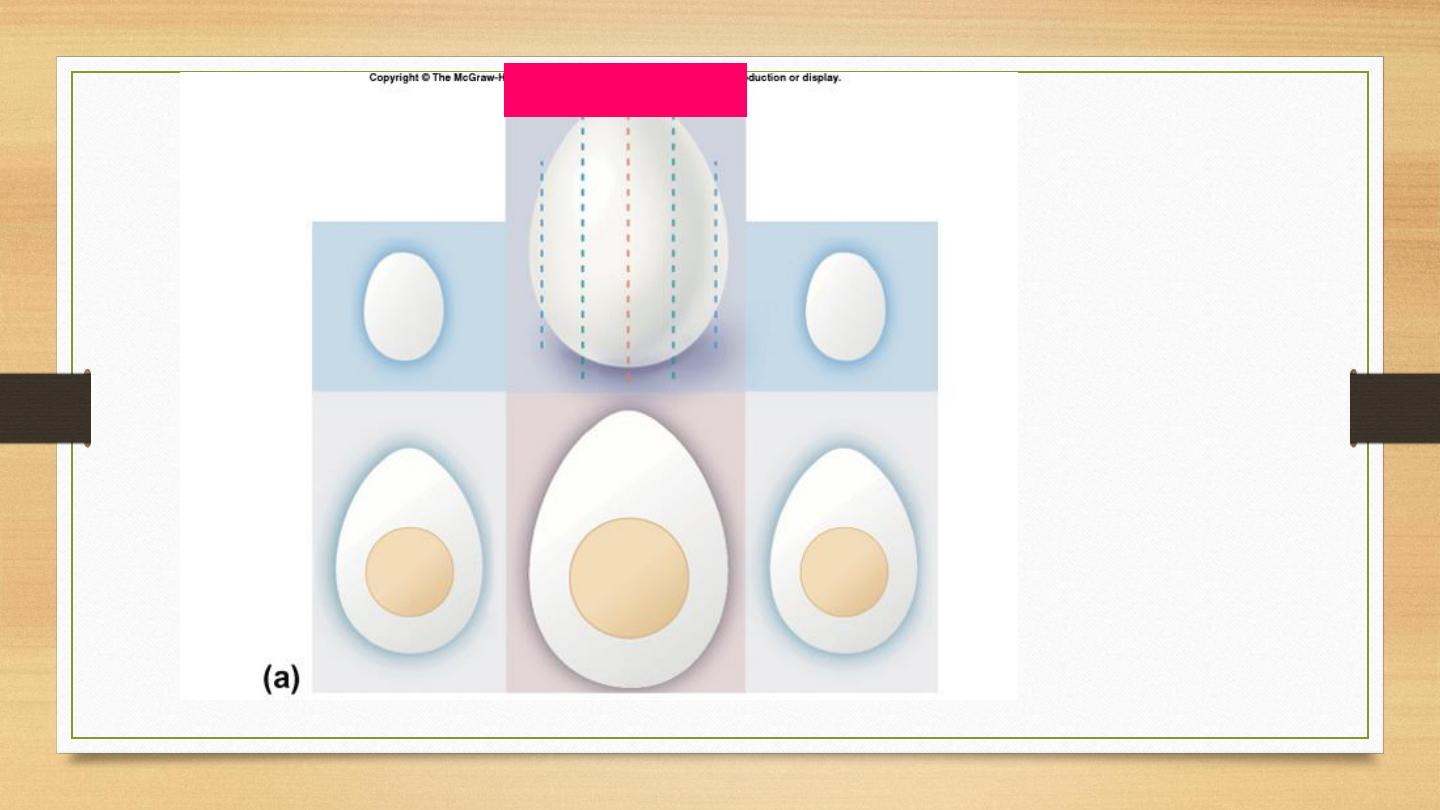
1 2 3 4 5
1
2
3
4
5
•Slices 1 & 5
miss the yolk
/ cell nucleus
•Cell nucleus
is smaller in
sections 2 &
4
Tissue Sectioning
8
9
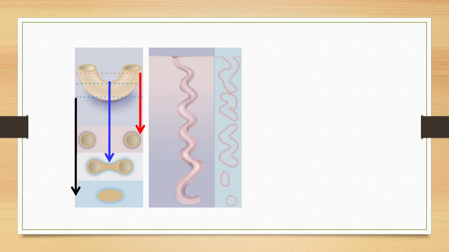
Tissue Sectioning
•
Image A
is a cross
section of elbow
macaroni, resembling a
blood vessel, piece of
gut, or other tubular
organ.
•
Image B
is a
longitudinal section of
a sweat gland. Notice
what a single slice
could look like
A
B
10
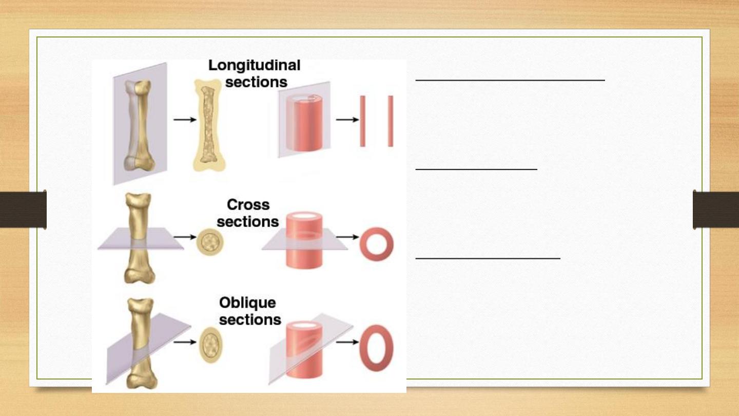
§ Types of Tissue Sections
•
Longitudinal section
•
tissue cut along the longest
direction of an organ
•
Cross section
•
tissue cut perpendicular to
the length of an organ
•
Oblique section
•
tissue cut at an angle
between a cross &
longitudinal section

Original 4 types of tissues:
Epithelial tissues
– surface coverage
Muscular tissues
– contractile property
Nervous tissues
– cells forming brain, spinal cord, and
nerves
Connective tissues
– to link or support other specialized
tissues

Epithelial tissue
A component of many organs specialized for absorption, secretion, and/or to act as a
barrier.
They may cover or form a lining for body surfaces.
May form functional secretory glands.
Firmly joined together by adhesion specialization:
To anchor the cytoskeleton of the neighboring epithelial cells together,
To anchor the epithelial cells to the underlying/surrounding extracellular
matrices.
Modified/specialized on the surface to fulfill their specific roles.
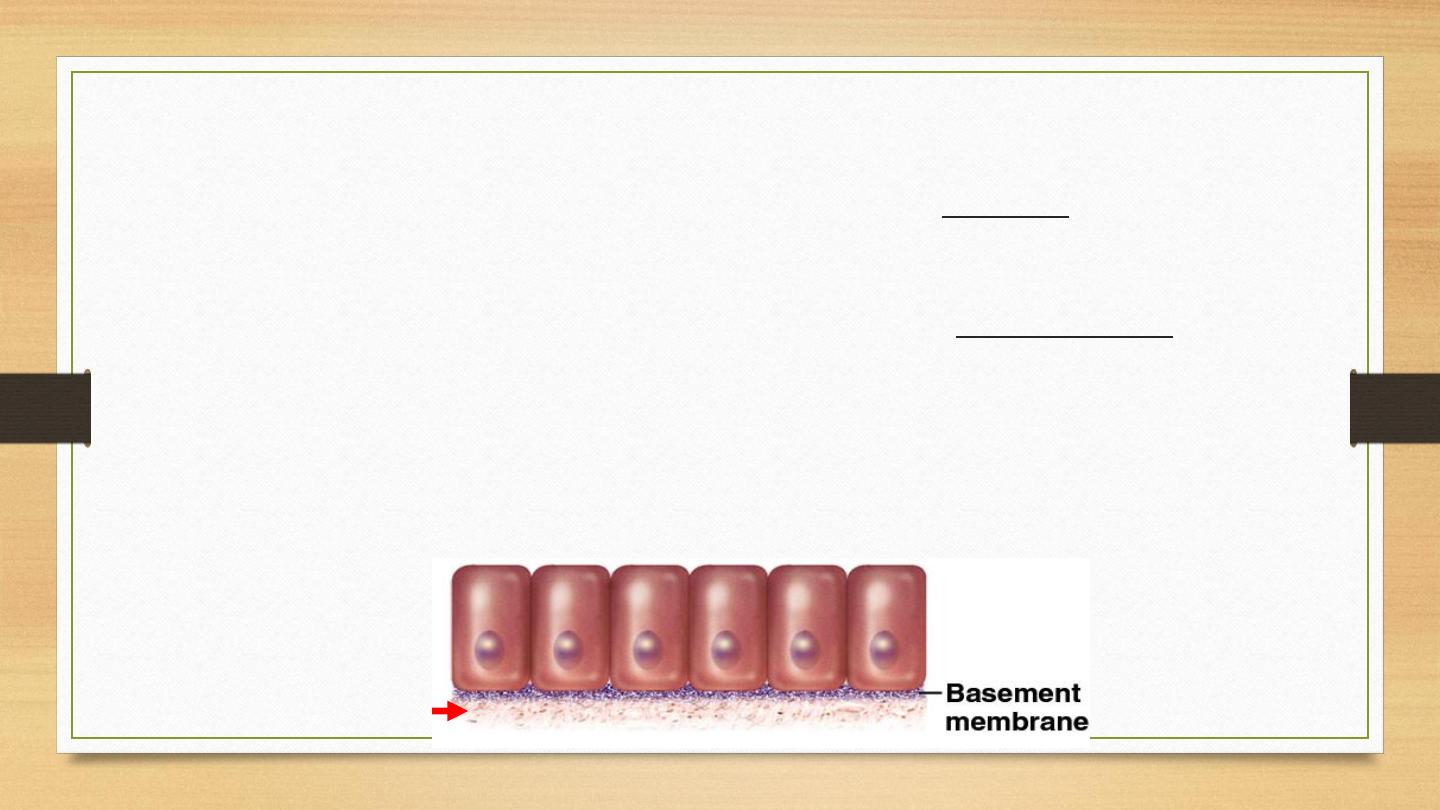
§ Epithelial Tissue Introduction
1.
One or more layers of closely adhering cells
2.
(Top) Forms a flat sheet with the upper (______) surface
exposed to the environment or an internal body cavity
3.
(Bottom) Sits on basement membrane (basal surface of
cells)
•
anchors epithelium to underlying connective tissue
4.
(Nourishment) No room for blood vessels; . . .
CT

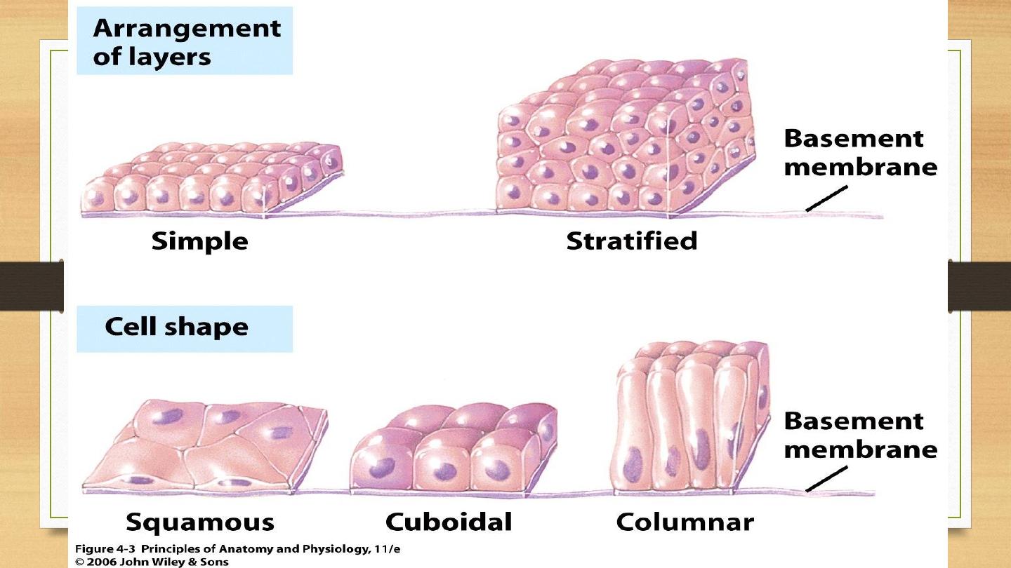
Classification of Epithelial cells: by their shape
and their stacking pattern
By shape (morphology):
•
Squamous (flat, plate-like)
•
Cuboidal (height and width similar)
•
Columnar (height = 2x – 5x greater than width)

Covering of external surfaces
Lining of cavities
Limiting structure
Control passage of substances
Variety of other functions
Compact sheets of cells
Very little intercellular substance
Basement membrane
Avascularity…
supporting tissue required.

By stacking:
•
Simple: forming a single layer, all the cells contact the underlying extracellular
matrix.
•
Stratified: multiple layer of cell stacking, where only the bottom layer is in
contact with the extracellular matrix.
•
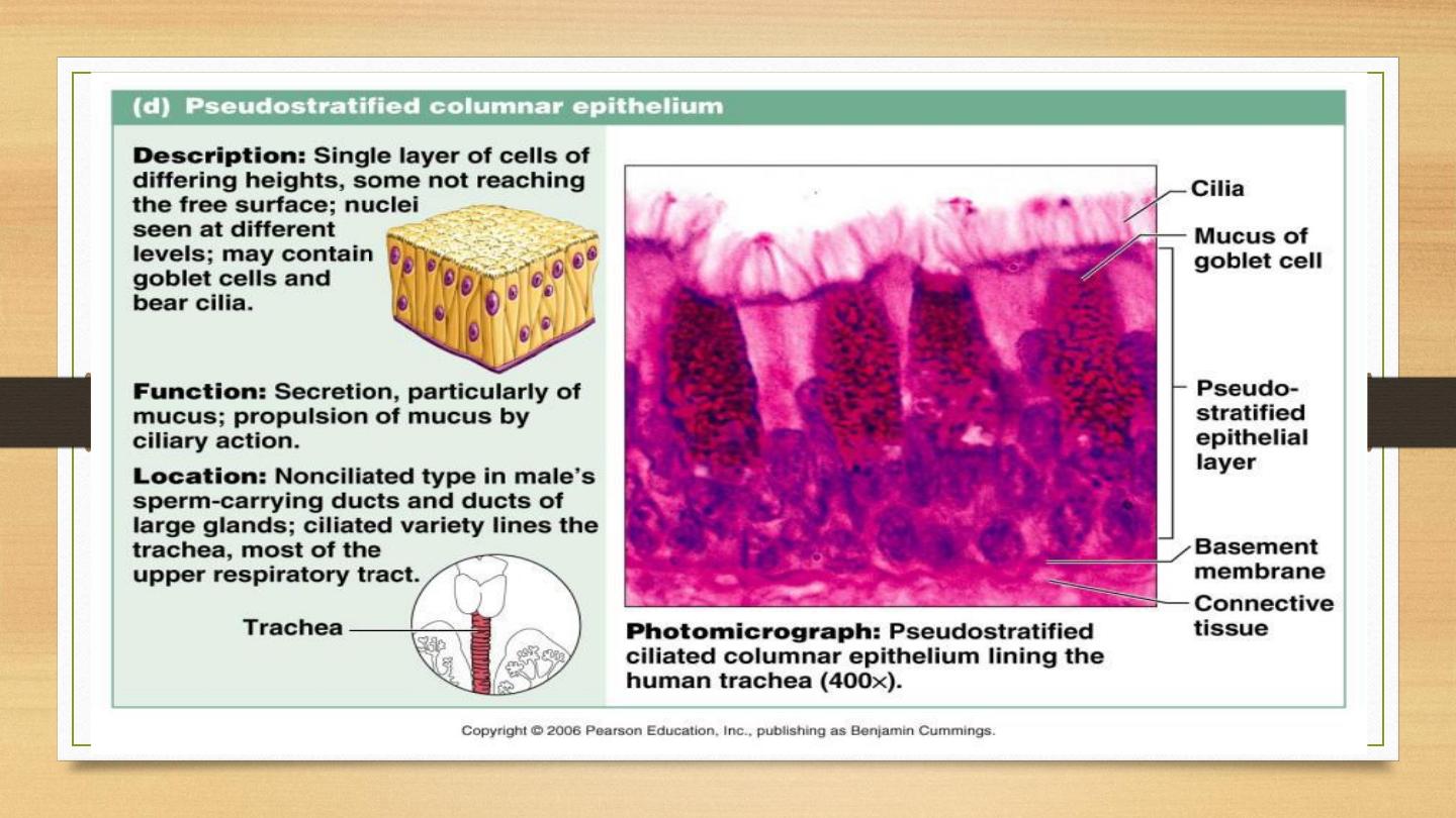
Pseudostratified: cells appear arranged in layers, but all in contact with the
extracellular matrix.
•
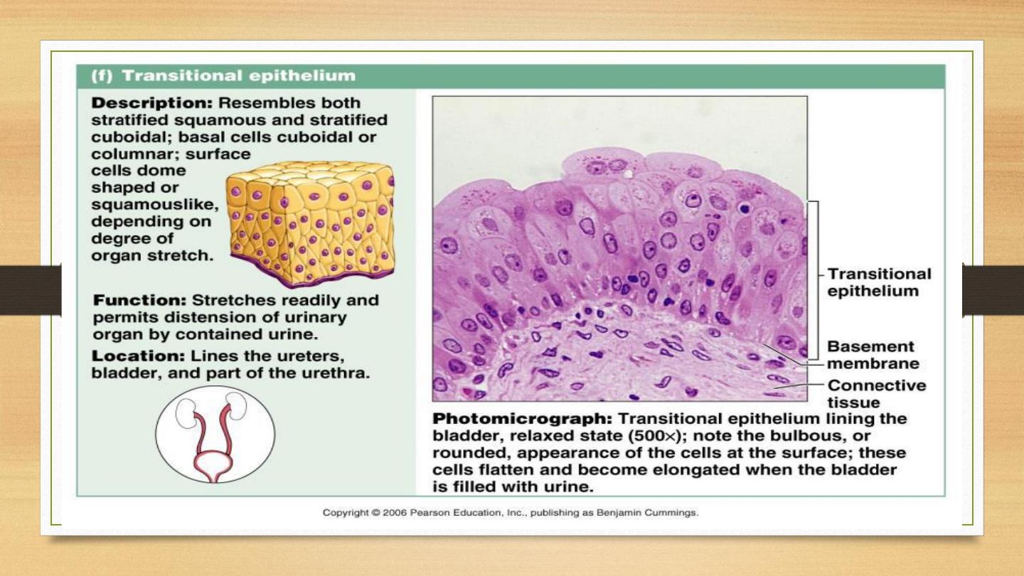
Transitional: specialized epithelium only in the urinary tract, varies between
cuboidal and squamous, depending on the degree of stretching.
17
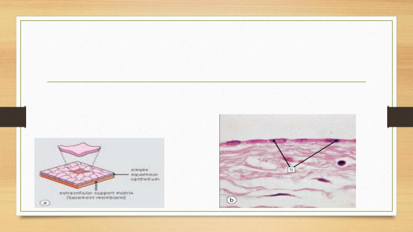
Simple squamous epithelium:
•
Consisted of a single layer of cells that are flat and plate like.
Many having such characteristics have specialized name, such as
endothelium.
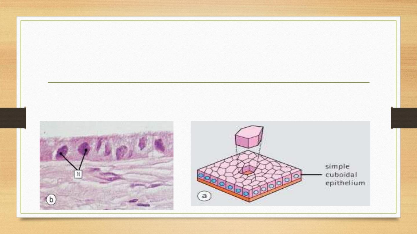
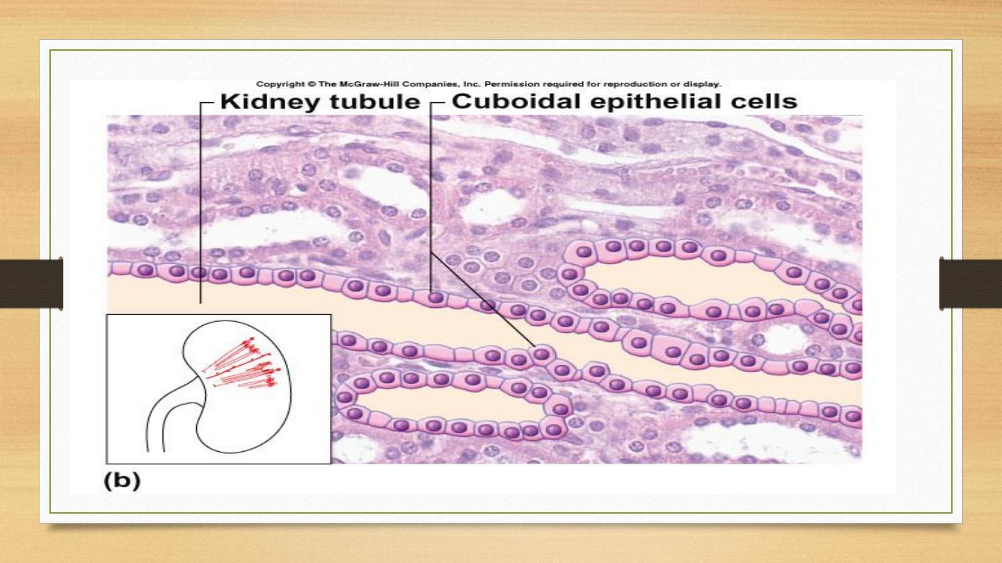
Simple cuboidal epithelium
•
A single layer of cells whose height, width, and depth are almost the same,
cells that have a basic cube shape.
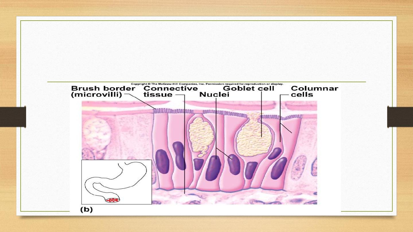
Simple columnar epithelium
•
A single layer of cells whose height is two to five times greater than its width.

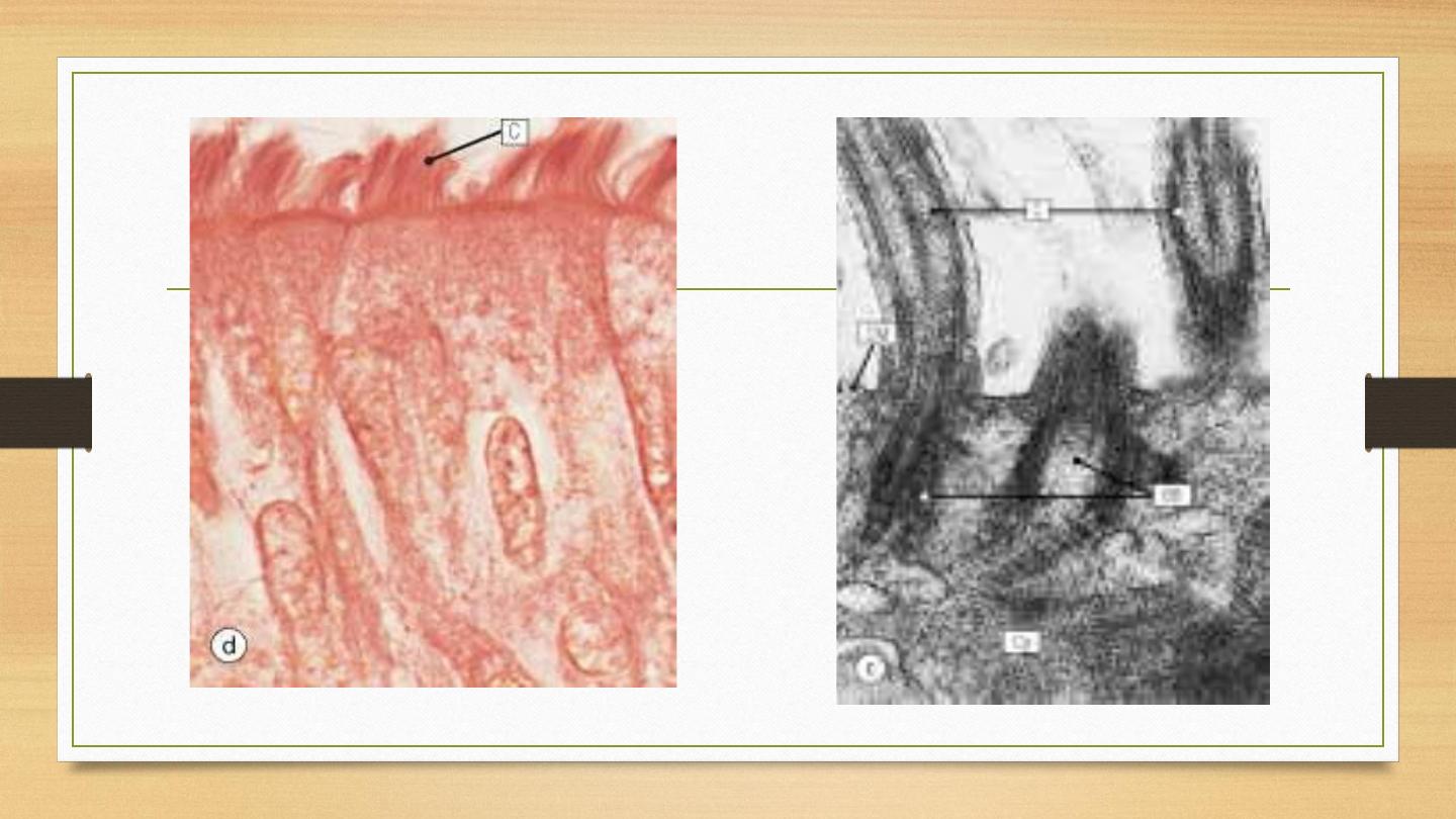
Special features of epithelium
•
Cilia- (singular= cilium, Latin= eyelash)- hair-like appendages attached to the apical surface
of cells that act as sensory structures or to produce movement.
•
Goblet cells- specialized cells that produce mucus to lubricate and protect the surface of an
organ
•
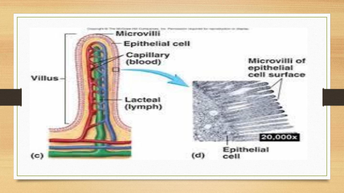
Villi- (singular= villus, Latin= shaggy hair)- finger-like projections that arise from the
epithelial layer in some organs. They help to increase surface area allowing for faster and more
efficient adsorption.
•
Microvilli- smaller projections that arise from the cell's surface that also increase surface area.
Due to the bushy appearance that they sometimes produce, they are sometimes referred to as
the brush border of an organ.

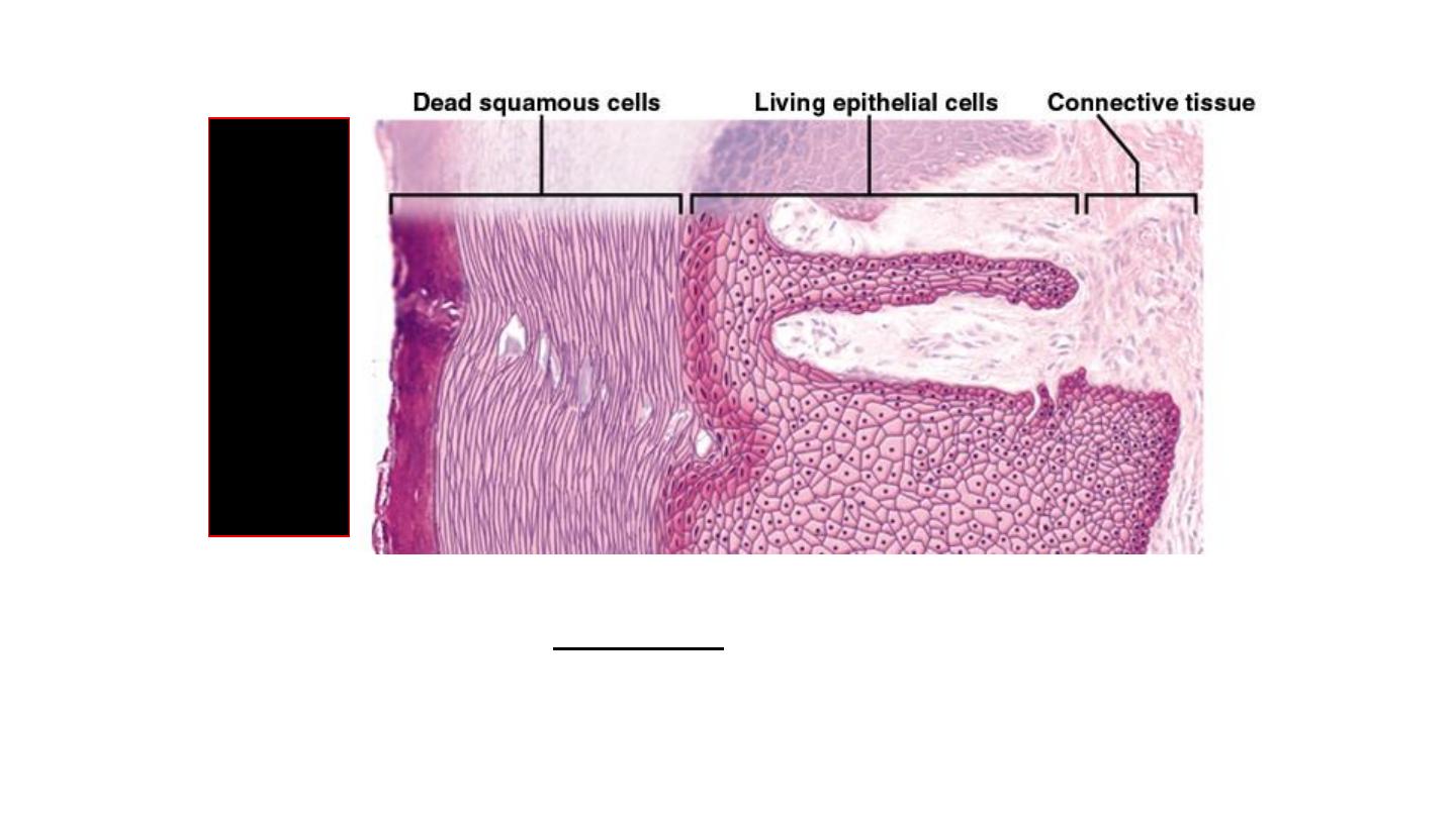
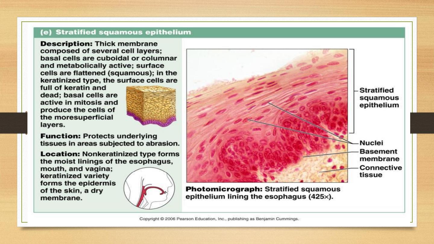
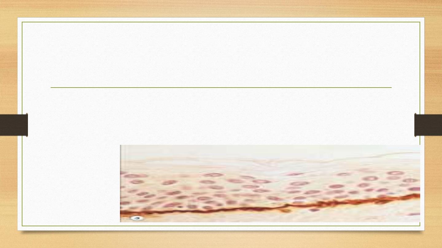
Stratified squamous epithelium
•
Multiple layers of stacked cells.
•
Upper layer: squamous (flattened) shape.
•
Middle and basal (bottom) layer: pyramidal or polygonal shape.
24
Keratinized Stratified Squamous
• Layers of epithelium covered with compact, ______
squamous cells (no nuclei) packed with protein keratin
• Retards water loss, prevents entrance of organisms
• Forms epidermal layer of skin (
esp.
soles and palms)
Fig.
5.
8
Sk
in fr
om
the sole of
the foot

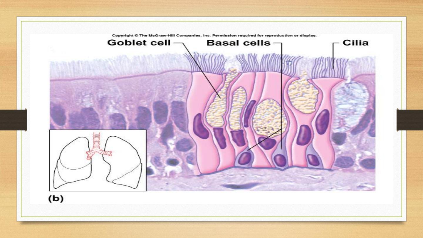
Pseudostratified columnar epithelium:
Multiple layers of nuclei, suggesting multiple layer of cells
But all the cells are in contact with the underlying extracellular matrix (basal
membrane).

Epithelial Cell Junctions:
•
Junction: specialized structures in the epithelia that link (adhere) individual
cells together to form a functional unit.
•
Two main systems involved in the cell adhesion:
1. Cell membrane proteins function as specialized cell adhesion molecules.
2. Specialized areas of cell membrane incorporated into cell junctions.

•
Three types of cell junctions:
1. Occluding junctions: Link cells to form impermeable barrier.
2. Anchoring junctions: Link cells to provide mechanical strength.
3. Communicating junctions: Allow movement of molecules between cells.

Occluding junctions
Function:
•
Prevention of diffusion of molecules between adjacent cells.
•
Prevention of lateral migration of specialized cell membrane proteins.
•
Delineating and maintaining specialized cell membrane domains.
•
Also known as tight junction ultrastructurally.
•
Well developed in the intestinal epithelia:
•
Prevent digested macromolecules from passing between the cells.
•
Confine specialized area of cell membrane involved in absorption or
•
secretion to the luminal side of the cell.

Occluding Junction: Also found in cells actively transport substances.
Prevent the back-diffusion of the transported substance.
Occludin and claudin are involved in the formation of occluding junctions.

Anchoring Junction
•
Provide mechanical stability to groups of epithelial cells.
•
Extracellular interaction may be mediated by additional extracellular proteins
or ions(such as cadherins).

Actin network interact with two types of junctions:
•
Adherent junctions link the actin filament network between adjacent cells.
•
Focal contacts link the actin filament network of a cells to the extracellular
matrix.

Adherent Junctions
•
Most common toward the apex of adjacent columnar and cuboidal
epithelial cells. Forms adhesion belt by linking the submembranous actin
bundles.
•
Prominent in the cells lining the small intestine, forming an eosinophilic
band
•
Transmit motile forces generated by the acting filaments across the whole
sheets ofcells.
•
Essential in mediating folding of epithelial sheet to form early organs in
the embryo
.

Intermediate filament network interact with two different
types of junctions:
•
Desmosomes that connect the intermediate filament
networks of adjacent cells.
•
Hemidesmosomes connect the intermediate filament
network of cells to
extracellular matrix.

Desmosomes
•
Very good characteristics of epithelial cells.
•
Provide mechanical stability in epithelial cells subject to tensile and
shearing
•
stresses.
•
Well developed in stratified squamous epithelium covering the skin.
•
A biomarker in differentiating the origin of the invasion in the malignant
tumors of uncertain nature.

Junctional complex
•
The close association of several types of junction
between adjacent epithelial cells.
•
A manifestation of the requirement for several types of
attachment between epithelial cells to maintain structural
and functional integrity.

Communication Junction (Gap Junction)
•
Allow selective diffusion of molecules between adhacent cells
and facilitate cell-cell direct communication.
•
Found mostly in embryogenesis.
•
In cardiac and smooth muscle: signal passage between cells.
•
In some cerebellar synapses: direct synapses.
Basement Membrane
•
Anchors epithelial cells to the underlying tissues.
•
Contains Type IV collagen synthesized by the epithelial cells.
•
Appears as a linear structure at the base of epithelia, can be stained with PAS
stain.

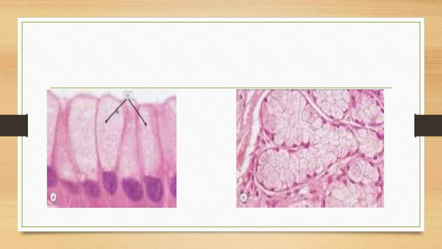
Microvilli
•
Finger-like projections of the apical cells surface.
•
Most developed in absorptive cells like kidney tubule cells and epithelia of small
intestine.
•
Morphology: maintained by bundle of actin filaments that anchored to the actin
cortex.
•
Surface of microvilli: specific cell surface glycoprotein and enzymes related to
absorption process.

Cilia
•
Hair-like projections, ~ 0.2 =m in diameter, arise from the surface of
certain specialized cells.
•
Involved in moving fluid over the surface of the cell or to give cells
motility.
•
Highly specialized extension of cytoskeleton (microtubules).
•
Microtubules bound with other proteins to produce energy-dependent
movement causing side-to-side beating.
•
Evident in respiratory tract epithelium (moving mucus), epithelium of
fallopian tube
(moving ova to the uterus)

Mucin-secreting epithelial cells: contains
greatly expanded Golgi system
•
Mucins: mixture of glycoproteins and proteoglycans.
Features:
•
Well-developed basal rER (stained faint blue) to the basal cytoplasm.
•
Well-developed supranuclear Golgi for protein glycosylation
•
Large secretory vesicles of mucins at cell apex impart an unstained
vacuolated appearance to the apical cell cytoplasm.
•
May be part of the surface epithelium which is called goblet cell.
•
May aggregate into specialized glands.

•
Four types of secretion by epithelial cells:
•
Exocrine secretion:
Merocrine, apocrine, and holocrine: deliver
through the apex of cell into a lumen.
•
Endocrine secretion: secretion from the side or the
base of cells into bloodstream
.

Epithelial cells grouped into secretory
glands:
•
Gland:
organized collection of secretory epithelial cells.
•
Invagination of surface epithelial cells to form the straight or coiled ducts,
or more complex, branched glands.
•
Regions of glands are divided into specialized zones for the secretion of
different products.