Dr. Zana
Nervous system HistologyIntroduction
The nervous system is designed to deliver rapid and precise communication between different parts of the body by the action of specialized nerve cells called neurons.
These highly specialized cells are interconnected and function to gather and process information and then generate appropriate response signals
The nervous system is divided into two main parts
The central nervous system (CNS) comprising the brain and spinal cord.
The peripheral nervous system (PNS) comprising the nerves which run between the CNS and other tissues, together with nerve 'relay stations' termed ganglia
Functionally, the nervous system is divided into the somatic nervous system which is involved in voluntary functions, and the autonomic nervous system which exerts control over many involuntary functions. Histologically, however, the entire nervous system merely consists of variations in the arrangement of neurons and their supporting tissues
The functions of the nervous system depend on a fundamental property of neurons called excitability. As in all cells, the resting neuron maintains an ionic gradient across its plasma membrane thereby creating an electrical potential.
Excitability involves a change in membrane permeability in response to appropriate stimuli such that the ionic gradient is reversed and the plasma membrane becomes depolarised
depolarized; a wave of depolarization, known as an action potential, then spreads along the plasma membrane. This is followed by the process of repolarization in which the membrane rapidly re-establishes its resting potential.
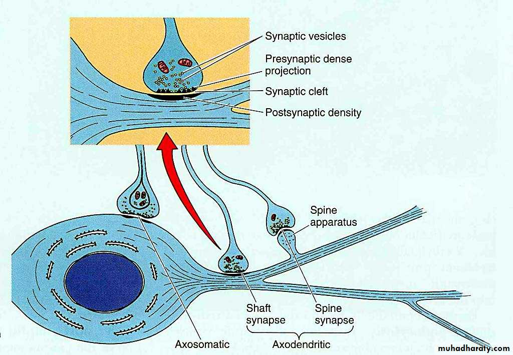
The sites of intercommunication between neurons are termed synapses. Depolarization of one neuron causes it to release chemical transmitter substances, neurotransmitters, which Initiate an action potential in the adjacent neuron.
Within the nervous system, neurons are arranged to form pathways for the conduction of action potentials from receptors to effector organs via integrating neurons.
neurotransmitters not only mediate neuron-to-neuron transmission but also act as chemical intermediates between the nervous system and effector organs which also exhibit the property of excitability
The Synapse
Concept: Synapses are highly specialized intercellular junctions which link the neurons of each nervous pathway.
Similar intercellular junctions link neurons and their effector cells such as muscle fibers; where neurons synapse with skeletal muscle they are referred to as neuromuscular junction or motor end plate.
Classification of synapses:
According the constitution:axodendritic synapse
axosomatic synapse
axoaxonal synapse
dendro-axonic
dendro-dendritic
somato-somatic synapse
somato-dendritic synapse
The effector organs of voluntary nervous pathways are generally skeletal muscle while those of involuntary pathways are usually smooth muscle, cardiac muscle and muscle-like epithelial cells (myoepithelial cells) within some exocrine glands.
Neuron
Despite great variation in size and shape in different parts of the nervous system, all neurons have the same basic structure.
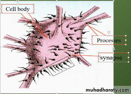
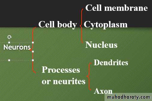
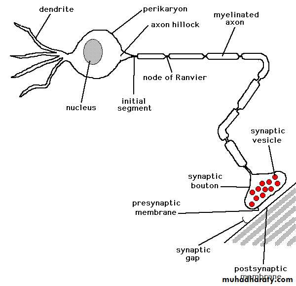
The neuron consists of a large cell body containing the nucleus surrounded by cytoplasm known as the perikaryon. Processes of two types extend from the cell body, namely a single axon and one or more dendrites
Cell body: Perikaryon
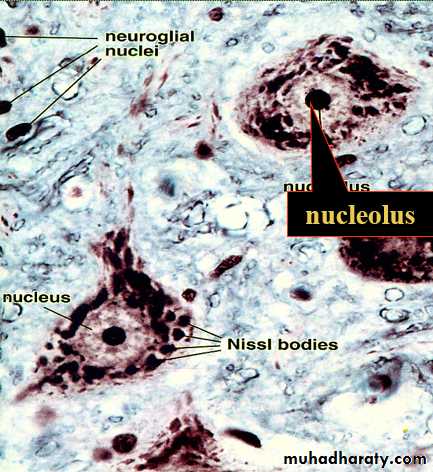
The cell body, soma, is the part of neuron that contains nucleus and surrounding cytoplasm, also called perikaryon. It is the trophic center of the neuron. The protein and enzymes synthesis in this area.
Position: only in grey matter in CNS which also contains dendrites and axons starting from or ending on the cell bodies,ganglia in PNS
Shape:They can be pyramidal, spherical, ovoid or pear-shaped.
Size: Measuring 5-150 um in diameter.
(1)Cell membrane: the structure is as the same as the normal cell. It functions in getting the stimuli and integration and conducting the nerve impulse.
(2) The nucleus is large and pale with H-E stain, prominent nucleoli are very clear.
(3)Cytoplasm: the cytoplasm has some distinctive characteristics not seen in other cells. The cytoplasm is basophilic and full of neurofibrils.
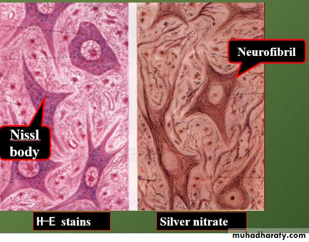
Nissl bodies: The cytoplasm shows the presence of a granular material that stains intensely with basic dyes; this material is the Nissl substance (also called Nissl bodies or granules).
Neurofibrils are thin black fibers observed in LM with silver nitrate slides, which is composed of microtubule and filaments in EM.
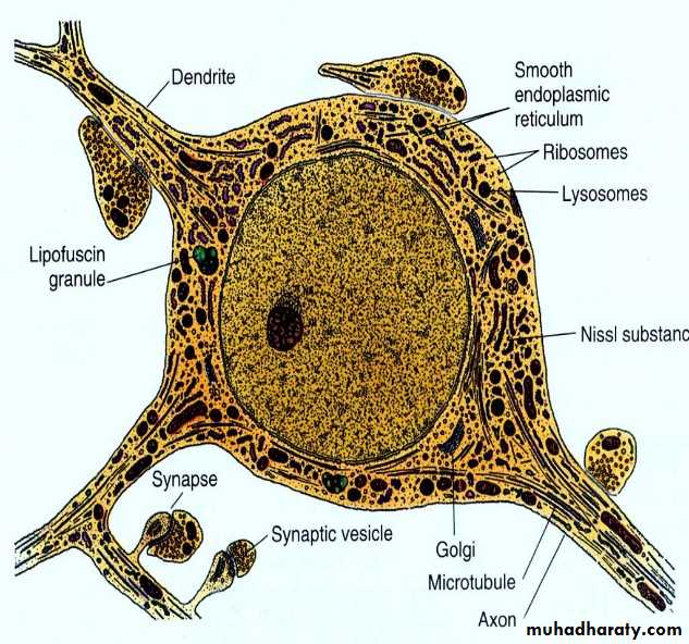
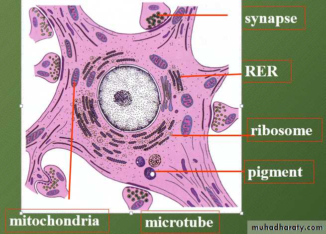
EM: rough surfaced endoplasmic reticulum.
The presence of abundant granular endoplasmic reticulum is an indication of the high level of protein synthesis in neurons. Mitochondria, SER, lysosomes, Golgi complexes, ribosome etc.The proteins are needed for maintenance and repair, and for production of neurotransmitters and enzymes.
Dendrites are highly branched, tapering processes which either end in specialized sensory receptors (as in primary sensory neurons) or form synapses with neighboring neurons from which they receive stimuli. In general, dendrites function as the major sites of information input into the neuron
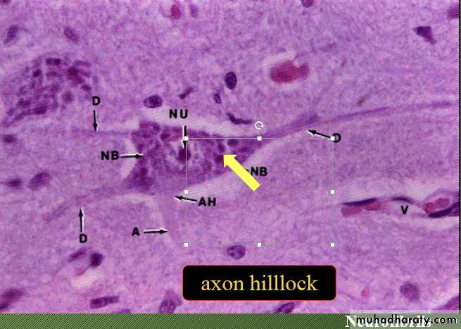
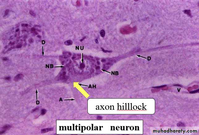
Each neuron has a single axon arising from a cone-shaped portion of the cell body called the axon hillock The axon is a cylindrical process up to 1 meter in length terminating on other neurons or effector organs by way of a variable number of small branches which end in small swellings called terminal boutons
An axon may have not much branches than that of dendrites. If branches, that arise near the cell body and lie at right angles to the axon are called collaterals. At its termination the axon breaks up into a number of fine branches called telodendria which may end in small swellings (terminal boutons.
The axon is identified according to the axon hillock with LM.The part of the axon just beyond the axon hillock is called the initial segment.
Neurites or processes
Dendrites Axonsmany one
short long
irregular in thickness uniform in diameter
Nissl granules No Nissl substance
spines axon hillock
impulse towards the soma away from the cell body
Action potentials arise in the cell body as a result of integration of afferent (incoming) stimuli; action potentials are then conducted along the axon to influence other neurones or effector organs.
Axons are commonly referred to as nerve fibres
In general, the cell bodies of all neurones are located in the central nervous system; exceptions are the cell bodies of most primary sensory neurones and the terminal effector neurones of the autonomic nervous system where, in both cases, the cell bodies lie in aggregations called ganglia in peripheral sites
Basic neuron types
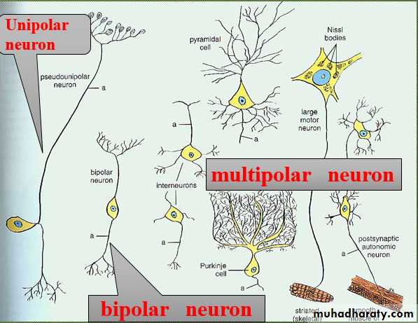
Throughout the nervous system, neurons have a wide variety of shapes which fall into three main patterns according to the arrangement of the axon and dendrites with respect to the cell body.The most common form is the multipolar neuron (90% of neurons) in which numerous dendrites project from the cell body; the dendrites may all arise from one pole of the cell body or may extend from all parts of the cell body
In general, intermediate, integratory and motor neurons conform to this pattern.
Bipolar neurons have only a single dendrite which arises from the pole of the cell body opposite to the origin of the axon. These unusual neurons act as receptor neurons for the senses of smell, sight and balance.
Most other primary sensory neurons are described as pseudo-unipolar neurons since a single dendrite and the axon arise from a common stem of the cell body; this stem is formed by the fusion of the first part of the dendrite and axon of a bipolar type of neuron during embryological development
According to the size of cell body and the length of axon:
Golgi type I neurons: long axonGolgi type II neurons: short axon
According to their function:
Sense (afferent) neuronsInterneurons
Motor (efferent) neurons
According to the neurotransmitter they release
Cholinergic neurons: acetylcholin
Aminergic neurons: adrenaline, non
Peptidergic neurons: neuropeptids
Neuroglia or glia: neurons are supported by a special kind of connective tissue within the brain and spinal cord, that is called neuroglia,it also located in the PNS.
Neuroglia:
Within the central nerve system:
Oligodendrocytes
astrocytes
microglia
ependymal cells
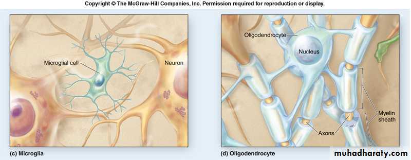
Oligodendrocytes
small cells that are active in the formation and maintenance of myelin in the CNS.
Round bullous cells with slender cytoplasm wrap around nerve axon.
Smaller than astrocytes with fewer processes.
found in both grey and white matter of CNS
particularly in white matter, processes from these cells form the myelin sheaths that are around many axons
analogous to Schwann cells of peripheral nervous system
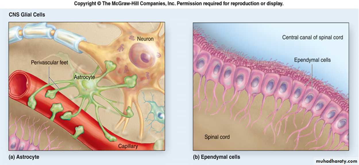
Astrocytes
Star shape and most numerous typeprovide physical support for neurons
store glycogen
isolate synaptic areas from one and other
In the brain, processes abut against the basement membrane of capillary endothelium (pedicles) forming the blood-brain barrier
other processes are closely applied to neurons (pedicles)
may form a conduit for nutrients from blood vessels to neurons
Two types of astrocytes
1. Protoplasmic astrocytes
granular cytoplasm, many branches on short processes
found mainly in gray matter
2. Fibrous Astrocytes
have longer slender processes
found mainly in white matter (but also occur in gray matter).
Microglia
small cell body that is usually elongated and stains denselysometimes an elongate nucleus with mostly heterochromatin (Other glia have spherical nucleus)
many of what were thought to be microglia under the light microscope, have turned out to be oligodendroglia when cells were examined with the electron microscope.
microglial cells are derived from mesoderm and originate from monocytes.
microglial cells function in phagocytosis - components of immune system, act as brain macrophages.
known to migrate and accumulate at the site of nerve damage within the central nervous system.
Ependymal cells
ciliated cells forming single layer of simple cuboidal to low columnar epithelium that lines the entire neurocoelEpithelial cells that line ventricles and central cavities of brain and spinal cord-secrete CSF
ciliary action acts to circulate cerebral spinal fluid.