
Physiology excitable tissues
( Nerve Fibers )
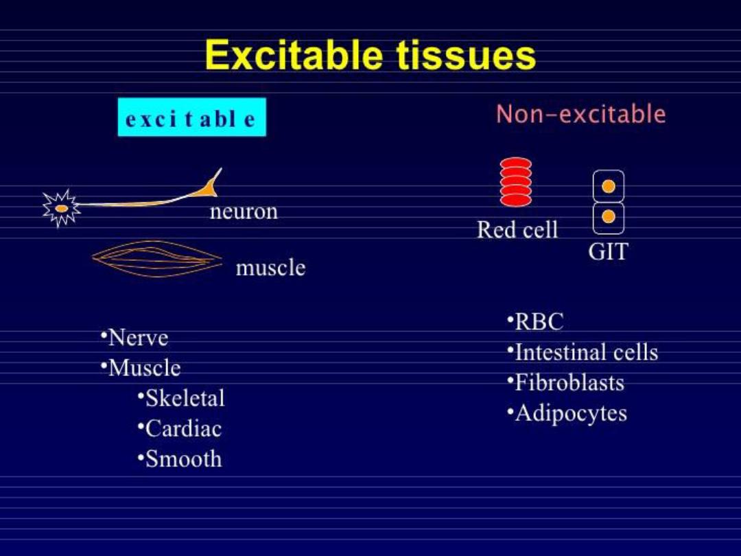

EXCITABLE TISSUE
:
• When an external stimulation (electrical ,
chemical ,mechanical ,physical ) is applied
, an electrical activity is generated and
conducted along their fibers

Neurons and Neuroglia
The human central nervous system (CNS)
contains about
100 billion
neurons
(electrical impulse conducting cells)
. It
also contains
10-50 times
this number of
glial cells (supporting cells)
.
The neurons are the basic building blocks
of the nervous system, their specialized
function is to integration and
transmission of nerve impulses.
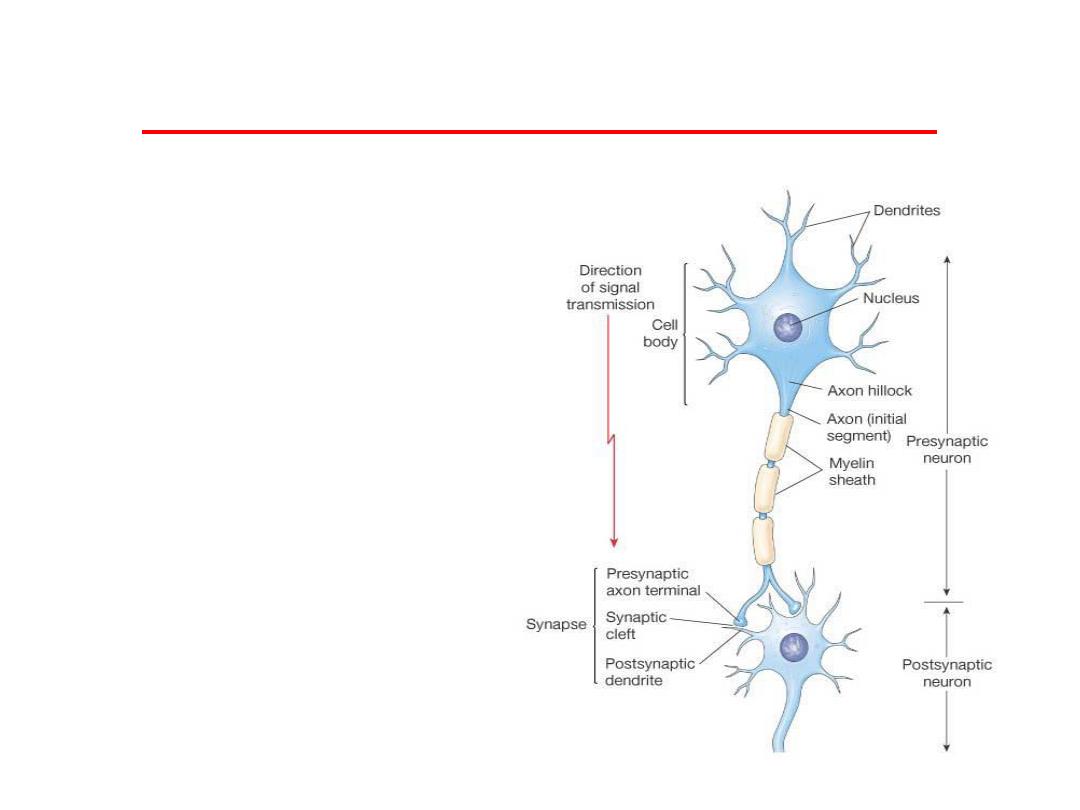
Morphology of the nerve cell
Neurons in the CNS have
different shapes and size;
however most of them
have the same parts:
A. Dendrites
B. Cell Body ( soma )
C. Axon
D. Nerve ending
E. Myelin

• Cell body:
contains nucleus,
cytoplasm and cell organelle.
• Dendrites:
are multiple small
projections from the cell body, their
function is acting as receptors of
nerve impulses from another nerve
cell and transmit the impulses
toward the cell body.

• Axon:
originate from thickened area
of the cell body called axon hillock.
the first part of the axon is called
initial segment, the axon ends by
dividing into terminal branches each
ending by a number of synaptic
knobs .The function of the axon is
transmission of nerve impulses to
the nerve ending.

• Nerve ending:
terminal parts of
the axon, they divide into a
number of synaptic knobs is
called (terminal buttons) contain
granules or vesicles in which the
synaptic transmitters secreted by
the nerve are stored.

• Myelin:
is a sheath of protein-lipid
complex that wrapped around the axon
of many nerve fiber (myelinated).In the
CNS it is formed by the oligodendrocyte
and in the PNS is formed by shwan cells.
*the myelin sheath envelops the axon
except at their ending and at Ranvier
nodes which are a periodic constrictions.
The myelin sheath is considers as
electrical insulator.

*some nerves are unmyelinated thus
simply surrounded by shwan cells
without the wrapping that forms myelin.
*in the multiple sclerosis crippling
autoimmune disease patchy destruction
of myelin occur in CNS.
*the loss of myelin is associated with
delayed or blocked conduction in the
demyelinated axon.

Functional organization of neurons
There are 4 functional zones in neurons :
• 1) Receptor Zone : it is represented by soma
and dendrites , where nervous impulses are
received ,integrated and multiple graded
electro genesis occurs
• 2) Initial Segment Zone : represented by the
initial segment of the axon where origination
of conducted impulses occurs

• 3) Axonal Zone : transmission & conduction of
impulses , represented by axon and its
branches
• 4) Nerve Ending Zone: where impulses causes
secretion of synaptic neurotransmitters to
affect other tissues (gland ,skeletal muscles)
,represented by synaptic vesicles

Protein Synthesis and Axoplasmic
Transporort
• Neurons are secretory cells that synthesize
proteins in cell body by endoplasmic reticulum
and Golgi apparatus, then, the proteins are
secreted from axonal endings ( synaptic
vesicles ) , therefore ,there should be a method
of transportation between cell body and
terminals called " AXOPLAXMIC TRANSPORT "
,it occurs along the microtubules , the
axoplamic transport divided in to two types :

• 1) Anterograded Transport : transporting substances
from soma to axonal endings , occurs at 2 speeds :
• A) Fast : occurs at rate (400 mm\day) , transports cell
organelles ( synaptic vesicles ) mediated by Kinesin
(microtubular protein)
• B) Slow : occurs at rate ( 0.5- 10 mm\day ), it involes
the polymerization and depolymerization of
cytoskeleton
• 2 ) Retrograde Transport : the transport from axon
terminal to soma , at rate of ( 200 mm\day) , involoves
the transport of used vesicles, viruses , Nerve Growth
Factors (NGF) , mediated by cytoplasmic Dyenin
(microtubular protein )

Excitation and neuronal conduction
Nerve cells have a low threshold for
excitation. The stimulus may be electrical,
chemical, or mechanical. Two types of
physicochemical disturbances are
produced:-
1.
Local, non propagated
potentials called
generator, or
electrotonic potentials
. (CNS)
2
. Propagated disturbances
, the
action
potentials
(or nerve impulses- PNS).

Resting Membrane Potential
Distribution of ions
( intracellular and extracellular) :
• Na+ (outside): 142 mEq/L
• Na+ (inside): 14 mEq/L
• K+ (outside): 4 mEq/L
• K+ (inside): 140 mEq/L
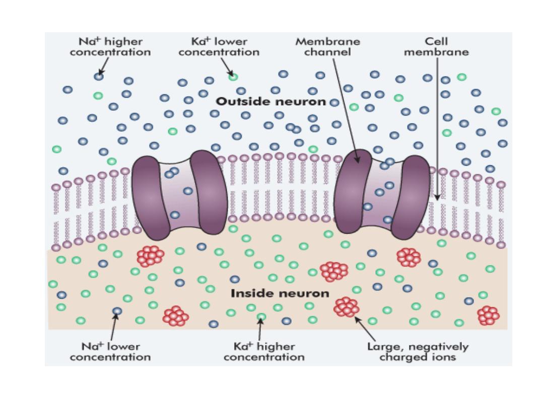

THE IONIC BASIS OF RMP
• How The RMP is maitaned nearly -70 mv ??
• There are three primary factors :
• 1) Active (Na+ – K+) pump
• 2) Membrane permeability (difference
between Na , K)
• 3) Presence of anions inside the cell

• In neurons, like other cells , there is an active
( Na+ - K+) pump system that pushs Na+ actively
to the outside of the cell and draws K+ actively to
the inside of the cell.
• As a result, Na+ will accumulates outside the cell
while K+ inside the cell, Na+ ions tries to enter
inside the cell passively down its concentration
gradient ,

but since the cell membrane is much more
permeable (50-100) times to K+ than Na+, so
passive K+ efflux (from inside to outside ) is more
than the passive Na+ influx ( from outside to insid
) leading to accumulation of more positive charges
outside the cell than the inside.

• Moreover, there are proteins (negatively
charged) inside the cell that cannot get out
increasing the negativity inside , all the three
factors cause the cell (neuron) to be polarized
in the resting state, it's RMP.

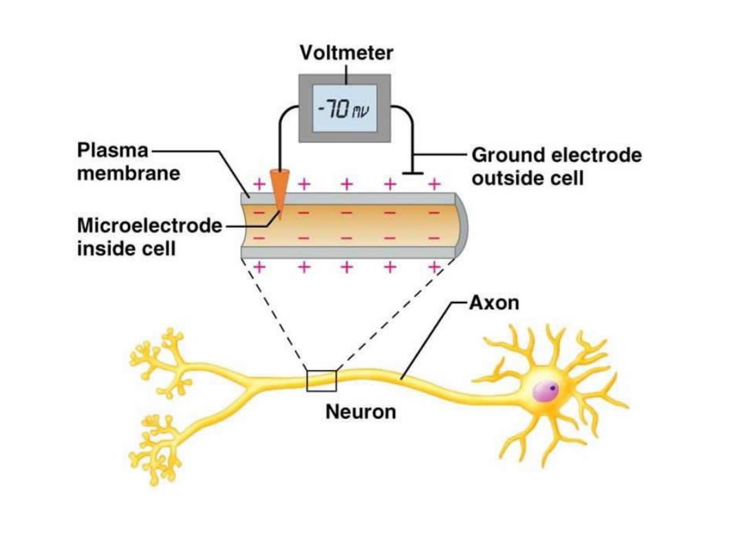
Resting Membrane Potential of Nerves
The resting membrane potential of large
nerve fibers when not transmitting nerve
signals is about
–70 millivolts. That is, the
potential inside the fiber is 70 millivolts
more negative than the potential in the
extracellular fluid on the outside of the
fiber.

• Resting Stage.
This is the resting membrane potential
before the action potential begins.
The membrane is said to be
polarized duri g this stage e ause
of the
–70 millivolts negative
membrane potential that is present.

• Nerve Action Potential
Nerve signals are transmitted by
action potential
s, which are
rapid
changes in the membrane potential
that spread rapidly along the nerve
fiber membrane.
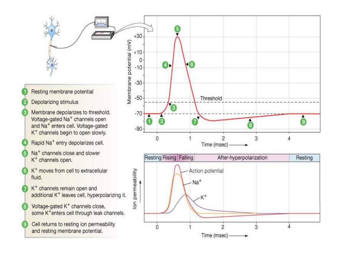

• These are the only electrical responses
of neurons and other excitable tissues,
and they are the main language of the
nervous system. They are due to
changes in the conduction of ions
across the cell membrane that are
produced by alterations in ion channels.
• The impulse is normally transmitted
(conducted) a long the axon to its
termination.

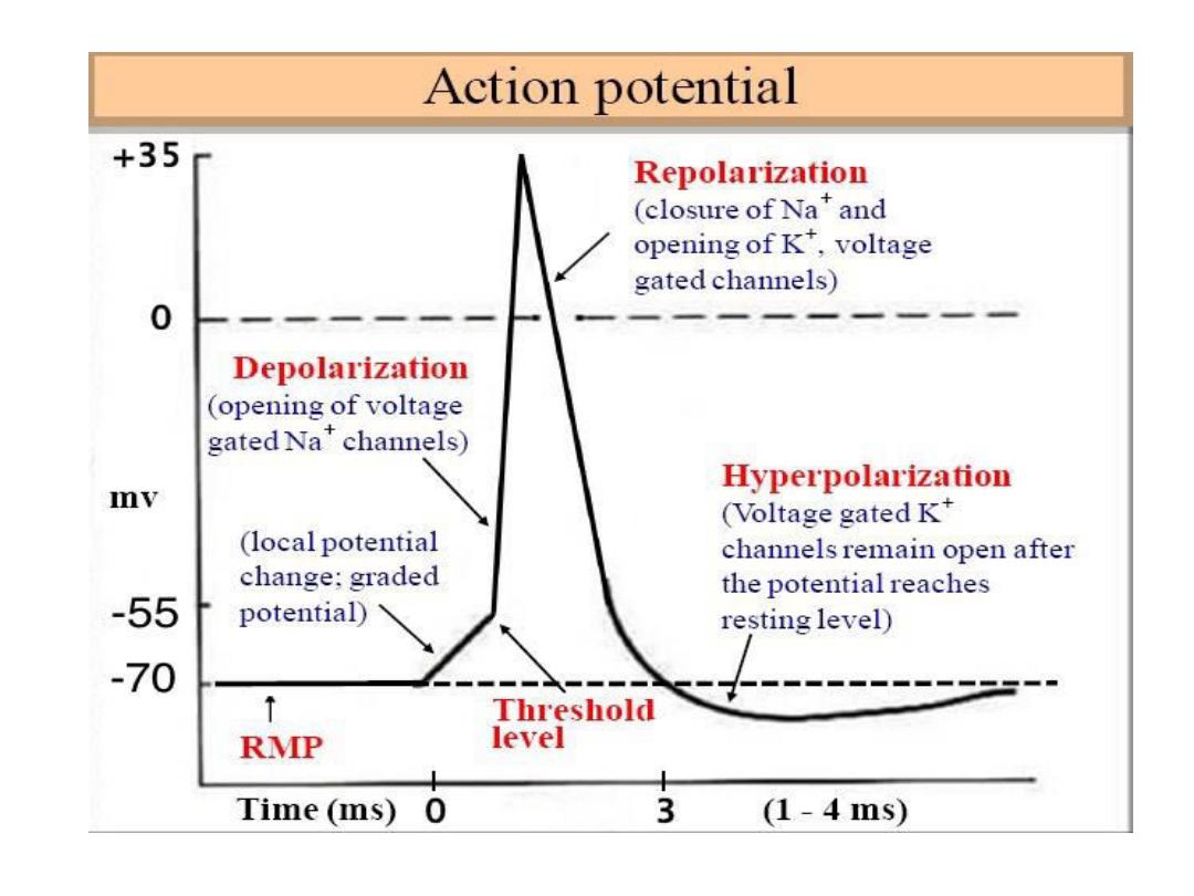
• Each action potential begins with a sudden
change from the normal resting negative
membrane potential to a positive potential
and then ends with an almost equally rapid
change back to the negative potential.
• To conduct a nerve signal, the action
potential moves along the nerve fiber until
it o es to the fi er’s e d. The su essive
stages of the action potential are as follows

• Depolarization Stage.
At this time, the membrane suddenly
becomes very permeable to sodium ions,
allowing tremendous numbers of
positively charged sodium ions to diffuse
to the interior of the axon. The normal
polarized state of –70 millivolts is
immediately neutralized by the inflowing
positively charged sodium ions, with the
potential rising rapidly in the positive
direction. This is called depolarization

• In large nerve fibers, the great excess of
positive sodium ions moving to the inside
causes the membrane potential to
a tually overshoot eyo d the zero
level and to become somewhat positive.
In some smaller fibers, as well as in many
central nervous system neurons, the
potential merely approaches the zero
level and does not overshoot to the
positive state.

• Repolarization Stage.
Within a few 10,000ths of a second
after the membrane becomes highly
permeable to sodium ions, the
sodium channels begin to close and
the potassium channels open more
than normal. Then, rapid diffusion of
potassium ions to the exterior

• re-establishes the normal negative resting
membrane potential. This is called
repolarization of the membrane. To explain
more fully the factors that cause both
depolarization and repolarization, we need
to describe the special characteristics of
two other types of transport channels
through the nerve membrane: the voltage-
gated sodium and potassium channels.

The Events That Cause the Action Potential
During the resting state, before the action
potential begins, the conductance for
potassium ions is 50 to 100 times as great as
the conductance for sodium ions. This is
caused by much greater leakage of potassium
ions than sodium ions through the leak
channels.

• However, at the onset of the action
potential, the sodium channels
instantaneously become activated and
allow up to a 5000-fold increase in
sodium conductance. Then the
inactivation process closes the sodium
channels within another fraction of a
millisecond.

• The onset of the action potential also
causes voltage gating of the potassium
channels, causing them to begin opening
more slowly a fraction of a millisecond after
the sodium channels open. At the end of
the action potential, the return of the
membrane potential to the negative state
causes the potassium channels to close
back to their original status, but again, only
after an additional millisecond or more
delay.
Initiation of the action potential
A Positive-Feedback Vicious Cycle Opens the
Sodium Channels.
As long as the membrane of the nerve fiber remains
undisturbed, no action potential occurs in the
normal nerve. However, if any event causes
enough initial rise in the membrane potential
from
–90 millivolts toward the zero level, the
rising voltage itself causes many voltage-gated
sodium channels to begin opening.

This allows rapid inflow of sodium
ions,which causes a further rise in the
membrane potential,

Thus, opening still more voltage-gated
sodium channels and allowing more
streaming of sodium ions to the interior
of the fiber. This process is a positive-
feedback vicious cycle that, once the
feedback is strong enough, continues
until all the voltage-gated sodium
channels have become activated
(opened).
Threshold for Initiation of the Action
Potential
An action potential will not occur until the
initial rise in membrane potential is great
enough to create the vicious cycle
described in the preceding paragraph.. A
sudden rise in membrane potential of 15 to
30 millivolts usually is required.

Therefore, a sudden increase in the
membrane potential in a large nerve
fiber from
–90 millivolts up to about –65
millivolts usually causes the explosive
development of an action potential. This
level of
–65 millivolts is said to be the
threshold for stimulation.