
Physiology of nerve Fiber
Lecture 2
Continued

Properities of action potential
1 )
the threshold
:
It's the minimum intensity of stimulus needed to
excite the neuron and produce an action
potential.
At sub threshold ( sub minimal)
intensity,
there is no action potential
.
The threshold depends on :
A ) type of the axon
B ) temperature

2 ) self reinforcement
The action potential conducts
electrical impulse with the same
strength along whole neuron nerve
fiber )
without the need for external
amount of energy

3)
The all or non law
:
The action potential occur in a constant
size (amplitude) and constant shape (form)
regardless the strength of stimulation is at
or above the threshold value and ,therefore
,the action potential of single nerve fiber is
independent on the stimulation energy
above the threshold value and obey the all
or non-law

• Once an action potential has been
elicited at any point on the membrane of
a normal fiber, the depolarization
process travels over the entire
membrane if conditions are right, or it
does not travel at all if conditions are
not right. This is also called the all-or-
nothing principle, and it applies to all
normal excitable tissues

4)
the refractory period
:
The time interval during which the nerve
fiber is incapable of firing or producing a
second potential after the first one when
the second stimulus is applied ( the period
of beginning of depolarization until the
end of the first third of repolarization ,the
membrane is completely refractory to
further stimulation ) .this is called absolute
refractory period

• No matter how strong the stimulus is
applied , it can't induce second
action potential ,the reason is that
during this period almost all Na +
channels are inactivated and no
stimulation can reopen them.

Propagation of action potential
An Action Potential elicited at any
point on the excitable membrane
usually excites adjacent portion of
the membrane resulting in
propagation of the A.P along the
membrane .
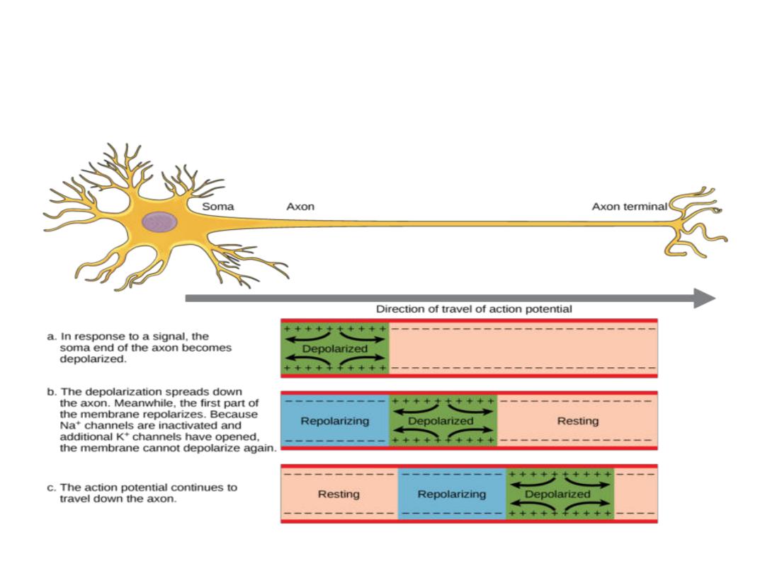
continuous conduction in Non
myelinated fibers

Saltatory
Conduction in Myelinated
Fibers from Node to Node.
• Even though almost no ions can flow
through the thick myelin sheaths of
myelinated nerves, they can flow with
ease through the nodes of Ranvier.
Therefore, action potentials occur only
at the nodes. Yet the action potentials
are conducted from node to node, as
shown in next Figure
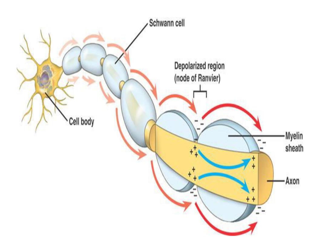

• This is called saltatory conduction
That is, electrical current flows
through the surrounding
extracellular fluid outside the
myelin sheath as well as through
the axoplasm inside the axon
from node to node, exciting
successive nodes one after
another.

• Thus, the nerve impulse jumps
down the fiber, which is the
origi of the ter saltatory.
Saltatory conduction is of value
for two reasons.
First
, by causing
the depolarization process to
jump long intervals along the
axis of the nerve fiber,

This mechanism increases the velocity of
nerve transmission in myelinated fibers
as much as 5- to 50-fold.
Second
,
saltatory conduction conserves energy
for the axon because only the nodes
depolarize ,allowing perhaps 100 times
less loss of ions than would be necessary
,and therefore requiring little
metabolism for reestablishing the
sodium and potassium concentration
differences across the membrane after a
series of nerve impulses.
Velocity of Conduction in Nerve Fibers.
• The velocity of conduction in nerve
fibers varies from as little as 0.25
m/sec in very small unmyelinated
fibers to as great as 100 m/sec (the
length of a football field in 1 second)
in very large myelinated fibers.

• Excitation—The Process of Eliciting the
Action Potential
• Any factor that causes sodium ions to
begin to diffuse inward through the
membrane in sufficient numbers can set
off automatic regenerative opening of the
sodium channels. This can result from
mechanical disturbance of the membrane,
chemical effects on the membrane, or
passage of electricity through the
membrane.

• All these are used at different points in the
body to elicit nerve or muscle action
potentials:
1. Mechanical pressure to excite sensory nerve
endings in the skin,
2. Chemical neurotransmitters to transmit
signals from one neuron to the next in the
brain, and
3. Electrical current to transmit signals between
successive muscle cells in the heart and
intestine.

Inhibition of
E ita ilit “ta ilizers
and Local Anesthetics
In contrast to the factors that increase nerve
excitability, still others, called membrane-
stabilizing factors, can decrease excitability.
For instance, a high extracellular fluid
calcium ion concentration decreases
membrane permeability to sodium ions
and simultaneously reduces excitability.
Therefore, calcium ions are said to be a
stabilizer
.

Local Anesthetics.
Among the most important stabilizers are
the many substances used clinically as
local anesthetics, including procaine and
tetracaine. Most of these act directly on
the activation gates of the sodium
channels, making it much more difficult
for these gates to open, thereby
reducing membrane excitability.

Electronic potentials
Although
sub threshold sti ulatio does ’t
produce action potentials, they affect the
membrane potential called electronic potentials.
These potentials can be recorded if we put the
recording electrodes too close to the stimulation

These potentials
characterized by
:
1) Local depolarizing potentials.
2) Non propagated.
3) Rise rapidly decay exponentially.
4) Produced by sub threshold.
5) Their size is proportional to the
intensity of stimulation.

6)
They are either :
A) Cathodal : depolarizing potential leads to
excitation
B) Anodal : hyper polarizing , drag the
membrane more (-ve) and causes inhibition
Thus these fibers either excite or inhibit the
potential of the membrane. Electronic potentials
arise in CNS and eye where large no. of
information sent between adjacent cells and
allgebric summation of these potantials
determine the excitability of the neuron.
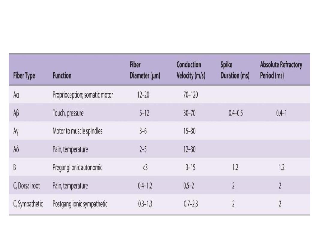
Types of Nerve fibers
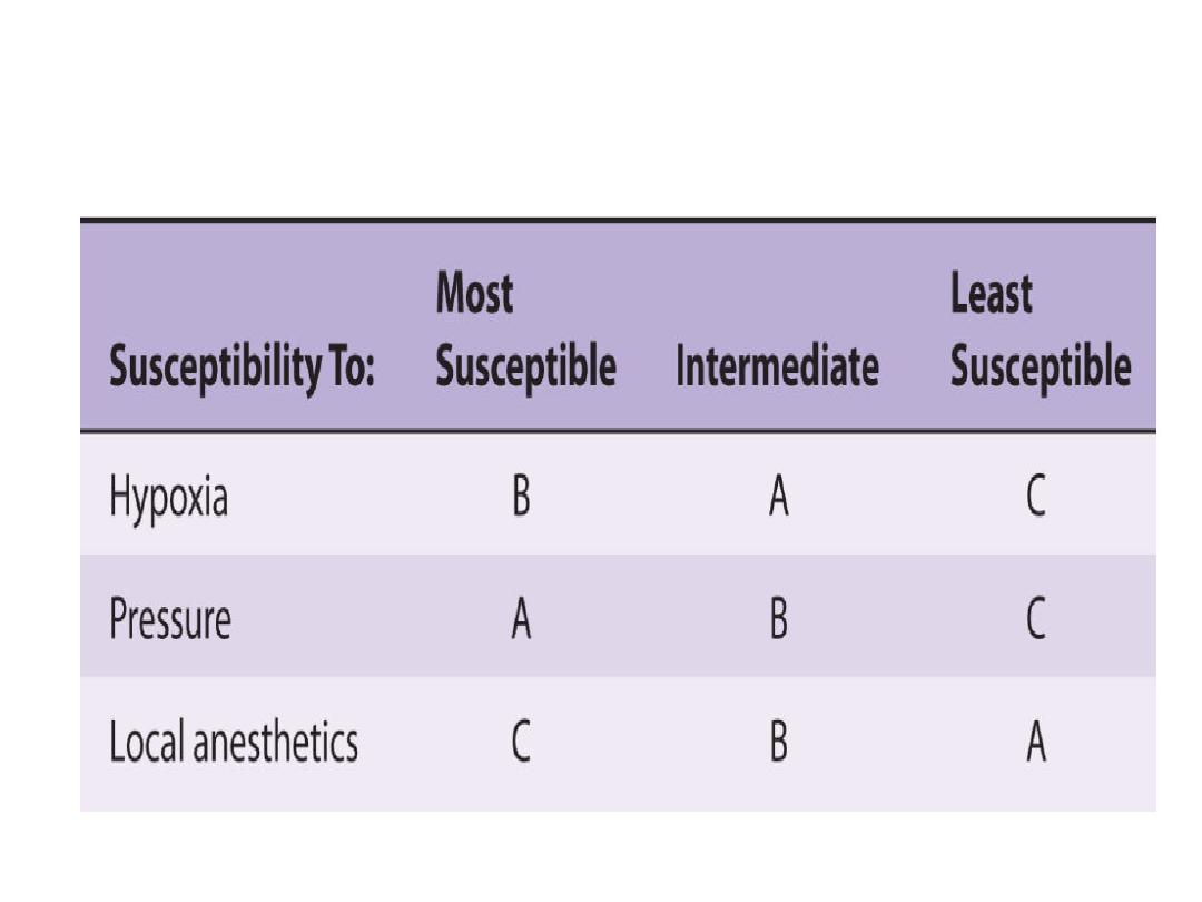
Relative susceptility of mammalian A, B, & C nerve
fibers to conduction block produce by various agents

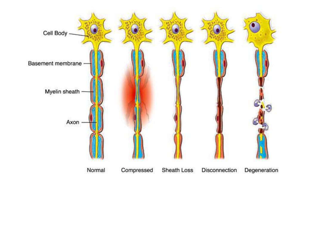
Nerve Injury
In 1943,seddon described three
basic types of peripheral nerve
injury that include:
• Neuropraxia (Class 1)
• Axonotmesis (Class 2)
• Neurotmesis (class 3)
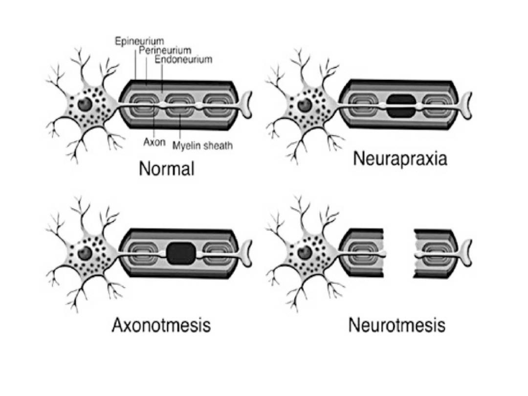

1. Neurapraxia : It is a transient
episode of
little or
no sensory
Neurapraxia describes nerve
damage in which there is no
disruption of the nerve or its
sheath.

• In this case there is an
interruption in conduction of the
impulse down the nerve fiber,
and complete recovery takes
place, &
does not occur.

• This is the mildest form of nerve
injury. This is probably a biochemical
lesion caused by concussion or
shock-like injuries to the fiber. In
the case of the role nerve,
neurapraxia is brought about by
compression or relatively mild, blunt
blows, including some low-velocity
missile injuries close to the nerve.

• There is a temporary loss of
function which is reversible
within hours to months of the
injury ( the average is 6
–8 weeks)
. There is frequently greater
involvement of motor than
sensory function with autonomic
function being retained.

• The
, and the
are intact.
• In neuropraxia, conduction is intact in
the distal segment and proximal
segment, but no conduction occurs
across the area of injury.
• Recovery of nerve conduction deficit is
full,and requires days to weeks.
• EMG shows lack of fibrillation potentials
(FP) and positive sharp waves.

2. Axonotmesis is a disruption of nerve
cell axon, with
slightly proximal to the site of injury.
If
are
damaged, but
and
remain intact is called
axonotmesis

The prognosis is usually good in terms
of recovery. Rate of recovery
depends on the distance from the
site of injury, with axonal
regeneration occurring at 1 to 4
mm/day. Peripheral nerves
regeneration may take several
months.
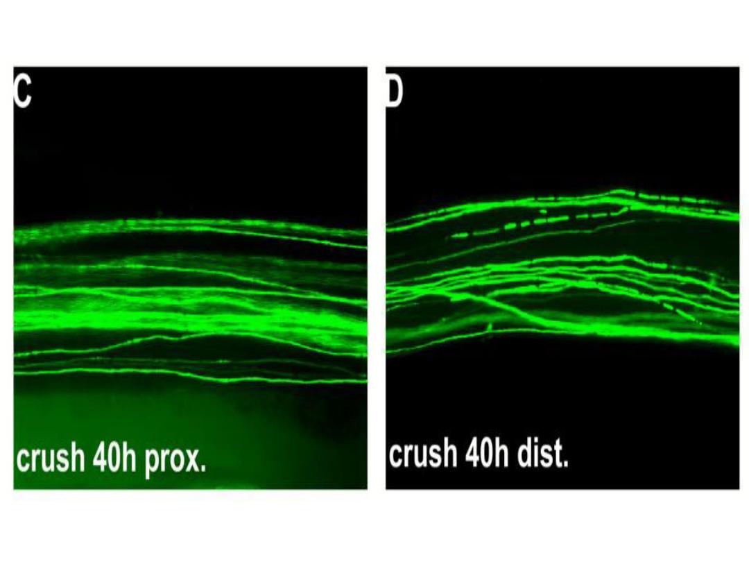
• Wallerian degeneration is a
process that results when a
is cut or crushed, in which
from the
degenerates.
• A related process known as
'Wallerian like degeneration' occurs
in many neurodegenerative diseases,
especially those where
is impaired.
studies suggest that a failure
to deliver sufficient quantities of the
essential axonal protein
is a
key initiating event.
Wallerian degeneration occurs after
axonal injury in both the
(CNS). It occurs in the
axon stump distal to a site of injury and
usually begins within 24-36 hours of a
lesion. Prior to degeneration distal axon
stumps tend to remain electrically
excitable. After injury, the axonal
skeleton disintegrates and the axonal
membrane breaks apart.
• The axonal degeneration is followed
by degradation of the
and infiltration by
. The
macrophages, accompanied by
, serve to clear the
debris from the degeneration

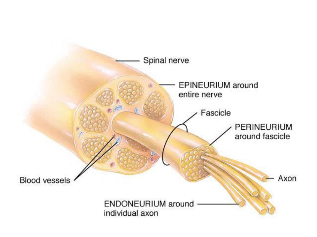
• 3. Neurotmesis:-is a total disruption of
the entire nerve fiber .A peripheral
nerve fiber contains an axon (Or long
dendrite),myelin sheath(if existence),
their schwann cells, and the
endoneurium. Neurotmesis may be
partial or complete.
In this type of injury, both the
and
partial recovery may occur, complete
recovery is impossible

Other characteristics:
• Wallerian degeneration occurs below
to the site of injury.
• There is connective tissue lesion that
may be partial or complete.
• Sensory-motor problems and
autonomic function defect are
severe.

• There is not nerve conduction
distal to the site 0f injury (3 to 4
days after lesion).
• EMG and NCV findings are as
axonotmesis.
• Because of lack of nerve repair,
surgical intervention is necessary.
Wallerian degeneration