
Synaptic Transmission
&
Neuromuscular Junction
Dr. Mahmood Ibrahim
Clinical Neurophysiologist

• A
synapse
is connection between
neuron axon and
dendrites
or
soma
or
even
axon
of another neuron :
• Axo
dendritic
• Axo
somatic
• Axo
axonic
There are electrical & chemical synapses

Physiologic Anatomy of the Synapse
Synapses consist of 3 parts:
presynaptic membrane:
this is the branching
terminal part of an axon which is enlarged at its
end.
Synaptic cleft:
the space which separates the
presynaptic from postsynaptic membrane, the
space is (20-40 nm) wide.
Postsynaptic membrane:
this is the thickened part
of the membrane of the dendrites, cell body or
even axon of postsynaptic neuron.

• It contains receptor proteins to which
neurotransmitters bind.
• In the cerebral cortex 98% of the synapses
are on the dendrites and only 2% are on
the cell bodies, but in the spinal cord the
proportion of ending on dendrites is less for
example: in the anterior horn cells 80-85%
of the terminals lies on dendrites and the
rest on the cell body.


Electrical synapses :-
are direct, ion-conducting cell
–cell
junctions ( bridging) through channels ,
these called (connexions) in the region of
gap junctions. They are responsible for the
conduction of impulses between neurons
in the retina and in the CNS) and ensure
also communication between neighbouring
epithelial or glial cells.

Chemical synapses :-
utilize neurotransmitters for the transmission
of information and provide not only simple 1 : 1
connections, but also serve as switching
elements for the nervous system. They can
facilitate or inhibit the neuronal transmission
of information or process them with other
neuronal input. At the chemical synapse, the
arrival of an action potential (AP) in the axon
triggers the release of the transmitter from the
presynaptic axon terminals.

The transmitter then diffuses across the
narrow synaptic cleft to bind
postsynaptically to receptors in the
subsynaptic membrane of a neuron or of a
glandular or muscle cell. Depending on the
type of transmitter and receptor involved,
the effect on the postsynaptic membrane
may either be excitatory or inhibitory

Properities of synaptic transmission
1) One way conduction :synapse permits
conduction in one direction ( from pre
– to post
synaptic cells
2) Convergence and divergence
• Convergence : when processes of many
presynaptic neurons terminate on a single
postsynaptic neuron
• Divergence : when axon of a single presynaptic
neuron terminates on many postsynaptic
neurons


3) Postsynaptic potentials are
electronic potentials
4) Postsynaptic potentials could be
either excitatory or inhibitory . if the
neurotransmitter causes the opening of
Na+ channels, the Na+ influx occurs and
depolarization happens( excitatory post
synaptic potentials - EPSP

• While if the neurotransmitter opens
K+ channels , hyperpolarization
occurs ( inhibitory postsynaptic
potentials
– IPSP) , if Cl-channels are
opened , also inhibitory post
synaptic potentials occurs

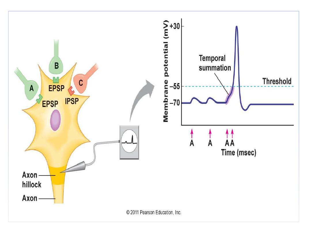
5) summation of postsynaptic
potentials net , we have two types of
summation
A) Spatial : where the effect of
multiple excitatory and inhibitory
postsynaptic potentials occurring at
the same time, if the net results
reaches the firing level , an action
potential is initiated

• B) Temporal summation ; this
happens if repeated stimuli causing
postsynaptic potentials before decay
of previous one ,this occurs where
there's a very short time interval
between the repeated stimuli.so the
effect of first stimulus will be added to
the next and so on until the firing level
is reached


6) Fatigue of synapse : in excitatory
synapses ,repeated stimulation at high
rate causes impairment of synaptic
transmission due to :
• A) Depression of stores of
neurotransmitters
• B) Inactivation of postsynaptic
receptors
• C) Slow build up of Ca+

• 7) synaptic delay : impulse
transmission is delayed at synapse ,
because time is consumed for the
release of neurotransmitter, binding
to receptors, activation and change
in the membrane permeability and
ionic fluxes until the firing level is
reached

The neuromuscular junction
• When axon reaches muscle fiber, it loses its
myelin and divides into a number of branches
(terminal endings)
• Each terminal nerve ending supplies single
muscle fiber ,so each nerve fiber (single
axon), after entering the muscle belly,
normally stimulates from three to several
hundred skeletal muscle fibers
.

•
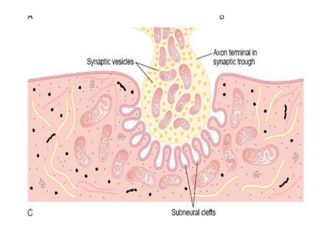
The terminal buttons ( synaptic knobs) lies
in invagination in the muscle fiber
membrane called synaptic gutter
• The muscle fiber is thickened at this area
forming the motor end plate
• The whole structure is covered by one or
more d Schwan cells to isolate it from the
surroundings

• The space between terminal nerve
ending and muscle fiber membrane is
called synaptic cleft
• At the bottom of synaptic gutter the
muscle fiber shows numerous folding
forming subneural clefts which serves to
increase the surface area at which the
neurotransmitter acts
• The muscle fibers at this point ( end
plate) contains large amount ( 50 ) million
specific Acetyl-Cholin ( A-Ch) nicotinic
receptors

•
In the axon terminal ,there are large
amounts of mitochondria which supply
energy for synthesis of A-Ch
• A-Ch is synthesized in the cytoplasm of
nerve terminal and rapidly absorbed and
stored in synaptic vesicle
• There are about 300,000 synaptic
vesicle per each nerve ending, arranged in
the form of rows in the nerve terminal in
an area called Active Zone

• On the side of neuronal membrane ,there
are proteins particles called ( linear dense
bars) ,on each side of them there are other
proteins penetrate the membrane, called
(Voltage Gated Calcium Channels)
• At the neuronal ( synaptic cleft), there's
an enzyme ( A-Ch esterase) which destroys
A-Ch into acetate and cholin

The sequence of events during
neuromuscular transmission
• When action potential is initiated, it's
conducted and reaches the nerve terminal ,it will
activate the voltage gated calcium channels
• (Ca++ ) enters nerve ending, binding to a
specific protein molecule at the inner surface of
synaptic membrane called release site.
• This binding leads to activation of enzyme
(calmodulin Kinase II ) which is going to loosen
the binding of synaptic vesicle to cytoskeleton.


•
The vesicle then moves towards the
membrane, fusing with it and discharging its
contents of A-Ch by process called exocytosis
• Acetyl Cholin (A-Ch) diffuses at the
subneural clefts and binds with nicotinic
receptors at the muscle fiber membrane
• This complex ( A-Ch & its receptor ) actives
and opens ligand gated sodium channels
• Rapid influx of sodium ions causes a
depolarization of muscle fiber membrane

•
This depolarization called motor end plate
potential ( electronic potentials )
• Once threshold values reaches, an action
potential is generated and conducted along
the muscle fiber
• Each nerve impulse release about 60
synaptic vesicle

• each vesicle contains 10 thousands
molecules of A-Ch, this amount is 10 times
enough to activate the number of receptors
required to produce a full end plate
potential
• So there's a safety factor of 10 folds of
neurotransmitter

Destruction of the Released
Acetylcholine by Acetyl cholinesterase
The acetylcholine, once released into the synaptic
cleft, is removed rapidly by two means:
(1) Most of the acetylcholine is destroyed by the
enzyme acetyl cholinesterase, which is attached
mainly to the spongy layer of fine connective
tissue that fills the synaptic space.
(2) A small amount of acetylcholine diffuses out of
the synaptic space.

Drugs That Stimulate the Muscle Fiber by
Acetylcholine-Like Action.
Many compounds, including methacholine,
carbachol, and nicotine, have the same effect on
the muscle fiber as does acetylcholine. The
difference between these drugs and
acetylcholine is that the drugs are not destroyed
by cholinesterase or are destroyed so slowly that
their action often persists for many minutes to
several hours.

Drugs That Stimulate the Neuromuscular
Junction by Inactivating Acetylcholinesterase
.
Three particularly well known drugs, neostigmine,
physostigmine,
inactivate the acetylcholinesterase
in the synapses
so that it no longer hydrolyzes
acetylcholine.
Therefore, with each successive nerve impulse,
additional acetylcholine accumulates and
stimulates the muscle fiber repetitively. This causes
muscle spasm when even a few nerve impulses
reach the muscle.

Unfortunately, it also can cause death due
to laryngeal spasm, which smothers the
person.
Neostigmine and physostigmine combine
with acetylcholinesterase to inactivate it
for up to several hours, after which
these drugs are displaced from the
acetylcholinesterase so that the esterase
once again becomes active.

Drugs That Block Transmission at the
Neuromuscular Junction
A group of drugs known as curariform drugs can
prevent passage of impulses from the nerve
ending into the muscle. For instance, D-
tubocurarine blocks the action of acetylcholine
on the muscle fiber acetylcholine receptors.

Myasthenia Gravis
Myasthenia gravis, which occurs in about 1
in every 20,000 persons, causes muscle
paralysis because of inability of the
neuromuscular junctions to transmit
enough signals from the nerve fibers to
the muscle fibers.
Myasthenia gravis is an autoimmune
disease in which the patients have
developed immunity against their own
acetylcholine receptors.

Regardless of the cause, the end plate potentials
that occur in the muscle fibers are mostly too
weak to stimulate the muscle fibers. If the
disease is intense enough, the patient dies of
paralysis
—in particular, paralysis of the
respiratory muscles .The disease usually can be
ameliorated for several hours by administering
neostigmine or some other anticholinesterase
drug, which allows larger than normal amounts
of acetylcholine to accumulate in the synaptic
space.