
544
CHAPTER 11
The Head and Neck
Cerebral hemorrhage
The pituitary gland, which lies medially in the sella
nerve (Fig. 11.12).
the ophthalmic and maxillary divisions of the 5th cranial
In the lateral wall, the 3rd and 4th cranial nerves, and
which travel through it (Fig. 11.12)
The internal carotid artery and the 6th cranial nerve,
the Cavernous Sinuses
Important Structures Associated with
connect the two cavernous sinuses through the sella turcica.
through the superior petrosal sinus. Intercavernous sinuses
ina. The sinus drains posteriorly into the transverse sinus
the inferior ophthalmic vein and the central vein of the ret
the sphenoid bone (Fig. 11.9). Anteriorly, the sinus receives
lies on the lateral side of the body of
cavernous sinus
Each
through the foramen magnum and the transverse sinuses.
falx cerebelli. It communicates with the vertebral veins
lies in the attached margin of the
occipital sinus
The
jugular vein (Fig. 11.30).
skull through the jugular foramen to become the internal
mastoid antrum of the temporal bone and then leaves the
transverse sinuses. Each sinus turns downward behind the
are a direct continuation of the
sigmoid sinuses
The
the sigmoid sinus.
tentorium cerebelli, and they end on each side by becoming
11.10). Each sinus lies in the lateral attached margin of the
usually a continuation of the straight sinus (Figs. 11.9 and
left transverse sinus
of the superior sagittal sinus; the
begins as a continuation
right transverse sinus
The
vein, it drains into the left transverse sinus.
union of the inferior sagittal sinus with the great cerebral
ebri with the tentorium cerebelli (Fig. 11.9). Formed by the
lies at the junction of the falx cer
straight sinus
The
veins from the medial surface of the cerebral hemisphere.
vein to form the straight sinus (Fig. 11.9). It receives cerebral
the falx cerebri. It runs backward and joins the great cerebral
lies in the free lower margin of
inferior sagittal sinus
The
superior cerebral veins.
lacunae (Fig. 11.2). The superior sagittal sinus receives the
Numerous arachnoid villi and granulations project into the
venous lacunae.
sinus communicates on each side with the
becomes continuous with the right transverse sinus. The
der of the falx cerebri (Fig. 11.9). It runs backward and
lies in the upper fixed bor
superior sagittal sinus
The
diploë of the skull, the orbit, and the internal ear.
have no valves. They receive tributaries from the brain, the
of fibrous tissue; they have no muscular tissue. The sinuses
are lined by endothelium. Their walls are thick and composed
situated between the layers of the dura mater (Fig. 11.2); they
The venous sinuses of the cranial cavity are blood-filled spaces
The Venous Blood Sinuses
the
great cerebral veins,
Bleeding then takes place from the
anteroposterior compression of the head often tears the ante
occur from the cerebral veins or the venous sinuses. Excessive
diately loses consciousness, and the paralysis is evident when
is generally caused by rupture of the
thin-walled lenticulostriate artery, a branch of the middle cere-
bral artery. The hemorrhage involves the vital corticobulbar
and corticospinal fibers in the internal capsule and produces
hemiplegia on the opposite side of the body. The patient imme-
consciousness is regained.
Intracranial Hemorrhage in the Infant
Intracranial hemorrhage in the infant may occur during birth and
may result from excessive molding of the head. Bleeding may
-
rior attachment of the falx cerebri from the tentorium cerebelli.
straight sinus, or the inferior sagittal sinus.
-
-
is
-
■
■
■
■
■
■
turcica (Fig.11.12)
The veins of the face, which are connected with
■
■
the cavernous sinus via the facial vein and inferior
ophthalmic vein, are an important route for the spread
of infection from the face (Fig. 11.9)
described.
the following account, only the main parts of the brain are
brain, a textbook of neuroanatomy should be consulted. In
For a detailed description of the gross structure of the
gland is vital to life and is fully described on page 652.
tion in the sella turcica of the sphenoid bone. The pituitary
11.12). The gland is well protected by virtue of its loca
(Fig.
infundibulum
the undersurface of the brain by the
The pituitary gland is a small, oval structure attached to
Pituitary Gland (Hypophysis Cerebri)
the temporal bone (Fig. 11.9)
along the upper and lower borders of the petrous part of
which run
inferior petrosal sinuses,
superior
The
■
■
and
-
Parts of the Brain
Major Parts of the Brain
Cavities of the Brain
Forebrain
Cerebrum
Diencephalon
Right and left lateral
ventricles
Third ventricle
Midbrain
Hindbrain
Pons
Medullaoblongata
Cerebellum
Cerebral aqueduct
Fourth ventricle
and central
canal
bones; above the anterior and middle cranial fossae; and,
Each hemisphere extends from the frontal to the occipital
(Fig. 11.13).
corpus callosum
of white matter called the
connected by a mass
cerebral hemispheres
sists of two
is the largest part of the brain and con
cerebrum
The
cord through the foramen magnum.
lies inside the cranial cavity. It is continuous with the spinal
The brain is that part of the central nervous system that
Cerebrum
-
posteriorly, above the tentorium cerebelli. The hemispheres
(Fig. 11.2). The cerebral
gray matter
and is composed of
cortex
The surface layer of each hemisphere is called the
(Fig. 11.13).
falx cerebri
which projects the
into
longitudinal fissure,
are separated by a deep cleft, the
Basic Anatomy
545
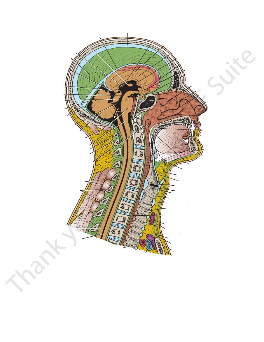
superior sagittal sinus
inferior sagittal sinus
interthalamic connection
thalamus
falx cerebri
great
cerebral vein
pineal
cerebral
aqueduct
midbrain
tentorium
cerebelli
straight
sinus
fourth ventricle
cerebellum
pons
medulla oblongata
atlas
ligamentum nuchae
opening of auditory tube
postvertebral muscles
cervical spines
spinal cord
central canal
brachiocephalic artery
left brachiocephalic vein
manubrium sterni
remnants of thymus
jugular arch
investing layer of
deep cervical fascia
isthmus of thyroid gland
esophagus
trachea
cricothyroid ligament
thyroid cartilage
vocal fold
vestibular fold
epiglottis
thyrohyoid membrane
hyoid bone
tonsil
mylohyoid
geniohyoid
genioglossus
tongue
soft palate
hard palate
inferior
concha
vestibule
of nose
middle concha
agger nasi
superior concha
frontal sinus
hypophysis cerebri
optic nerve
anterior cerebral artery
interventricular foramen
septum pellucidum
corpus callosum
fornix
suprasternal space
arch of cricoid cartilage
1
2
3
4
5
6
7
T1
T2
FIGURE 11.13
Sagittal section of the head and neck.
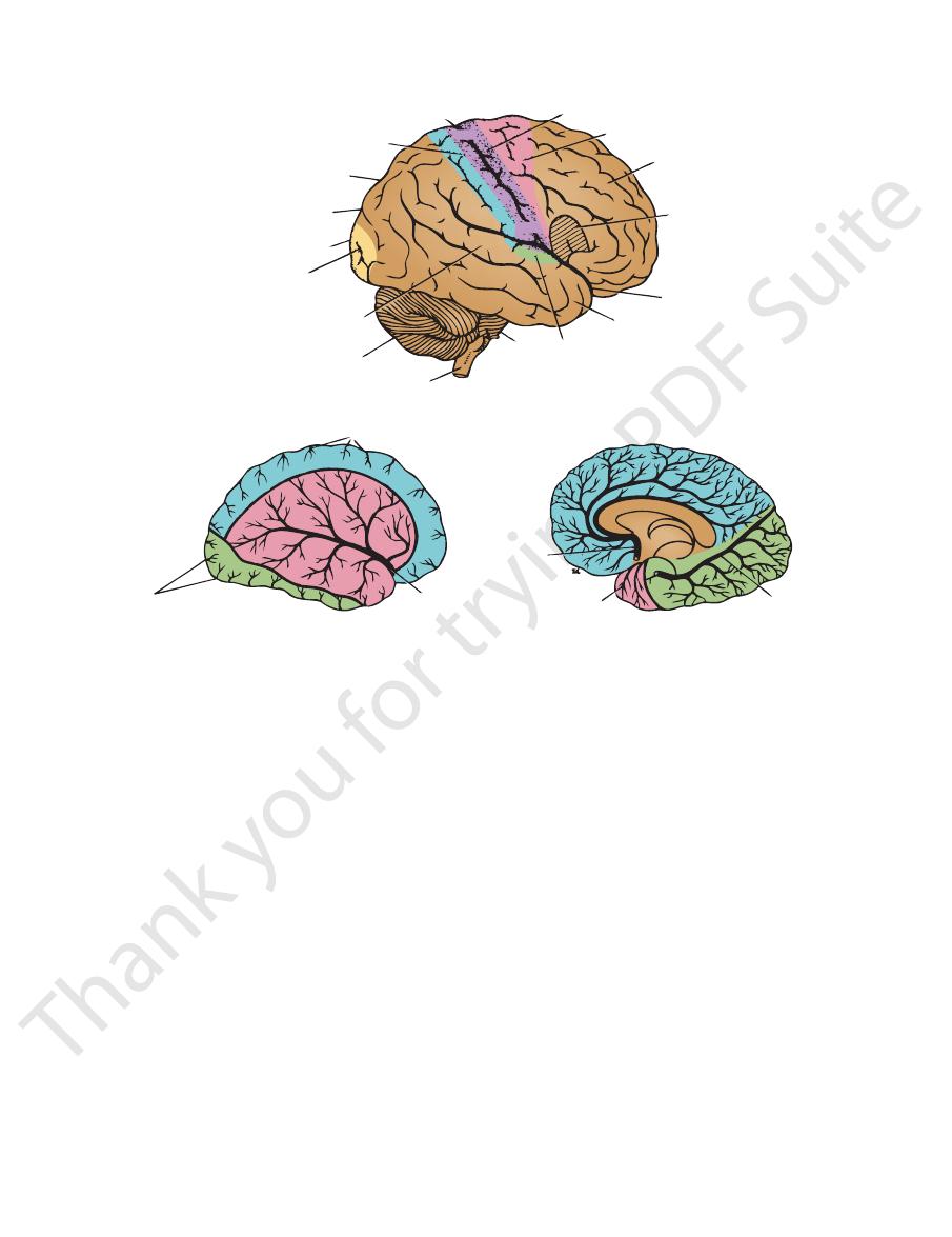
occipital lobe
sulcus. The
is situated behind the central sulcus and above the lateral
parietal lobe
The
lateral sulcus.
(Fig. 11.14) and above the
central sulcus
is situated in front of the
frontal lobe
The
(Fig. 11.14).
named for the bones of the cranium under which they lie
The lobes are
lobes.
the surface of each hemisphere into
increased. Several of the large sulci conveniently subdivide
By this means, the surface area of the cortex is greatly
sulci.
or
separated by fissures,
gyri,
cortex is thrown into folds, or
lies below the
position, with the nerve cells controlling the movements
In the motor area, the body is represented in an inverted
gata as they descend to the spinal cord.
fibers cross over to the opposite side in the medulla oblon
movements on the opposite side of the body. Most nerve
The large motor nerve cells in this area control voluntary
(Fig. 11.14).
motor area
central sulcus and is known as the
lies immediately anterior to the
precentral gyrus
The
lobe.
temporal
Below the lateral sulcus is situated the
sulcus.
parietooccipital
-
546
CHAPTER 11
The Head and Neck
central sulcus
sensory area
parietal lobe
parieto-occipital sulcus
occipital lobe
visual area
superotemporal gyrus
cerebellum
medulla oblongata
pons
auditory area
temporal lobe
lateral sulcus
anterior
motor speech area
(if right hemisphereis
dominant)
frontal lobe
premotor area
motor area
branches of anterior
cerebral artery
anterior
middle
cerebral
artery
branches of posterior
cerebral artery
anterior
cerebral
artery
branches of middle
cerebral artery
posterior
cerebral
artery
A
C
B
foot
face
FIGURE 11.14
A.
by the anterior cerebral artery; those colored red, by the middle cerebral artery; and those colored green, by the posterior
hemisphere showing areas supplied by the cerebral arteries. In this and the next figure, areas colored
Lateral surface of the cerebral
speech area is most commonly located in the left rather than the right cerebral hemisphere.
Right side of the brain showing some important localized areas of cerebral function. Note that the motor
B.
blue are supplied
cerebral artery.
the movements of the face and hands in the lower part
of the feet located in the upper part and those controlling
Medial surface of the cerebral hemisphere showing the areas supplied by the cerebral arteries.
C.
(Fig. 11.14).
The lateral ventri
lateral ventricle.
sphere is called the
The cavity present within each cerebral hemi
visual impressions.
(Fig. 11.14). It is the receiving area for
calcarine sulcus
medial aspect of the cerebral hemisphere in the region of
is situated on the posterior pole and
visual area
The
left-handed persons.
in right-handed persons and in the right hemisphere in
employed in speech. It is dominant in the left hemisphere
the lateral sulcus (Fig. 11.14). It controls the movements
lies just above
motor speech area,
or the
Broca’s area,
auditory area.
and is known as the
concerned with the reception and interpretation of sound
the lateral sulcus (Fig. 11.14). The middle of this gyrus is
lies immediately below
superior temporal gyrus
The
from the opposite side of the body.
pret sensations of pain, temperature, touch, and pressure
11.14). The small nerve cells in this area receive and inter
(Fig.
sensory area
the central sulcus and is known as the
lies immediately posterior to
postcentral gyrus
The
-
the
-
-
cles communicate with the third ventricle through the
the hindbrain (Fig. 11.13).
through the tentorial notch and connects the forebrain to
The midbrain is the narrow part of the brain that passes
posterior perforated substance.
mammillary bodies,
infundibulum,
cinereum
tuber
(Fig. 11.15), the
optic chiasma
before backward: the
tures are found in the floor of the third ventricle from
wall and floor of the third ventricle. The following struc
The hypothalamus forms the lower part of the lateral
pathway to the cerebral cortex.
tricle. It is the great relay station on the afferent sensory
mass of gray matter that lies on either side of the third ven
The thalamus is a large
hypothalamus.
11.13) and a ventral
(Fig.
thalamus
surface of the brain. It consists of a dorsal
The diencephalon is almost completely hidden from the
(Fig. 11.13).
interventricular foramina
Diencephalon
-
-
and the
the
and the
Midbrain
Basic Anatomy
547
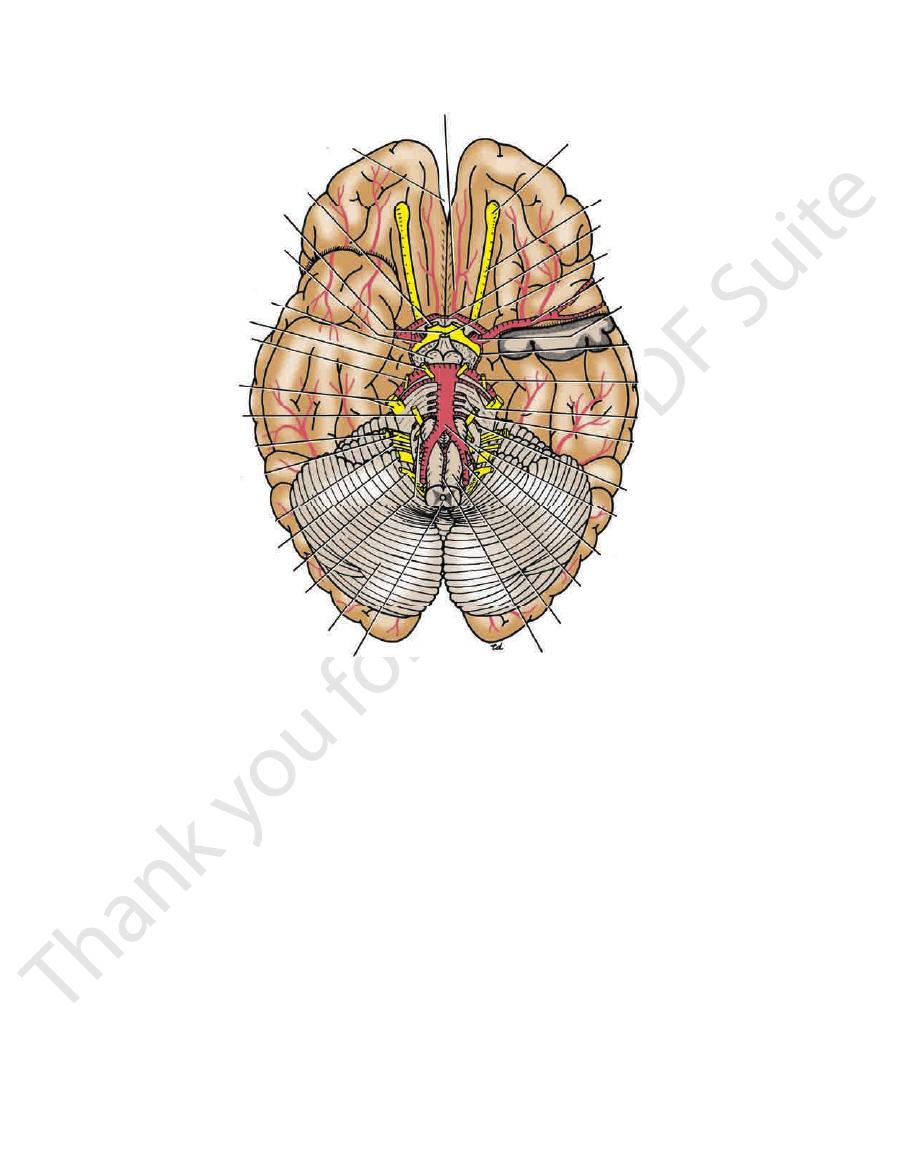
anterior cerebral artery
longitudinal cerebral fissure
optic nerve
optic chiasma
optic tract
mammillary body
oculomotor
nerve
trochlear
nerve
trigeminal
nerve
abducent
nerve
facial nerve
vestibulo-
cochlear nerve
vagus nerve
accessory nerve
(cranial part)
hypoglossal nerve
cerebellum
medulla oblongata
anterior spinal artery
pyramid
vertebral artery
basilar artery
olive
anteroinferior
cerebellar artery
pontine arteries
superior
cerebellar artery
posterior
cerebral artery
posterior
communicating artery
internal
carotid artery
middle cerebral artery
infundibulum
anterior
communicating artery
olfactory tract
olfactory bulb
glossopharyngeal
nerve
FIGURE 11.15
ace of the brain. To show the course of the middle cerebral
Arteries and cranial nerves seen on the inferior surf
produced
cuneate tubercles,
gracile
oblongata are the
On the posterior surface of the inferior part of the medulla
which connect the medulla to the cerebellum.
cles,
inferior cerebellar pedun
11.15). Behind the olives are the
(Fig.
olivary nuclei
elevations produced by the underlying
which are oval
olives,
Posterior to the pyramids are the
decussation of the pyramids.
the opposite side, forming the
below, and here most of the descending fibers cross over to
precentral gyrus of the cerebral cortex. The pyramids taper
dles of nerve fibers that originate in large nerve cells in the
(Fig. 11.15). The pyramids are composed of bun
pyramid
medulla, and on each side of this is a swelling called the
is present on the anterior surface of the
median fissure
nects the pons above to the spinal cord below (Fig. 11.13).
is conical in shape and con
medulla oblongata
The
cranial nerve nuclei.
within the pons serve as relay stations, whereas others form
the midbrain, and the spinal cord. Some of the nerve cells
ascending and descending fibers connecting the forebrain,
connect the two halves of the cerebellum. It also contains
(Fig. 11.13). It is composed mainly of nerve fibers, which
lum below the midbrain and above the medulla oblongata
is situated on the anterior surface of the cerebel
pons
The
middle age, and thus it can be visualized on radiographs.
tricle (see also page 656). The pineal commonly calcifies in
a stalk to the region of the posterior wall of the third ven
between the superior colliculi (Fig. 11.13). It is attached by
is a small glandular structure that lies
pineal body
The
lum and the cerebral hemispheres.
The colliculi are deeply placed between the cerebel
two inferior col
(Fig. 11.12) and
two superior
namely, the
to the cerebral aqueduct; it has four small surface swellings,
is the part of the midbrain posterior
tectum
ventricles. The
which connects the third and fourth
cerebral aqueduct,
(Fig. 11.12). The narrow cavity of the midbrain is the
nigra
substantia
by a pigmented band of gray matter, the
tegmen
and a posterior part, the
crus cerebri;
rior part, the
each of these is divided into an ante
cerebral peduncles;
The midbrain comprises two lateral halves called the
artery, the anterior pole of the left temporal lobe has been removed.
-
-
tum,
-
liculi.
-
-
Hindbrain
-
-
A
-
-
and

548
CHAPTER 11
by the medially placed underlying
The Head and Neck
nucleus gracilis
sinus (Fig. 11.9).
and drains into the straight
internal cerebral veins
the two
is formed by the union of
great cerebral vein
present. The
Cerebral and cerebellar veins and veins of the brainstem are
brain and drain into the cranial venous sinuses (Fig. 11.2).
walls, and they possess no valves. They emerge from the
The veins of the brain have no muscular tissue in their thin
Veins of the Brain
the circle of Willis are fully described on pages 598 and 599.
The internal carotid arteries, the vertebral arteries, and
(circulus arteriosus).
circle of Willis
inferior surface of the brain and form the
two vertebral arteries. The four arteries anastomose on the
The brain is supplied by the two internal carotid and the
Arteries of the Brain
(Figs 11.124, 11.125, and 11.126)
phy (CT) scans and magnetic resonance imaging (MRI).
cles may be visualized clinically using computed tomogra
fourth ventricle. The size and shape of the cerebral ventri
of the two lateral ventricles, the third ventricle, and the
choroid plexuses
spinal fluid, which is produced by the
subarachnoid space. The ventricles are filled with cerebro
cord and, through the three foramina in its roof, with the
of the spinal
central canal
is continuous with the narrow
The fourth ventricle, in turn,
cerebral aqueduct.
by the
third ventricle communicates with the fourth ventricle
(Fig. 11.13); the
interventricular foramina
through the
third ventricle
communicate with the
lateral ventricles
cles, the third ventricle, and the fourth ventricle. The two
The ventricles of the brain consist of the two lateral ventri
Ventricles of the Brain
a median and two lateral openings.
space through three openings in the lower part of the roof:
of the spinal cord. It communicates with the subarachnoid
aqueduct, and below it is continuous with the central canal
cle is connected above to the third ventricle by the cerebral
and the cerebellum. The fourth ventri
rior medullary vela
infe
superior
medulla oblongata and behind by the
(Fig. 11.13). This is bounded in front by the pons and the
The cavity of the hindbrain is the fourth ventricle
the same side of the body.
muscle tone and the coordination of muscle movement on
The cerebellum plays an important role in the control of
dentate nucleus.
matter; the largest of these is known as the
in the interior of the cerebellum, embedded in the white
transverse fissures. Certain masses of gray matter are found
separated by closely set
folia,
tex is thrown into folds, or
is composed of gray matter. The cerebellar cor
cortex,
The surface layer of each cerebellar hemisphere, called
inferior cerebellar peduncles.
and to the medulla by the
middle cerebellar peduncles,
to the pons by the
superior cerebellar peduncles,
by the
The cerebellum is connected to the midbrain
vermis.
sists of two hemispheres connected by a median portion,
posterior to the pons and the medulla oblongata. It con
beneath the tentorium cerebelli (Fig. 11.13). It is situated
lies within the posterior cranial fossa
cerebellum
The
nucleus cuneatus.
laterally placed underlying
and the
-
the
the
-
and
-
-
-
-
-
-
Blood Supply of the Brain
may produce severe cerebral damage, stretching and distor
Injuries of the brain are produced by displacement and distor
Brain Injuries
-
tion of the neuronal tissues at the moment of impact. The brain
may be likened to a log soaked with water floating submerged
in water. The brain is floating in the cerebrospinal fluid in the
subarachnoid space and is capable of a certain amount of
anteroposterior movement, which is limited by the attachment
of the superior cerebral veins to the superior sagittal sinus.
Lateral displacement of the brain is limited by the falx cerebri.
The tentorium cerebelli and the falx cerebelli also restrict dis-
placement of the brain.
It follows from these anatomic facts that blows on the front
or back of the head lead to displacement of the brain, which
-
tion of the brainstem, and stretching and even tearing of the
commissures of the brain. The terms concussion, contusion,
and laceration are used clinically to describe the degrees of
brain injury.
Blows on the side of the head produce less cerebral
displacement, and the injuries to the brain consequently tend
to be less severe.
C L I N I C A L N O T E S
The Cranial Nerves in the Cranial Cavity
The 12 pairs of cranial nerves are named as follows:
I. Olfactory (sensory)
II. Optic (sensory)
III. Oculomotor (motor)
IV. Trochlear (motor)
V. Trigeminal (mixed)
VI. Abducent (motor)
VII. Facial (mixed)
VIII. Vestibulocochlear (sensory)
IX. Glossopharyngeal (mixed)
X. Vagus (mixed)
XI. Accessory (motor)
XII. Hypoglossal (motor)
skull are summarized in Table 11.6.
tion, and the openings through which they exit from the
The cranial nerves, their component parts, their func
on page 605.
origins and courses of the cranial nerves are described
entirely motor; and the remaining nerves are mixed. The
lear, abducent, accessory, and hypoglossal nerves are
lear nerves are entirely sensory; the oculomotor, troch
and abdomen. The olfactory, optic, and vestibulococh
the vagus, which also supplies structures in the thorax
All the nerves are distributed in the head and neck except
through foramina and fissures in the base of the skull.
The nerves emerge from the brain and are transmitted
-
-
-