
Basic Anatomy
forms the spiral organ of Corti and contains
membrane
The highly specialized epithelium that lies on the
ductus reuniens.
and is connected to the saccule by the
is triangular in cross section
duct of the cochlea
The
semicircular ducts.
is detected in the sensory receptors in the ampullae of the
to that of the walls of the semicircular ducts. This change
semicircular ducts changes its speed of movement relative
the head accelerates or decelerates, the endolymph in the
begins or ceases to move, or whenever a movement of
so that all three planes are represented. Whenever the head
figuration. They are arranged at right angles to each other
diameter than the semicircular canals, have the same con
although much smaller in
semicircular ducts,
The
tation of the head to gravity or other acceleration forces.
cialized sensory receptors, which are sensitive to the orien
Located on the walls of the utricle and saccule are spe
face of the petrous part of the temporal bone.
(Fig. 11.30). This lies beneath the dura on the posterior sur
saccus endolymphaticus
end in a small blind pouch, the
being joined by the ductus utriculosaccularis, passes on to
as described previously. The ductus endolymphaticus, after
is globular and is connected to the utricle,
saccule
The
ductus utriculosaccularis.
lymphaticus by the
indirectly connected to the saccule and the ductus endo
is the larger of the two vestibular sacs. It is
utricle
The
All these structures freely communicate with one another.
the duct of the cochlea, which lies within the bony cochlea.
lar ducts, which lie within the bony semicircular canals; and
which are lodged in the bony vestibule; the three semicircu
rounded by perilymph. It consists of the utricle and saccule,
labyrinth (Fig. 11.30). It is filled with endolymph and sur
The membranous labyrinth is lodged within the bony
Membranous Labyrinth
fenestra cochleae.
middle ear by the secondary tympanic membrane at the
The perilymph in the scala tympani is separated from the
stapes and the anular ligament at the fenestra vestibuli.
tibuli is separated from the middle ear by the base of the
below. The perilymph within the scala ves
scala tympani
above and the
scala vestibuli
the cochlear canal into the
the spiral lamina to the outer bony wall, thus dividing
stretches from the free edge of
basilar membrane
it. The
jects into the interior of the canal and partially divides
winds around the modiolus and pro
spiral lamina,
rated by branches of the cochlear nerve. A spiral ledge,
the bottom of the internal acoustic meatus. It is perfo
The modiolus has a broad base, which is situated at
wall of the middle ear.
lea is responsible for the promontory seen on the medial
base faces posteromedially. The first basal turn of the coch
structure is conical. The apex faces anterolaterally and the
successive turn is of decreasing radius so that the whole
low bony tube makes two and one half spiral turns. Each
around which a hol
modiolus,
sists of a central pillar, the
anterior part of the vestibule (Fig. 11.30). Basically, it con
resembles a snail shell. It opens into the
cochlea
The
the mastoid antrum, above the facial nerve canal.
zontal position, and it lies in the medial wall of the aditus to
axis of the petrous bone. The lateral canal is set in a hori
569
-
-
-
-
-
the
-
-
-
-
-
-
-
-
-
basilar
the sensory receptors for hearing. For a detailed
ion
descript
of the spiral organ, a textbook of histology should be
consulted.
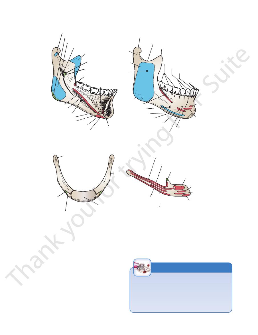
sion on the base, on either side of the symphysis menti
is a small, roughened depres
digastric fossa
The
The lower border of the body of the mandible is called
roots of the teeth.
in the adult, it contains 16 sockets for the
alveolar part;
The upper border of the body of the mandible is called
part of the mylohyoid line (Fig. 11.32).
for the sublingual gland, lies above the anterior
gual fossa,
sublin
below the posterior part of the mylohyoid line. The
superficial part of the submandibular salivary gland, lies
for the
submandibular fossa,
the third molar tooth. The
the area of the mental spines to an area below and behind
as an oblique ridge that runs backward and laterally from
can be seen
mylohyoid line
muscles below (Fig. 11.31). The
gin to the genioglossus muscles above and the geniohyoid
these give ori
mental spines;
the median plane are seen the
On the medial surface of the body of the mandible in
alveolar nerve and vessels.
lar tooth; it transmits the terminal branches of the inferior
can be seen below the second premo
mental foramen
The
symphysis menti.
two halves during development at the
midline, has a faint ridge indicating the line of fusion of the
on its external surface in the
body of the mandible,
The
(Fig. 11.32).
angle of the mandible
on each side at the
The body of the mandible meets the ramus
rami.
a pair of
body
The mandible consists of a horseshoe-shaped
mandibular joint.
of the face, and it articulates with the skull at the temporo
The mandible or lower jaw is the largest and strongest bone
organ of Corti.
spiral
branches of this nerve pass from the ganglion to the
modiolus in the base of the spiral lamina. The peripheral
that is lodged in a canal winding around the
ganglion
spiral
glion of this nerve takes the form of an elongated
foramina at the base of the modiolus. The sensory gan
divides into branches, which enter
cochlear nerve
The
the saccule, and the ampullae of the semicircular ducts.
the membranous labyrinth, where they supply the utricle,
end of the internal acoustic meatus and gain entrance to
The branches of the nerve then pierce the lateral
ganglion.
vestibular
is expanded to form the
vestibular nerve
The
lear portions (Fig. 11.28).
(see page 613), the nerve divides into vestibular and coch
On reaching the bottom of the internal acoustic meatus
Vestibulocochlear Nerve
-
-
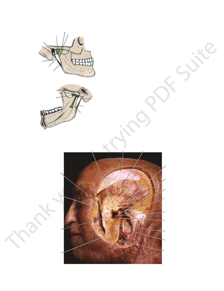
The Mandible
-
and
-
-
-
the
the base.
-
(Fig. 11.32). It is in these fossae that the anterior bellies of
On the lateral surface of the ramus are markings for the
(Fig. 11.32).
mandibular notch
the two processes are separated by the
head;
or
process,
condyloid
and a posterior
coronoid process
an anterior
is vertically placed and has
ramus of the mandible
The
the digastric muscles are attached.
attachment of the masseter muscle. On the medial
e is
surfac
for the inferior alveolar nerve and
mandibular foramen
the
570
CHAPTER 11
The Head and Neck
of pharynx
medial pterygoid
lateral pterygoid
stylomandibular ligament
mandibular foramen
temporalis
lingula
sphenomandibular
ligament
superior constrictor
sublingual fossa
mental spines
digastric fossa
anterior belly of digastric
geniohyoid
genioglossus
submandibular fossa
mylohyoid muscle
mylohyoid line
neck
masseter
ramus
angle
buccinator
body
base of body
platysma
depressor anguli oris
mental foramen
alveolar part of body
depressor
labii
inferioris
mentalis
incisor teeth
canine tooth
premolar teeth
molar teeth
coronoid process
mandibular notch
condyloid process (head)
medial aspect (left side)
lateral aspect (right side)
greater horn (cornu)
stylohyoid
ligament
lesser horn (cornu)
body
hyoglossus
middle constrictor
stylohyoid ligament
geniohyoid
mylohyoid
omohyoid
sternohyoid
digastric and stylohyoid
thyrohyoid
right aspect
anterosuperior aspect
A
B
FIGURE 11.32
A.
ligament
sphenomandibular
for the attachment of the
vessels. In front of the foramen is a projection of bone, called
Hyoid bone.
Mandible. B.
the lingula,
(Figs. 11.32 and 11.33). The foramen leads into the
mandibular canal,
which opens on the lateral surface of the
(see above). The
mental foramen
body of the mandible at the
mandible are shown in Figure 11.32.
The important muscles and ligaments attached to the
(Fig. 11.32).
neck
is a short
head,
or
process,
condyloid
attachment of the temporalis muscle. Below the
receives on its medial surface the
coronoid process
The
canal beyond the mental foramen and below the incisor teeth.
is a continuation forward of the mandibular
incisive canal
Fractures of the Mandible
The mandible is horseshoe shaped and forms part of a bony
ring with the two temporomandibular joints and the base of
the skull. Traumatic impact is transmitted around the ring,
causing a single fracture or multiple fractures of the mandible,
often far removed from the point of impact.
C L I N I C A L N O T E S
Basic Anatomy
571
articular
tubercle
zygomatic
arch (cut)
zygomatic
process
tendon of
temporalis
coronoid
process
of
mandible
head of
mandible
ramus of
mandible
neck of
mandible
angle of
mandible
external
carotid
artery
temporal fascia
temporalis
mandibular
fossa
sternocleido-
mastoid
external
auditory
meatus
mandibular
notch
The temporomandibular joint is synovial. The articular disc
Type of Joint
are covered with fibrocartilage.
dible below (Figs. 11.33 and 11.34). The articular surfaces
bone above and the head (condyloid process) of the man
anterior portion of the mandibular fossa of the temporal
Articulation occurs between the articular tubercle and the
Articulation
Temporomandibular Joint
-
divides the joint into upper and lower cavities (Fig. 11.35).
side of the joint (Fig. 11.33). It is a thin band that is
lies on the medial
sphenomandibular ligament
The
direction and thus protects the external auditory meatus.
ligament limits the movement of the mandible in a posterior
lateral surface of the neck of the mandible (Fig. 11.33). This
backward from the tubercle on the root of the zygoma to the
lateral aspect of the capsule, and its fibers run downward and
strengthens the
lateral temporomandibular ligament
The
Ligaments
fossa and below to the neck of the mandible.
the articular tubercle and the margins of the mandibular
The capsule surrounds the joint and is attached above to
Capsule
external auditory
meatus
tympanic
plate
mastoid
process
styloid process
stylomandibular
ligament
capsule
temporomandibular
ligament
articular tubercle
capsule
styloid
process
sphenomandibular
ligament
A
B
spine of
sphenoid
stylomandibular
ligament
FIGURE 11.33
Temporomandibular joint as seen from the
lateral (A) and medial (B) aspects.
FIGURE 11.34
ve been
A dissection of the left temporomandibular joint. The capsule and lateral temporomandibular ligament ha
of the mandible. The articular disc is present within the joint cavity on the upper surface of the head of the mandible.
removed to reveal the interior of the joint. Note the articular tubercle and mandibular fossa of the temporal bone and the head
572
CHAPTER 11
The Head and Neck
articular
disc
articular
cartilage
mandibular
fossa
articular tubercle
lateral
pterygoid
muscle
head
of mandible
synovial
membrane
temporalis
lateral pterygoid
medial pterygoid
digastric
(anterior belly)
sternohyoid
omohyoid
(superior belly)
thyrohyoid
masseter
digastric
(posterior belly)
A
B
C
neck of mandible
FIGURE 11.35
Temporomandibular joint with mouth closed
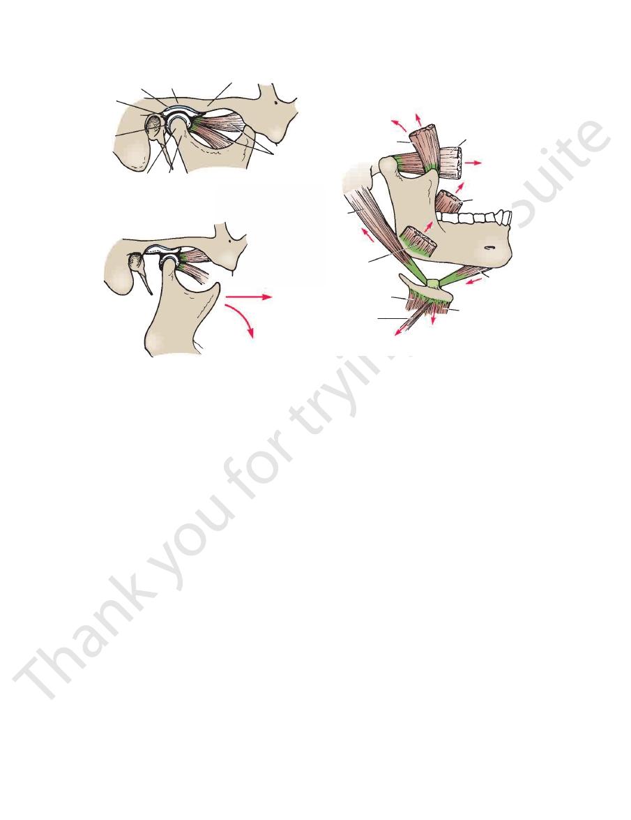
ular disc so that the latter moves onto the articular tubercle
which pulls forward the neck of the mandible and the artic
plished by the contraction of the lateral pterygoid muscle,
muscle, the mandible is pulled forward. This is accom
essarily on the parotid gland and the sternocleidomastoid
tal axis. To prevent the angle of the jaw impinging unnec
on the undersurface of the articular disc around a horizon
As the mouth is opened, the head of the mandible rotates
contact.
slightly apart. On closure of the jaws, the teeth come into
position of rest, the teeth of the upper and lower jaws are
retracted. Rotation can also occur, as in chewing. In the
The mandible can be depressed or elevated, protruded or
Movements
dibular nerve
Auriculotemporal and masseteric branches of the man
Nerve Supply
joint (Fig. 11.35).
This lines the capsule in the upper and lower cavities of the
Synovial Membrane
the lower surface is concave to fit the head of the mandible.
shape of the articular tubercle and the mandibular fossa;
the disc is concavoconvex from before backward to fit the
tion and retraction of the mandible. The upper surface of
backward with the head of the mandible during protrac
ble. These bands ensure that the disc moves forward and
muscle and by fibrous bands to the head of the mandi
also attached in front to the tendon of the lateral pterygoid
lage that is attached circumferentially to the capsule. It is
lower cavities (Fig. 11.35). It is an oval plate of fibrocarti
divides the joint into upper and
articular disc
The
(Fig. 11.33).
apex of the styloid process to the angle of the mandible
of thickened deep cervical fascia that extends from the
to the joint and some distance from it. It is merely a band
lies behind and medial
stylomandibular ligament
The
remains of the first pharyngeal arch in this region.
to the lingula of the mandibular foramen. It represents the
attached above to the spine of the sphenoid bone and below
arrows
tion to the mandible. The
The attachment of the muscles of mastica
the mandible and articular disc in relation to the articular tubercle in each case.
(A) and with the mouth open (B). Note the position of the head of
C.
-
indicate the direction of their actions.
-
-
-
-
Depression of the Mandible
-
-
-
-
(Fig. 11.35). The forward movement of the disc is limited
disc to the temporal bone posteriorly.
pulled backward by the fibroelastic tissue, which tethers the
the posterior fibers of the temporalis. The articular disc is
goids. The head of the mandible is pulled backward by
tion of the temporalis, the masseter, and the medial ptery
Elevation of the mandible is brought about by contrac
and then the head rotates on the lower surface of the disc.
First, the head of the mandible and the disc move backward,
The movements in depression of the mandible are reversed.
mandible forward.
the lateral pterygoids play an important role by pulling the
tion of the digastrics, the geniohyoids, and the mylohyoids;
Depression of the mandible is brought about by contrac
disc to the temporal bone posteriorly.
by the tension of the fibroelastic tissue, which tethers the
-
Elevation of the Mandible
-
-
Basic Anatomy
The muscles of mastication are summarized in
in unison.
cles responsible on both sides work alternately and not
place, a certain amount of rotation occurs, and the mus
retracting the mandible on each side. For this to take
These are accomplished by alternately protruding and
Lateral Chewing Movements
about by contraction of the posterior fibers of the temporalis.
backward into the mandibular fossa. Retraction is brought
The articular disc and the head of the mandible are pulled
Retraction of the Mandible
cles of both sides, assisted by both medial pterygoids.
brought about by contraction of the lateral pterygoid mus
lower teeth are drawn forward over the upper teeth, which is
takes place in the upper cavity of the joint. In protrusion, the
carrying the head of the mandible with it. All movement thus
The articular disc is pulled forward onto the anterior tubercle,
Protrusion of the Mandible
573
-
-
Table 11.4. See also Figure 11.35.
nerve and artery (Fig. 11.36)
The mandibular notch and the masseteric
Anteriorly:
Important Relations of the Temporomandibular Joint
■
■
Muscles of the Head
T A B L E 1 1 . 4
Muscle
Origin
Insertion
Nerve
Supply
Action
Muscle of Scalp
Occipitofrontalis
Occipital belly
Frontal belly
Highest nuchal line of
occipital bone
Skin and superficial fascia
of eyebrows
Epicranial aponeurosis
Facial nerve Moves scalp on skull and raises
eyebrows
Muscles of Facial Expression
Orbicularis oculi
Palpebral part
Medial palpebral ligament
Lateral palpebral raphe
Facial nerve Closes eyelids and dilates lacrimal
sac
Orbital part
See Table 11.5
Wrinkles skin of nose
Vertical wrinkles of forehead, as in
Medial palpebral ligament
and adjoining bone
Loops return to origin
Facial nerve Throws skin around orbit into folds
to protect eyeball
Corrugator supercilii
Superciliary arch
Skin of eyebrow
Facial nerve
frowning
Compressor nasi
Frontal process of maxilla
Aponeurosis of bridge
of nose
Facial
nerve
Compresses mobile nasal
cartilages
Dilator naris
Maxilla
Ala of nose
Facial nerve Widens nasal aperture
Procerus
Nasal bone
Skin between eyebrows
Facial nerve
Orbicularis oris
Maxilla, mandible, and skin Encircles oral orifice
Facial nerve Compresses lips together
Dilator Muscles of Lips
Levator labii superioris
alaeque nasi
Levator labii superioris
Zygomaticus minor
Zygomaticus major
Levator anguli oris
Risorius
Depressor anguli oris
Depressor labii inferioris
Mentalis
Buccinator
Platysma
Arise from bones and fas-
cia around oral aperture
and insert into substance
of lips
Outer surface of alveolar
margins of maxilla and
mandible and ptery-
gomandibular ligament
Facial nerve
Facial nerve
Separate lips
Compresses cheeks and lips
against teeth
(continued)
574
CHAPTER 11
The Head and Neck
Muscles of the Head (continued )
T A B L E 1 1 . 4
Laterally:
parotid gland
tory meatus (Fig. 11.33) and the glenoid process of the
The tympanic plate of the external audi
Posteriorly:
Tuberosity of maxilla and
Temporalis
Muscle
Origin
Insertion
Nerve
Supply
Action
Muscles of Mastication
Masseter
Zygomatic arch
Lateral surface
ramus of mandible
Mandibular
division of
trigeminal
nerve
Elevates mandible to occlude teeth
Floor of temporal fossa
Coronoid process of
mandible
Mandibular
division of
trigeminal
nerve
Anterior and superior fibers
elevate mandible; posterior
fibers retract mandible
Lateral pterygoid (two
heads)
Greater wing of sphenoid
and lateral pterygoid
plate
Neck of mandible and
articular disc
Mandibular
division of
trigeminal
nerve
Pulls neck of mandible forward
Medial pterygoid (two
heads)
lateral pterygoid plate
Medial surface of angle
of mandible
Mandibular
division of
trigeminal
nerve
Elevates mandible
■
■
-
■
■
The parotid gland, fascia, and skin (see Fig. 11.85)
lotemporal nerve
The maxillary artery and vein and the auricu
Medially:
■
■
-
Clinical Significance of the Temporomandibular
of the temporomandibular joint may
articular disc
Joint
The temporomandibular joint lies immediately in front of the
external auditory meatus. The great strength of the lateral
temporomandibular ligament prevents the head of the mandi-
ble from passing backward and fracturing the tympanic plate
when a severe blow falls on the chin.
The
become partially detached from the capsule, and this results
in its movement becoming noisy and producing an audible
click during movements at the joint.
Dislocation of the Temporomandibular Joint
Dislocation sometimes occurs when the mandible is
depressed. In this movement, the head of the mandible and
the articular disc both move forward until they reach the sum-
mit of the articular tubercle. In this position, the joint is unsta-
ble, and a minor blow on the chin or a sudden contraction of
the lateral pterygoid muscles, as in yawning, may be sufficient
C L I N I C A L N O T E S
(continued)
to pull the disc forward beyond the summit. In bilateral cases,
lateral margins of the aponeurosis are attached to
occipitofrontalis muscle (Figs. 11.37 and 11.38). The
sheet that unites the occipital and frontal bellies of the
poneurosis (epicranial), which is a thin, tendinous
arteries, and a free anastomosis takes place between them.
arteries are branches of the external and internal carotid
Numerous arteries and veins are found in this layer. The
aponeurosis of the occipitofrontalis muscle (Fig. 11.37).
the fibrous septa uniting the skin to the underlying
onnective tissue beneath the skin, which is fibrofatty,
numerous sebaceous glands
kin, which is thick and hair bearing and contains
layer of the scalp.
to denote the
the scalp, use each letter of the word
To assist one in memorizing the names of the five layers of
intimately bound together and move as a unit (Fig. 11.37).
The scalp consists of five layers, the first three of which are
dislocation is easily achieved by pressing the gloved thumbs
the mouth is fixed in an open position, and both heads of the
mandible lie in front of the articular tubercles. Reduction of the
downward on the lower molar teeth and pushing the jaw back-
ward. The downward pressure overcomes the tension of the
temporalis and masseter muscles, and the backward pressure
overcomes the spasm of the lateral pterygoid muscles.
The Scalp
Structure
SCALP
■
■
S
■
■
C
■
■
A