Basic Anatomy
579
posterior
auricular vein
vertebral vein
internal jugular
vein
external
jugular
vein
right
brachiocephalic
vein
anterior jugular vein
facial vein
maxillary
vein
superficial
temporal
vein
subclavian
vein
retromandibular
vein
FIGURE 11.39
Main veins of the head and neck.
retroauricular
(mastoid nodes)
occipital
nodes
superficial
cervical
nodes
deep cervical nodes
jugular trunk
tracheal nodes
laryngeal nodes
anterior cervical
nodes
submandibular
nodes
submental nodes
buccal
nodes
parotid nodes
FIGURE 11.40
Lymph drainage of the head and neck.
area over the angle of the mandible and the parotid gland
divisions of the trigeminal nerve, except for the small
The skin of the face is supplied by branches of the three
they follow the wrinkle lines.
elasticity. Surgical scars of the face are less conspicuous if
contracting muscles, coupled with the loss of youthful skin
of the skin perpendicular to the long axis of the underlying
Wrinkle lines of the face result from the repeated folding
No deep fascia is present in the face.
of facial expression.
loose connective tissue, in which are embedded the muscles
ceous glands. It is connected to the underlying bones by
The skin of the face possesses numerous sweat and seba
The Face
Skin of the Face
-
Sensory Nerves of the Face
(Fig. 11.41), which is supplied by the great auricular nerve
process; the maxillary nerve serves the region developed
nerve supplies the region developed from the frontonasal
of dermatomes of the trunk and limbs. The ophthalmic
inal nerve is slight compared with the considerable overlap
(C2 and 3). The overlap of the three divisions of the trigem-
supratrochlear nerve
infratrochlear nerve
supraorbital
nerve
lacrimal nerve
zygomaticotemporal
nerve
auriculotemporal
nerve
infraorbital nerve
external nasal nerve
buccal nerve
great auricular nerve
mental nerve
cervical branch
mandibular
branch
buccal
branch
zygomatic
branch
temporal
branch
supratrochlear artery
supraorbital artery
zygomaticotemporal
artery
superficial temporal artery
lacrimal artery
zygomaticofacial artery
infraorbital artery
transverse facial artery
external nasal artery
facial artery
mental artery
external carotid artery
internal jugular vein
mental vein
facial vein
transverse facial
vein
infraorbital vein
zygomaticofacial
vein
supraorbital vein
supratrochlear vein
A
C
zygomaticofacial
nerve
lacrimal vein
superficial temporal
vein
zygomaticotemporal
vein
B
D
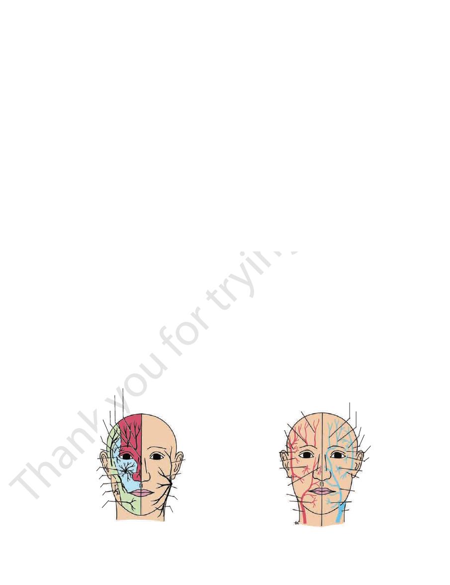
FIGURE 11.41
A.
Venous drainage of the face.
Arterial supply of the face.
expression.
Branches of the 7th cranial nerve to muscles of facial
Sensory nerve supply to the skin of the face. B.
C.
D.

580
CHAPTER 11
above the auricle (Fig. 11.41).
of the tympanic membrane, and the skin of the scalp
auricle, the external auditory meatus, the outer surface
poral vessels and the auricle. It supplies the skin of the
border of the parotid gland between the superficial tem
ascends from the upper
auriculotemporal nerve
The
a small area of the cheek (Fig. 11.41).
border of the masseter muscle and supplies the skin over
emerges from beneath the anterior
buccal nerve
The
chin (Fig. 11.41).
the mandible and supplies the skin of the lower lip and
emerges from the mental foramen of
mental nerve
The
branches of the nerve pass to the skin.
auricle. It then passes upward to the side of the scalp. Three
lower part of the face, the temporal region, and part of the
The mandibular nerve supplies the skin of the lower lip, the
Mandibular Nerve
temple (Fig. 11.41).
face of the zygomatic bone. It supplies the skin over the
ral fossa through a small foramen on the posterior sur
emerges in the tempo
zygomaticotemporal nerve
The
the cheek (Fig. 11.41).
matic bone. It supplies the skin over the prominence of
through a small foramen on the lateral side of the zygo
passes onto the face
zygomaticofacial nerve
The
(Fig. 11.41).
eyelid and cheek, the side of the nose, and the upper lip
out from the foramen and supply the skin of the lower
divides into numerous small branches, which radiate
face through the infraorbital foramen. It immediately
maxillary nerve. It enters the orbit and appears on the
is a direct continuation of the
infraorbital nerve
The
branches of the nerve pass to the skin.
upper lip, and the lateral side of the orbital opening. Three
part of the side of the nose, the lower eyelid, the cheek, the
The maxillary nerve supplies the skin on the posterior
Maxillary Nerve
the tip (Fig. 11.41).
supplies the skin on the side of the nose down as far as
between the nasal bone and the upper nasal cartilage. It
leaves the nose by emerging
external nasal nerve
The
and the adjoining part of the side of the nose (Fig. 11.41).
and conjunctiva on the medial part of the upper eyelid
ley of the superior oblique muscle. It supplies the skin
leaves the orbit below the pul
infratrochlear nerve
The
the median plane.
and the skin over the lower part of the forehead, close to
and conjunctiva on the medial part of the upper eyelid
(Fig. 11.41). It divides into branches that supply the skin
margin of the orbit medial to the supraorbital nerve
winds around the upper
supratrochlear nerve
The
supplies the skin of the forehead.
junctiva on the central part of the upper eyelid; it also
It divides into branches that supply the skin and con
gin of the orbit at the supraorbital notch (Fig. 11.41).
winds around the upper mar
supraorbital nerve
The
the lateral part of the upper eyelid (Fig. 11.41).
supplies the skin and conjunctiva of
lacrimal nerve
The
pass to the skin.
down to and including the tip. Five branches of the nerve
the upper eyelid, the conjunctiva, and the side of the nose
The ophthalmic nerve supplies the skin of the forehead,
Ophthalmic Nerve
sinuses.
supply to the mouth, teeth, nasal cavities, and paranasal air
of facial expression. They are, in addition, the sensory nerve
also supply proprioceptive fibers to the underlying muscles
These nerves not only supply the skin of the face, but
mandibular process of the first pharyngeal arch.
the mandibular nerve serves the region developed from the
from the maxillary process of the first pharyngeal arch; and
The Head and Neck
■
■
■
■
-
-
■
■
■
■
-
■
■
■
■
■
■
-
■
■
-
-
■
■
■
■
■
■
-
is a relatively
Trigeminal neuralgia
auricular nerve (C2 and 3).
area of skin over the angle of the jaw is supplied by the great
The facial skin receives its sensory nerve supply from the
Sensory Innervation and Trigeminal Neuralgia
three divisions of the trigeminal nerve. Remember that a small
common condition in which the patient experiences excru-
ciating pain in the distribution of the mandibular or maxillary
division, with the ophthalmic division usually escaping. A phy-
sician should be able to map out accurately on a patient’s face
the distribution of each of the divisions of the trigeminal nerve.
C L I N I C A L N O T E S
Arterial Supply of the Face
branches to the septum and ala of the nose.
the mouth. It runs medially in the upper lip and gives
arises near the angle of
superior labial artery
The
ses with its fellow of the opposite side.
mouth. It runs medially in the lower lip and anastomo
arises near the angle of the
inferior labial artery
The
skin of the chin and lower lip.
lower border of the body of the mandible. It supplies the
arises from the facial artery at the
submental artery
The
Branches
of the ophthalmic artery (Fig. 11.41).
of the eye, where it anastomoses with the terminal branches
cle and runs along the side of the nose to the medial angle
zygomaticus muscles and the levator labii superioris mus
sma and the risorius muscles. It then ascends deep to the
toward the angle of the mouth and is covered by the platy
(Fig. 11.132). It runs upward in a tortuous course
ily felt
It is here that the pulse can be eas
of the masseter muscle.
margin of the body of the mandible at the anterior border
submandibular salivary gland, it curves around the inferior
(Figs. 11.55 and 11.59). Having arched upward and over the
arises from the external carotid artery
facial artery
The
sensory nerves of the face.
supplemented by several small arteries that accompany the
sels: the facial and superficial temporal arteries, which are
The face receives a rich blood supply from two main ves-
-
-
-
■
■
■
■
-
■
■

Basic Anatomy
(Fig. 11.41).
of the ophthalmic artery, supply the skin of the forehead
branches
supratrochlear arteries,
supraorbital
The
(Fig. 11.41).
forward across the cheek just above the parotid duct
temporal artery, arises within the parotid gland. It runs
a branch of the superficial
transverse facial artery,
The
auricle to supply the scalp (see page 578).
mences in the parotid gland. It ascends in front of the
terminal branch of the external carotid artery, com
(Fig. 11.41), the smaller
superficial temporal artery
The
dorsum of the nose.
alongside the nose. It supplies the skin on the side and
arises from the facial artery
lateral nasal artery
The
581
■
■
■
■
-
■
■
■
■
and
of the masseter, are commonly used by the anesthetist to take
Blood Supply of the Facial Skin
The blood supply to the skin of the face is profuse so that it is
rare in plastic surgery for skin flaps to necrose in this region.
Facial Arteries and Taking the Patient’s Pulse
The superficial temporal artery, as it crosses the zygomatic
arch in front of the ear, and the facial artery, as it winds around
the lower margin of the mandible level with the anterior border
the patient’s pulse.
C L I N I C A L N O T E S
Venous Drainage of the Face
drains into the submandibular lymph nodes (Fig. 11.42).
Lymph from the forehead and the anterior part of the face
Lymph Drainage of the Face
vein within the parotid gland.
joins the superficial temporal
transverse facial vein
The
sinus by the superior ophthalmic vein.
and to the cavernous
deep facial vein
venous plexus by the
branches of the facial artery. It is joined to the pterygoid
The facial vein receives tributaries that correspond to the
Tributaries
ing into the internal jugular vein.
of the retromandibular vein. The facial vein ends by drain
submandibular gland and is joined by the anterior division
of the body of the mandible. It crosses superficial to the
vein descends behind the facial artery to the lower margin
of infection from the face to the cavernous sinus. The facial
cal importance because it provides a pathway for the spread
cavernous sinus (Fig. 11.9); this connection is of great clini
superior ophthalmic vein, the facial vein is connected to the
directly through the supraorbital vein. By means of the
(Fig. 11.41). It is connected to the superior ophthalmic vein
by the union of the supraorbital and supratrochlear veins
is formed at the medial angle of the eye
facial vein
The
-
-
Facial Infections and Cavernous Sinus Thrombosis
sinus thrombosis may be fatal unless adequately treated with
The area of facial skin bounded by the nose, the eye, and the
upper lip is a potentially dangerous zone to have an infection.
For example, a boil in this region can cause thrombosis of the
facial vein, with spread of organisms through the inferior oph-
thalmic veins to the cavernous sinus. The resulting cavernous
antibiotics.
C L I N I C A L N O T E S
A few buccal lymph nodes may be present along the course
maxilla.
of the
process of the frontal bone and below by the frontal process
The medial orbital margin is formed above the maxillary
maxilla.
zygomatic bone
margin is formed by the
and the inferior orbital
zygomatic bone
is formed by the
The lateral orbital margin
frontal air sinuses.
which con
frontal bone,
above them are formed by the
Figure 11.42. The superior orbital margins and the area
The bones that form the front of the skull are shown in
submental lymph nodes.
of the lower lip and the skin of the chin are drained into the
sels that end in the parotid lymph nodes. The central part
ing the lateral parts of the eyelids, is drained by lymph ves
of these lymph vessels. The lateral part of the face, includ-
-
Bones of the Face
-
tains the
and the
frontal
ethmoid
parietal
lesser wing of
sphenoid
greater wing of
sphenoid
squamous temporal
lacrimal
zygomatic
mastoid process
nasal
maxilla
mandible
submental
nodes
deep cervical
nodes
submandibular
nodes
buccal
node
parotid
nodes
A
B
FIGURE 11.42
A.
drainage of the face.
Lymph
Bones of the front of the skull. B.

582
CHAPTER 11
Buccal branch of the facial nerve
Nerve supply:
thus blends and forms part of the orbicularis oris muscle.
respectively, without intersecting. The buccinator muscle
and lowest fibers continue into the upper and lower lips,
and those from above entering the lower lip; the highest
ers decussate, those from below entering the upper lip
parotid duct. At the angle of the mouth the central fib
muscle layer of the cheek. The muscle is pierced by the
The muscle fibers pass forward, forming the
Insertion:
and from the pterygomandibular ligament (Fig. 11.38).
of the maxilla and mandible opposite the molar teeth
From the outer surface of the alveolar margins
Origin:
nerve
Buccal and mandibular branches of the facial
Nerve Supply
Mentalis
Depressor labii inferioris
Depressor anguli oris
Risorius
Levator anguli oris (deep to the zygomatic muscles)
Zygomaticus major
Zygomaticus minor
Levator labii superioris
Levator labii superioris alaeque nasi
are named as follows:
the mouth and then below the oral aperture, the muscles
of the lips. Traced from the side of the nose to the angle of
oral aperture and converge to be inserted into the substance
The muscles arise from the bones and fascia around the
usually accompanied by separation of the jaws.
and their action is to separate the lips; this movement is
The dilator muscles (Fig. 11.38) radiate out from the lips,
Compresses the lips together
Action:
facial nerve
Buccal and mandibular branches of the
Nerve supply:
muscle.
Many of the fibers are derived from the buccinator
mucous membrane lining the inner surface of the lips.
the deep surface of the skin and pass obliquely to the
above and the mandible below. Other fibers arise from
of the fibers arise near the midline from the maxilla
fice within the substance of the lips (Fig. 11.38). Some
The fibers encircle the oral ori
Origin and insertion:
from the lips.
muscles consist of a series of small muscles that radiate out
The sphincter muscle is the orbicularis oris. The dilator
shown in Table 11.4.
compressor naris, the dilator naris, and the procerus are
The origin, insertion, nerve supply, and action of the
tor muscle is the dilator naris (Fig. 11.38).
The sphincter muscle is the compressor naris and the dila
Muscles of the Nostrils
in Table 11.4.
orbicularis oculi and the corrugator supercilii are described
The origin, insertion, nerve supply, and action of the
page 575.
itofrontalis forms part of the scalp and is described on
palpebrae superioris is described on page 550. The occip
rioris and the occipitofrontalis (Fig. 11.38). The levator
and the dilator muscles are the levator palpebrae supe
The sphincter muscle of the eyelids is the orbicularis oculi,
Muscles of the Eyelids
supplied by the facial nerve.
face are developed from the second pharyngeal arch and are
modify the expression of the face. All the muscles of the
structures. A secondary function of the facial muscles is to
the facial muscles to serve as sphincters or dilators of these
eyelids, nostrils, and lips, respectively. It is the function of
namely, the orbit, nose, and mouth, are guarded by the
inserted into the skin (Fig. 11.38). The orifices of the face,
fascia, and most arise from the bones of the skull and are
The muscles of the face are embedded in the superficial
page 530).
bones of the face is given in the discussion of the skull (see
mandible, with its teeth. A more detailed account of the
air sinus. The bone of the lower third of the face is the
face is the maxilla, containing its teeth and the maxillary
The important central bone of the middle third of the
and lower plates of hyaline cartilage and small cartilages of
frontal bones. Anteriorly, the nose is completed by upper
which articulate below with the maxilla and above with the
nasal bones,
The root of the nose is formed by the
The Head and Neck
the ala nasi.
Muscles of the Face (Muscles of Facial
Expression)
-
-
-
Muscles of the Lips and Cheeks
Sphincter Muscle of the Lips: Orbicularis Oris
■
■
-
■
■
■
■
Dilator Muscles of the Lips
■
■
■
■
■
■
■
■
■
■
■
■
■
■
■
■
■
■
Muscle of the Cheek
Buccinator
■
■
■
■
-
■
■
cles of the lips and cheeks are shown in Table 11.4.
The origin, insertion, nerve supply, and action of the mus
Compresses the cheeks and lips against the teeth
Action:
■
■
-
essentially a lower motor neuron lesion. An upper motor neu
(by a tumor) or caused by lacerations of the face will cause
), or in the parotid gland
Bell’s palsy
nerve canal (perineuritis,
Facial Muscle Paralysis
The facial muscles are innervated by the facial nerve. Damage
to the facial nerve in the internal acoustic meatus (by a tumor),
in the middle ear (by infection or operation), in the facial
distortion of the face, with drooping of the lower eyelid, and
the angle of the mouth will sag on the affected side. This is
-
ron lesion (involvement of the pyramidal tracts) will leave the
upper part of the face normal because the neurons supply-
ing this part of the face receive corticobulbar fibers from both
cerebral cortices.
C L I N I C A L N O T E S

Basic Anatomy
emerges from the anterior
mandibular branch
The
nostril.
buccinator muscle and the muscles of the upper lip and
of the gland below the parotid duct and supplies the
emerges from the anterior border
buccal branch
The
oculi.
border of the gland and supplies the orbicularis
emerges from the anterior
zygomatic branch
The
supercilii.
frontalis, the orbicularis oculi, and the corrugator
auricular muscles, the frontal belly of the occipito
of the gland and supplies the anterior and superior
emerges from the upper border
temporal branch
The
terminal branches (Fig. 11.41).
parotid salivary gland (see page 630), it divides into its five
As the facial nerve runs forward within the substance of the
583
Facial Nerve
■
■
-
■
■
■
■
■
■
border of the gland and supplies the muscles of the
shown in Figure 11.67.
mary of the origin and distribution of the facial nerve is
the central nervous system via the trigeminal nerve. A sum
facial nerve in these communicating branches and pass to
proprioceptive nerve fibers of the facial muscles leave the
branches of the trigeminal nerve. It is believed that the
but its branches communicate with
not supply the skin,
It does
and supplies all the muscles of facial expression.
The facial nerve is the nerve of the second pharyngeal arch
the depressor anguli oris muscle.
the lower margin of the body of the mandible to supply
mandible to supply the platysma muscle; it may cross
of the gland and passes forward in the neck below the
emerges from the lower border
cervical branch
The
lower lip.
■
■
-
Development of the Face
ous processes that ultimately form the face unite during the
finally bury the premaxilla and fuse in the midline. The vari
extend medially, forming the upper jaw and the cheek, and
The maxillary processes
development, the maxillary processes grow medially and fuse
With further
medial
appear as depressions in the lower edge
oping brain, and this grows toward the stomodeum. Meanwhile,
together and fusion of several important processes, namely, the
area is a depression in the ectoderm known as the
Early in development, the face of the embryo is represented by
an area bounded cranially by the neural plate, caudally by the
pericardium, and laterally by the mandibular process of the first
pharyngeal arch on each side (Fig. 11.43). In the center of this
stomodeum.
In the floor of the depression is the buccopharyngeal membrane.
By the fourth week, the buccopharyngeal membrane breaks
down so that the stomodeum communicates with the foregut.
The further development of the face depends on the coming
frontonasal process, the maxillary processes, and the mandibu-
lar processes (Fig. 11.43). The frontonasal process begins as a
proliferation of mesenchyme on the ventral surface of the devel-
the maxillary process grows out from the upper end of each first
arch and passes medially, forming the lower border of the devel-
oping orbit. The mandibular processes of the first arches now
approach one another in the midline below the stomodeum and
fuse to form the lower jaw and lower lip (Fig. 11.43).
The olfactory pits
of the advancing frontonasal process, dividing it into a
nasal process and two lateral nasal processes.
with the lateral nasal processes and with the medial nasal pro-
cess (Fig. 11.43). The medial nasal process forms the philtrum
of the upper lip and the premaxilla.
-
second month.
The upper lip is formed by the growth medially of the maxillary
processes of the first pharyngeal arch on each side. Ultimately,
the maxillary processes meet in the midline and fuse with each
other and with the medial nasal process (Fig. 11.43). Thus, the
lateral parts of the upper lip are formed from the maxillary
processes, and the medial part, or philtrum, from the medial
remains and tethers each lip to the gum, thus forming the
the gums. In the midline, a short area of the labiogingival lamina
later degenerates. A deep groove thus forms between the lips and
which grows down into the underlying mesenchyme and
labiogingival
Each lip separates from its respective gum as the result of the
nasal process, with contributions from the maxillary processes.
The lower lip is formed from the mandibular process of the
first pharyngeal arch on each side (Fig. 11.43). These processes
grow medially below the stomodeum and fuse in the midline to
form the entire lower lip.
appearance of a linear thickening of ectoderm, the
lamina,
frenulum.
At first, the mouth has a broad opening, but later this dimin-
ishes in extent because of fusion of the lips at the lateral angles.
Sensory Nerve Supply to the Skin of the Developing Face
The area of skin overlying the frontonasal process and its deriva-
tives receives its sensory nerve supply from the ophthalmic divi-
sion of the trigeminal nerve, whereas the maxillary division of the
trigeminal nerve supplies the area of skin overlying the maxillary
process. The area of skin overlying the mandibular process is
supplied by the mandibular division of the trigeminal nerve.
Muscles of the Developing Face (Muscles of Facial
Expression)
The muscles of the face are derived from the mesenchyme of the
second pharyngeal arch. The nerve supply of these muscles is
the nerve of the second pharyngeal arch—namely, the seventh
cranial nerve.
Cleft Upper Lip
Cleft upper lip may be confined to the lip or may be associated
with a cleft palate. The anomaly is usually unilateral cleft lip and
is caused by a failure of the maxillary process to fuse with the
medial nasal process (Figs. 11.44 and 11.45). Bilateral cleft lip
is caused by a failure of both maxillary processes to fuse with
E M B R Y O L O G I C N O T E S
(continued)
584
CHAPTER 11
The Head and Neck
the medial nasal process, which then remains as a central flap
gery no later than 2 months after birth, provided the baby’s condi
Cleft lower lip is a rare condition. The cleft is exactly central and is
of tissue (Figs. 11.46 and 11.48). Median cleft upper lip is very
rare and is caused by the failure of the rounded swellings of the
medial nasal process to fuse in the midline.
Oblique Facial Cleft
Oblique facial cleft is a rare condition in which the cleft lip on
one side extends to the medial margin of the orbit (Figs. 11.44 and
11.47). This is caused by the failure of the maxillary process to
fuse with the lateral and medial nasal processes.
Cleft Lower Lip
caused by incomplete fusion of the mandibular processes (Fig. 11.44).
Treatment of Isolated Cleft Lip
The condition of isolated cleft lip usually is treated by plastic sur-
-
tion permits. The surgeon strives to approximate the vermilion
border and to form a normal-looking lip (Fig. 11.48A–C).
Macrostomia and Microstomia
The normal size of the mouth shows considerable individual vari-
ation. Rarely, there is incomplete fusion of the maxillary with the
mandibular processes, producing an excessively large mouth or
macrostomia. Very rarely, there is excessive fusion of these pro-
cesses, producing a small mouth or microstomia. These condi-
tions can easily be corrected surgically.
olfactory pit
olfactory pit
olfactory pit
frontonasal process
frontonasal process
mandibular process
maxillary process
lateral
nasal process
lateral
nasal process
medial
nasal process
medial nasal process
mandibular process
buccopharyngeal membrane
forming floor of
stomodeum
second pharyngeal
arch
mandible
nostril
future
external ear
future
external ear
philtrum
maxilla
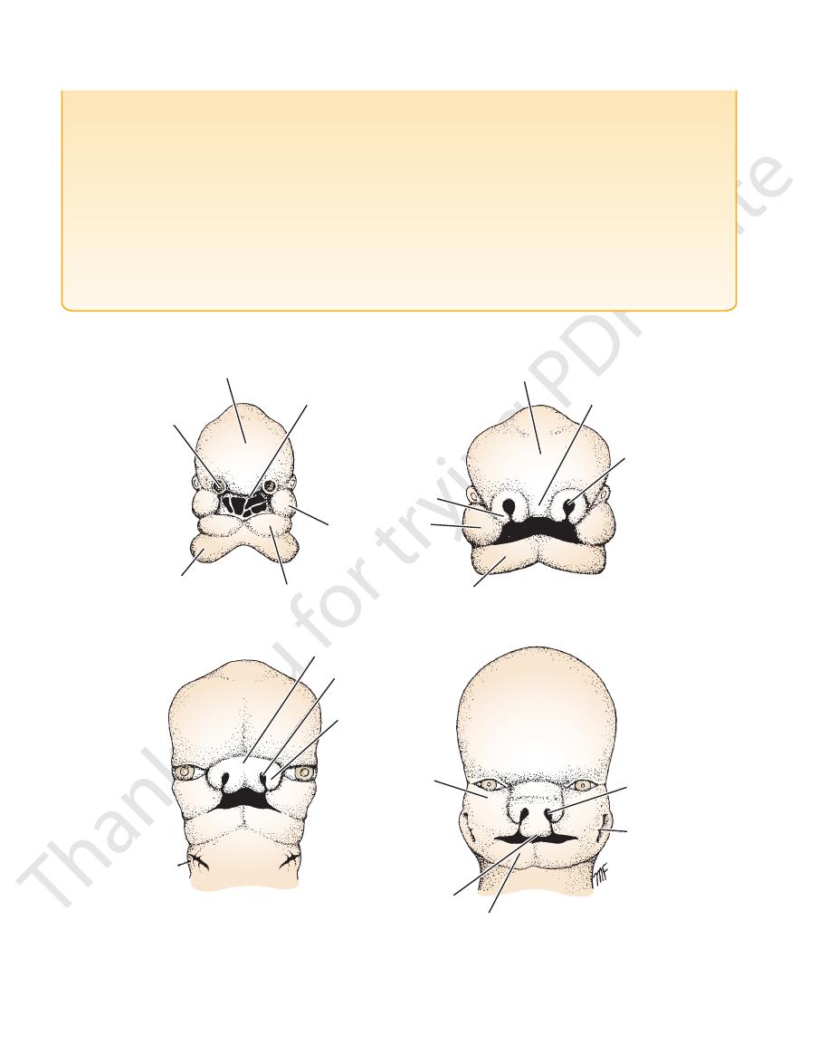
8 weeks
6.5 weeks
5.5 weeks
5 weeks
A
C
B
D
FIGURE 11.43
Different stages in development of the face.