662
CHAPTER 11
The Head and Neck
of the subclavian or internal jugular veins.
chiocephalic vein. It may, however, end in the terminal part
downward and drains into the beginning of the left bra
reaching the medial border of the scalenus anterior, it turns
bends laterally behind the carotid sheath (Fig. 11.57). On
transverse process of the seventh cervical vertebra. Here, it
margin of the esophagus until it reaches the level of the
the root of the neck, it continues to ascend along the left
passing upward along the left margin of the esophagus. At
the left. On reaching the superior mediastinum, it is found
through the posterior mediastinum, inclining gradually to
through the aortic opening in the diaphragm and ascends
of the cisterna chyli (see page XXX). It enters the thorax
The thoracic duct begins in the abdomen at the upper end
jugular vein to form the brachiocephalic vein.
medial border of the scalenus anterior, it joins the internal
rib as a continuation of the axillary vein (Fig. 11.57). At the
The subclavian vein begins at the outer border of the first
Subclavian Vein
been described on page 599.
The relations and branches of the subclavian arteries have
similar to that of the right subclavian artery (Fig. 11.57).
the root of the neck and then arches laterally in a manner
arises from the arch of the aorta in the thorax. It ascends to
rib it becomes the axillary artery. The left subclavian artery
The Thoracic Duct
-
extend up into the root of the neck on each side. Covered by
Pleura and Lung Injuries in the Root of the Neck
The cervical dome of the pleura and the apex of the lung
the suprapleural membrane, they lie behind the subclavian
artery. A penetrating wound above the medial end of the clav-
icle may involve the apex of the lung.
C L I N I C A L N O T E S
natomy
aphic
adiog
R
R
a
Radiographic Appearance of the
the choroid plexuses also become calcified frequently.
normal adults. It lies in the midline. The falx cerebri and
dition. The pineal gland, for example, is calcified in 50% of
structures may indirectly give evidence of a pathologic con
become calcified in the adult, and the displacement of such
mass. However, a few normal structures within the skull
muscles, tendons, and nerves blend into a homogeneous
centrates mainly on the bony structures because the brain,
Routine radiologic examination of the head and neck con
Head and Neck
-
-
scan (Figs. 11.125, 11.126, and 11.127).
matter in the brain, its use can be more revealing than a CT
it provides better differentiation between gray and white
lesions. MRI is absolutely safe to the patient, and because
MRI is also commonly used for detection of intracranial
ples of CT scans of the head can be seen in Figure 11.124.
lesions. It is safe and provides accurate information. Exam
CT is commonly used for the detection of intracranial
Computed Tomography Scans
be seen in Figures 11.120, 11.121, 11.122, and 11.123.
clots, or abscesses. Examples of cerebral arteriograms can
tion of space-occupying lesions such as tumors, blood
detect abnormalities of the cerebral arteries and localiza
The technique of cerebral arteriography can be used to
studied in Figures 11.116, 11.117, 11.118, and 11.119.
straight posteroanterior views and lateral views can be
The radiographic appearances of the skull as seen on
ods of studying the intracranial contents.
scans has provided physicians with safe and accurate meth
The introduction of CT and MRI
(cerebral arteriogram).
contrast media into the arterial system leading to the brain
The brain can be studied indirectly by the injection of
-
Radiographic Appearance of the
Skull
Cerebral Arteriography
-
-
Magnetic Resonance Imaging
natomy
face
s
uR
a
Surface Landmarks of the Head
right and left cerebral hemispheres.
which separates the
longitudinal cerebral fissure,
superior sagittal sinus,
falx cerebri,
of the underlying
the superior aspect of the head would indicate the position
ing the nasion to the external occipital protuberance over
the spinous processes of the cervical vertebrae. A line join
runs down the back of the neck, connecting the skull to
to the ligamentum nuchae, which is a large ligament that
at the junction of the head and neck and gives attachment
part of the occipital bone (Fig. 11.128). It lies in the midline
This is a bony prominence in the middle of the squamous
the nose (Fig. 11.128).
The nasion is the depression in the midline at the root of
Nasion
External Occipital Protuberance
-
the
and the

Surface Anatomy
Tympanic Membrane
pulled straight back or downward and backward.
cle upward and backward. In small children, the auricle is
otoscope, the tube may be straightened by pulling the auri
surface of the tympanic membrane in the adult with an
long and forms an S-shaped curve. To examine the outer
11.27). The external auditory meatus is about 1 in. (2.5 cm)
These structures lie in front of the mastoid process (Fig.
end of the second year.
her head. It can be recognized as a bony projection at the
pull of the sternocleidomastoid, as the child moves his or
in the newborn child and grows only as the result of the
behind the ear (Figs. 11.128 and 11.131). It is undeveloped
The mastoid process projects downward and forward from
Mastoid Process of the Temporal Bone
pezius and sternocleidomastoid muscles.
process of the temporal bone. It gives attachment to the tra
ally from the external occipital protuberance to the mastoid
The superior nuchal line is a curved ridge that runs later
frontal air sinuses.
11.128). Deep to these ridges on either side of the midline
frontal bones above the upper margin of the orbit (Fig.
The superciliary ridges are two prominent ridges on the
by the end of the first year.
of the two parietal bones (Fig. 11.128). It is usually closed
mous part of the occipital bone and the posterior borders
In the baby, the posterior fontanelle lies between the squa
18 months.
bones behind (Fig. 11.128). It is usually not palpable after
halves of the frontal bone in front and the two parietal
In the baby, the anterior fontanelle lies between the two
plane (Fig. 11.128).
The vertex is the highest point on the skull in the sagittal
Vertex
663
Anterior Fontanelle
Posterior Fontanelle
-
Superciliary Ridges
lie the
Superior Nuchal Line
-
-
Auricle and External Auditory Meatus
-
The tympanic membrane is normally pearly gray and is
on deep palpation when this muscle is relaxed.
mandible is covered by the masseter muscle and can be felt
through the skin. The outer surface of the ramus of the
by the parotid gland (Fig. 11.85), but below it is easily felt
The posterior border of the ramus is overlapped above
band on its medial side.
the pterygomandibular ligament can be palpated as a tense
can be felt with the gloved finger inside the mouth, and
masseter muscle. The coronoid process of the mandible
The anterior border of the ramus can be felt deep to the
ward below the tubercle of the zygomatic arch.
is opened, the head of the mandible rotates and moves for
front of the auricle (Fig. 11.128). Note that as the mouth
The temporomandibular joint can be easily palpated in
Temporomandibular Joint
mastoid antrum.
of the
meatus. The bony floor of the triangle forms the lateral wall
of the temporal bone, and below by the external auditory
external auditory meatus, above by the suprameatal crest
drawn vertically upward from the posterior margin of the
(Fig. 11.128). This is bounded behind by a line
triangle
suprameatal
the auricle, can be felt a small depression, the
Above and behind the external auditory meatus, deep to
geal artery.
anterior branch of the middle menin
beneath it lies the
eminence or a depression, but it is important because
zygomatic arch (Fig. 11.128), it is not marked by an
bone. Lying 1.5 in. (4 cm) above the midpoint of the
sphenoid meets the anteroinferior angle of the parietal
The pterion is the point where the greater wing of the
auricle (Fig. 11.128).
as it crosses the zygomatic arch, immediately in front of the
The pulsations of the superficial temporal artery can be felt
Superficial Temporal Artery
can be felt by clenching the teeth.
of both the temporalis and masseter muscles (Fig. 11.85)
Contraction
masseter muscle.
of the zygomatic arch is the
Attached to the lower margin
temporalis muscle.
with the
which is filled
temporal fossa,
the zygomatic arch is the
ends in front in the zygomatic bone (Fig. 11.128). Above
The zygomatic arch extends forward in front of the ear and
surface.
the attachment of the handle of the malleus on its medial
and is caused by
umbo
part of the concavity is called the
concave toward the meatus (Fig. 11.27). The most depressed
Zygomatic Arch
Pterion
-
-
Anterior Border of the Ramus of the
Mandible
Posterior Border of the Ramus of the
Mandible
Surface Anatomy
675
medial rectus
muscle
lateral
rectus
muscle
temporal
lobe
optic
chiasma
optic
nerve
eyeball
ethmoid
sinuses
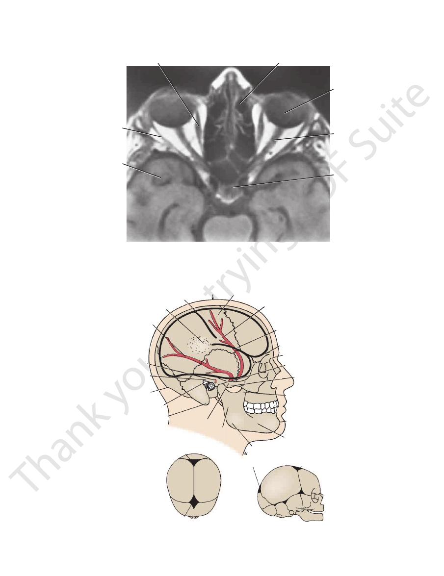
FIGURE 11.127
al and the cranial cavities. Note that the eyeballs, the
Axial (horizontal) MRI showing the contents of the orbit
optic nerves, the optic chiasma, and the extraocular muscles can be identified.
vertex
central sulcus
parietal eminence
posterior branch
of the middle meningeal artery
superficial temporal artery
suprameatal crest
external occipital protuberance
suprameatal triangle
mastoid process
zygomatic arch
ramus of mandible
angle of mandible
body of mandible
symphysis menti
coronoid process
articular tubercle
temporomandibular joint
nasion
superciliary ridge
pterion
anterior branch of the middle meningeal artery
motor area of cerebral cortex
posterior fontanelle
anterior fontanelle
posterior fontanelle
anterior fontanelle
A
B
FIGURE 11.128
A.
Superior aspect and right side of the neonatal skull. Note the positions of the anterior and posterior fontanelles.
Right side of the head showing relations of the middle meningeal artery and the brain to the surface of the
skull. B.

676
CHAPTER 11
the second, third, and fourth rings of the trachea
This lies in front of
Isthmus of the thyroid gland:
pation just above the isthmus of the thyroid gland.
This can be felt by gentle pal
First ring of the trachea:
trachea (Fig. 11.98).
val between the cricoid cartilage and the first ring of the
This structure fills in the inter
Cricotracheal ligament:
enters the thyroid gland.
glion, and at the level where the inferior thyroid artery
gus, at the level of the middle cervical sympathetic gan
the level of the junction of the pharynx with the esopha
vertebra, at the junction of the larynx with the trachea, at
(Fig. 11.129), this lies at the level of the 6th cervical
An important landmark in the neck
Cricoid cartilage:
lage (Fig. 11.130).
val between the cricoid cartilage and the thyroid carti
This structure fills in the inter
Cricothyroid ligament:
structure lies opposite the 4th cervical vertebra (Figs.
This notched
Upper border of the thyroid cartilage:
(Fig. 11.130).
between the hyoid bone and the thyroid cartilage
This fills in the interval
Thyrohyoid membrane:
cal vertebra (Figs. 11.13 and 11.129).
This lies opposite the 3rd cervi
Body of the hyoid bone:
are located in this triangle.
by the mylohyoid muscle. The submental lymph nodes
orly by the body of the hyoid bone. The floor is formed
by the anterior belly of the digastric muscle, and inferi
bounded anteriorly by the midline of the neck, laterally
menti and the body of the hyoid bone (Fig. 11.56). It is
This lies between the symphysis
Submental triangle:
midline (Figs. 11.129 and 11.130).
the two halves of the body of the mandible unite in the
The lower margin can be felt where
Symphysis menti:
palpated from above downward:
In the midline anteriorly, the following structures can be
lies deep to the superciliary ridge on each side (Fig. 11.97).
The frontal air sinus is situated within the frontal bone and
(Fig. 11.97).
bone and lies below the infraorbital foramen on each side
The maxillary air sinus is situated within the maxillary
plies the skin of the face.
The infraorbital nerve emerges from the foramen and sup
premolar teeth.
supraorbital notch to the interval between the two lower
of the orbit (Fig. 11.1), on a line drawn downward from the
The infraorbital foramen lies 5 mm below the lower margin
against the bone (Fig. 11.18).
which can be rolled
supraorbital nerve,
It transmits the
and intermediate thirds of the upper margin of the orbit.
If present, the notch can be felt at the junction of the medial
maxillary bones (Fig. 11.18).
The orbital margin is formed by the frontal, zygomatic, and
mouth opposite the upper second molar tooth (Fig. 11.72).
der of the masseter as it turns medially and opens into the
be rolled beneath the examining finger at the anterior bor
fingerbreadth below the zygomatic arch (Fig. 11.132). It can
The parotid duct runs forward from the parotid gland one
clenching the teeth.
The anterior border of the masseter can be easily felt by
rior border of the masseter muscle (Fig. 11.132).
the lower margin of the body of the mandible, at the ante
The pulsations of the facial artery can be felt as it crosses
angle of the mandible (Fig. 11.128).
sis menti, in the midline anteriorly, as far backward as the
it is possible to examine the mandible from the symphy
finger inside the mouth and another on the outside. Thus,
The body of the mandible is best examined by having one
The Head and Neck
Body of the Mandible
-
Facial Artery
-
Anterior Border of the Masseter
Parotid Duct
-
Orbital Margin
Supraorbital Notch
Infraorbital Foramen
Infraorbital Nerve
-
Maxillary Air Sinus
Frontal Air Sinus
Surface Landmarks of the Neck
Anterior Aspect
■
■
■
■
-
■
■
-
■
■
■
■
11.13 and 11.129).
■
■
-
-
■
■
-
-
■
■
-
■
■
-
■
■
(Figs. 11.129 and 11.130).
lar veins just above the suprasternal notch (Fig. 11.13).
This vein connects the two anterior jugu
Jugular arch:
gland, from the brachiocephalic artery (Fig. 11.110).
in front of the trachea to the isthmus of the thyroid
When present, this artery ascends
Thyroidea ima artery:
(Fig. 11.110).
front of the fifth, sixth, and seventh rings of the trachea
The inferior thyroid veins lie in
Inferior thyroid veins:
■
■
■
■
■
■
-
Surface Anatomy
677
angle of mandible
anterior
triangle of neck
posterior
triangle of neck
trapezius
sternocleidomastoid
suprasternal notch
symphysis menti
body of hyoid
thyroid cartilage
cricoid cartilage
isthmus of
thyroid gland
trachea
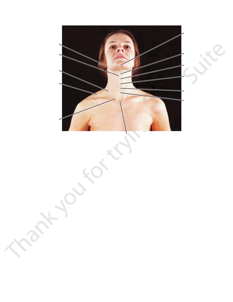
FIGURE 11.129
-old woman. Note that the atlanto-occipital joints and the cervi
Anterior view of the head and neck of a 29-year
In the adult, the trachea may measure as much as 1 in.
vertebra.
the lower border of the body of the 2nd thoracic
rior border of the manubrium sterni and lies opposite
rior ends of the clavicles (Fig. 11.129). It is the supe
This can be felt between the ante
Suprasternal notch:
cal part of the vertebral column are partially extended for full exposure of the front of the neck.
-
■
■
-
-
(2.5 cm) in diameter, whereas in a baby it may be nar
cleidomastoid, and the midline (Fig. 11.56). The posterior
neck is bounded by the body of the mandible, the sterno
rior and posterior triangles. The anterior triangle of the
The sternocleidomastoid divides the neck into ante
tract, and its anterior and posterior borders will be defined.
carried out against resistance, the muscle will be felt to con
looks upward toward the opposite side. If the movement is
side and at the same time rotate the head so that the face
patient to approximate the ear to the shoulder of the same
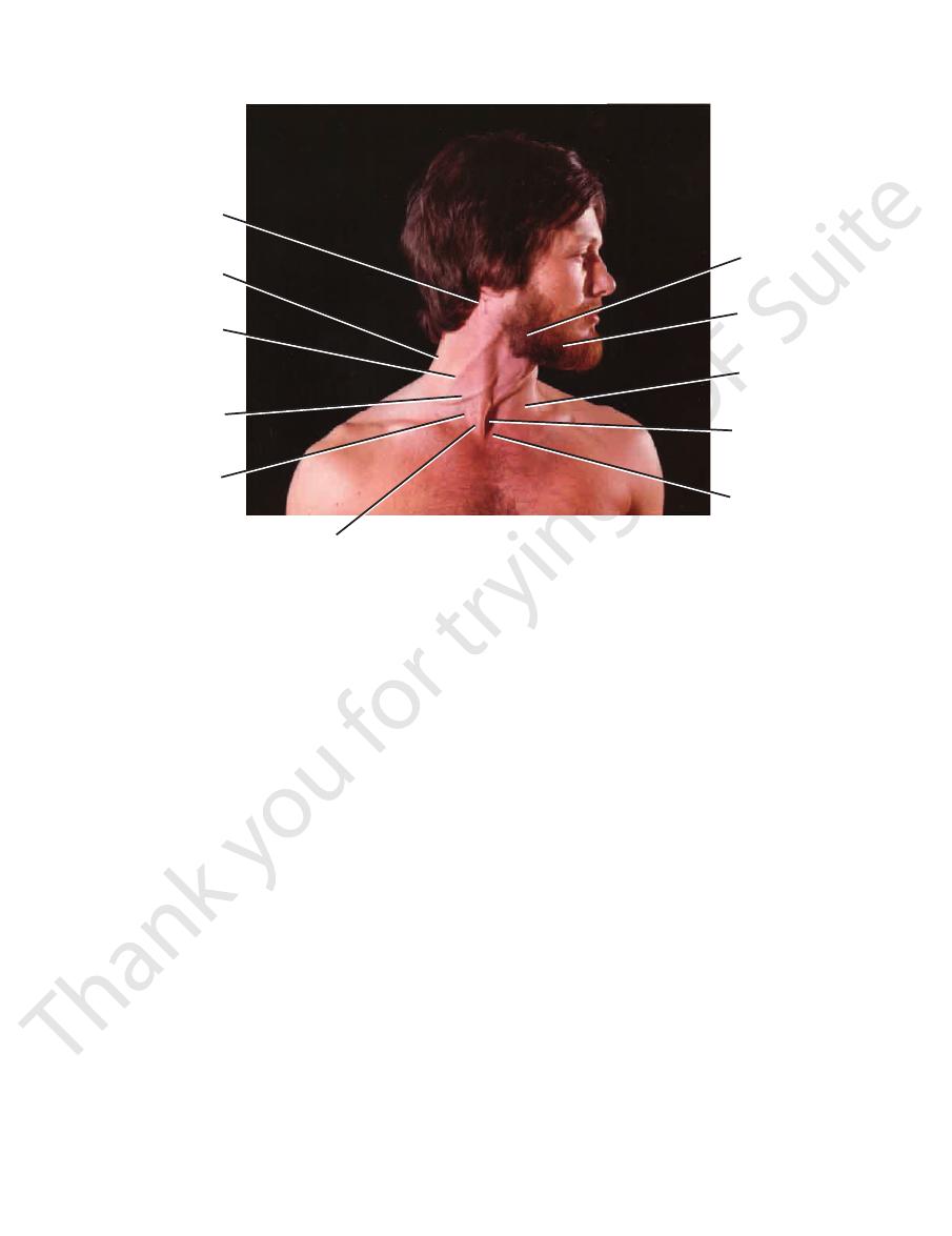
11.132). The muscle can be made to stand out by asking the
sternum and clavicle to the mastoid process (Figs. 11.131 and
pated throughout its length as it passes upward from the
On the side of the neck, the sternocleidomastoid can be pal
Sternocleidomastoid Muscle
ligamentum nuchae.
Cervical spines one to six are covered by
bra prominens).
7th cervical vertebra (verte
process to be felt is that of the
The first spinous
nuchal groove.
drawn downward in the
index finger is placed on the skin in the midline, it can be
at the junction of the head and neck (Fig. 11.132). If the
lies in the midline
external occipital protuberance
The
palpated from above downward.
In the midline posteriorly, the following structures can be
suprasternal notch.
and the left brachiocephalic vein may protrude above the
mus of the thyroid gland, and the brachiocephalic artery
may extend above the suprasternal notch as far as the isth
rower than a pencil. In young children, the thymus gland
-
-
Posterior Aspect
-
the
Lateral Aspect
-
-
-
-
triangle is bounded by the anterior border of the trapezius,
border of the lateral third of the clavicle.
occipital bone, downward and forward to the posterior
will be seen to extend from the superior nuchal line of the
can be felt by asking the patient to shrug the shoulders. It
The anterior border of the trapezius muscle (Fig. 11.129)
Trapezius Muscle
the sternocleidomastoid, and the clavicle (Fig. 11.56).
678
CHAPTER 11
The Head and Neck
symphysis menti
body of hyoid bone
angle of mandible
superior belly of
the omohyoid muscle
sternohyoid muscle
thyroid cartilage
thyroid gland
sternothyroid muscle
isthmus of thyroid gland
suprasternal notch
trachea
trapezius muscle
inferior belly of the
omohyoid muscle
cricothyroid ligament
sternocleidomastoid muscle
thyrohyoid membrane
posterior belly of the
digastric muscle
submandibular salivary gland
mylohyoid muscle
anterior belly of
the digastric muscle
FIGURE 11.130
Surface anatomy of the neck from in front.
seated patient and asking the patient to flex the neck for
This is most easily carried out by standing behind the
be palpated deep to the sternocleidomastoid muscles.
and 11.130). The lateral lobes of the thyroid gland can
ond, third, and fourth rings of the trachea (Figs. 11.129
The isthmus of the thyroid gland lies in front of the sec
Anterior Triangle of the Neck
be identified.
sternoclavicular joint
medial end of the clavicle, the
lateral extremity with the acromion of the scapula. At the
can be easily palpated (Fig. 11.132). It articulates at its
clavicle is subcutaneous throughout its entire length and
midline anteriorly (see page 677) and the clavicles. Each
suprasternal notch
At the root of the neck are the
Root of the Neck
the anterior thoracic wall (Fig. 11.51).
the body of the mandible downward over the clavicle onto
patient to clench the jaws firmly. The muscle extends from
The platysma can be seen as a sheet of muscle by asking the
Platysma Muscle
in the
can
-
-
Surface Anatomy
679
suprasternal notch
sternocleidomastoid
sternal head of
triangle of neck
anterior
body of mandible
(third part)
site of subclavian artery
brachial plexus
triangle of neck
posterior
ternal
mastoid process
trapezius
ex
jugular vein
site of
angle of mandible
FIGURE 11.131
ear-old man. Note that the head has been laterally rotated to the left at the
Anterior view of the neck of a 27-y
duct is given on page 631.
muscle (Fig. 11.85). The surface marking of the parotid
dible and the anterior border of the sternocleidomastoid
gland lies below the ear in the interval between the man
The three large salivary glands can be palpated. The parotid
Salivary Glands
vian vein.
fascia just above the clavicle and drains into the subcla
clavicle (Figs. 11.131 and 11.132). It perforates the deep
region of the angle of the mandible to the middle of the
cia deep to the platysma. It passes downward from the
The external jugular vein lies in the superficial fas
External Jugular Vein
enter the neck.
The subclavian vein lies behind the clavicle and does not
surface of the 1st rib, that its pulsations can be felt easily.
of the clavicle. It is here, where the artery lies on the upper
about 0.5 in. (1.3 cm) and then downward to the middle
which passes upward from the sternoclavicular joint for
and 11.132). Its course may be indicated by a curved line,
lower anterior angle of the posterior triangle (Figs. 11.131
The third part of the subclavian artery also occupies the
Third Part of the Subclavian Artery
middle of the clavicle.
by a line drawn from the cricoid cartilage downward to the
and 11.132). The upper limit of the plexus can be indicated
lower anterior angle of the posterior triangle (Figs. 11.131
The roots and trunks of the brachial plexus occupy the
Roots and Trunks of the Brachial Plexus
angle; the second line indicates the course of the nerve.
and extend the second line downward across the posterior tri
to the tip of the mastoid process. Bisect this line at right angles
cated as follows: Draw a line from the angle of the mandible
trapezius (Fig. 11.132). The course of this nerve may be indi
ward and backward to pass beneath the anterior border of the
posterior border of the sternocleidomastoid and runs down
is relatively superficial as it emerges from the
accessory nerve
At the posterior triangle of the neck, the spinal part of the
Posterior Triangle of the Neck
ies can be felt at this level.
carotid arteries (Fig. 11.132). The pulsations of these arter
mon carotid artery bifurcates into the internal and external
level of the upper border of the thyroid cartilage, the com
the mastoid process and the angle of the mandible. At the
sternoclavicular joint to a point midway between the tip of
cal lymph nodes, can be marked out by a line joining the
internal jugular vein, the vagus nerve, and the deep cervi
The carotid sheath, which contains the carotid arteries, the
Carotid Sheath
the fingers of both hands.
then examine both lobes simultaneously with the tips of
ward and so relax the overlying muscles. The observer can
atlantoaxial joints and at the joints of the cervical part of the vertebral column.
-
-
-
-
-
-
-
-
-
680
CHAPTER 11
The Head and Neck
mastoid process
angle of
mandible
external occipital
protuberance
spinal part
of the accessory nerve
external jugular vein
trapezius
inferior belly of omohyoid
acromion
brachial plexus
clavicle
subclavian artery
sternocleidomastoid
isthmus of thyroid gland
trachea
cricoid cartilage
thyroid cartilage
common carotid artery
hyoid bone
digastric muscle
external
carotid artery
facial artery
masseter muscle
internal jugular vein
parotid duct
zygomatic arch
head of mandible
FIGURE 11.132
Surface anatomy of the neck from the lateral aspect.

Surface Anatomy
beneath the lower margin of the body of the mandi
superficial and deep parts. The superficial part lies
The submandibular gland can be divided into
681
-
ble (Fig.
eep part of the submandibular
11.86). The d
tongue (Fig. 11.72).
opens into the mouth on the side of the frenulum of the
the tongue and the lower jaw. The submandibular duct
covering the floor of the mouth in the interval between
gland can be palpated through the mucous membrane
gland, the submandibular duct, and the sublingual
www.thePoint.lww.com/Snell9e.
Clinical Cases
and
Review Questions
are available online at