136
CHAPTeR 4
umn to the same side.
tion (see page 77) and laterally flexes the vertebral col
It fixes or depresses the 12th rib during respira
Action:
plexus.
This muscle is supplied by the lumbar
Nerve supply:
umbar ligament.
and below to form the
lateral arcuate ligament
covered by lumbar fascia, which is thickened above to form
four lumbar vertebrae. The anterior surface of the muscle is
of the 12th rib and the transverse processes of the upper
upward and medially and are inserted into the lower border
the lower lumbar vertebrae (see Fig. 4.33). The fibers run
the iliac crest, and the tips of the transverse processes of
below from the iliolumbar ligament, the adjoining part of
muscle that lies alongside the vertebral column. It arises
The quadratus lumborum is a flat, quadrilateral-shaped
thigh, as in sitting up from a lying position.
trunk, or if the thigh is fixed, it flexes the trunk on the
The psoas flexes the thigh at the hip joint on the
Action:
plexus.
This muscle is supplied by the lumbar
Nerve supply:
medial arcuate ligament.
thickened above to form the
sheath that is derived from the lumbar fascia. The sheath is
trochanter of the femur. The psoas is enclosed in a fibrous
the inguinal ligament. The muscle is inserted into the lesser
and leave the abdomen to enter the thigh by passing behind
vertebrae (Fig. 4.33). The fibers run downward and laterally
intervertebral discs, from the 12th thoracic to 5th lumbar
verse processes, the sides of the vertebral bodies, and the
arises from the roots of the trans
The psoas muscle
Muscles of the Posterior Abdominal Wall
The Abdomen: Part I—The Abdominal Wall
Psoas Major
†
-
■
■
■
■
Quadratus Lumborum
the
iliol-
■
■
■
■
-
-
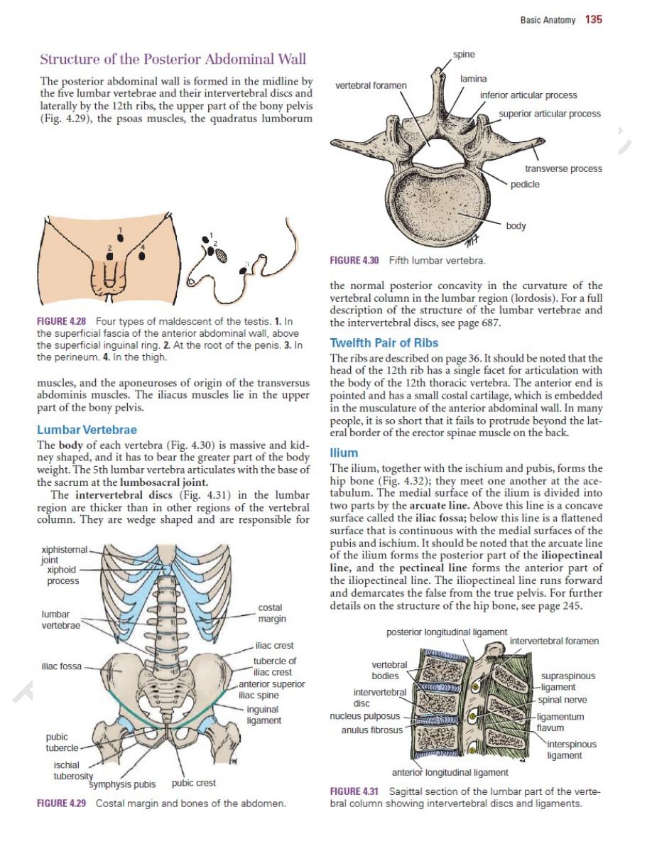
iliacus muscle
iliac crest
anterior superior
iliac spine
arcuate line
anterior inferior iliac
spine
iliopubic eminence
pectineal line
pubic tubercle
pubic crest
obturator foramen
ischial
tuberosity
ischial spine
iliopectineal line
quadratus
lumborum muscle
articular surface
for sacrum
FIGURE 4.32
Internal aspect of the right hip bone.
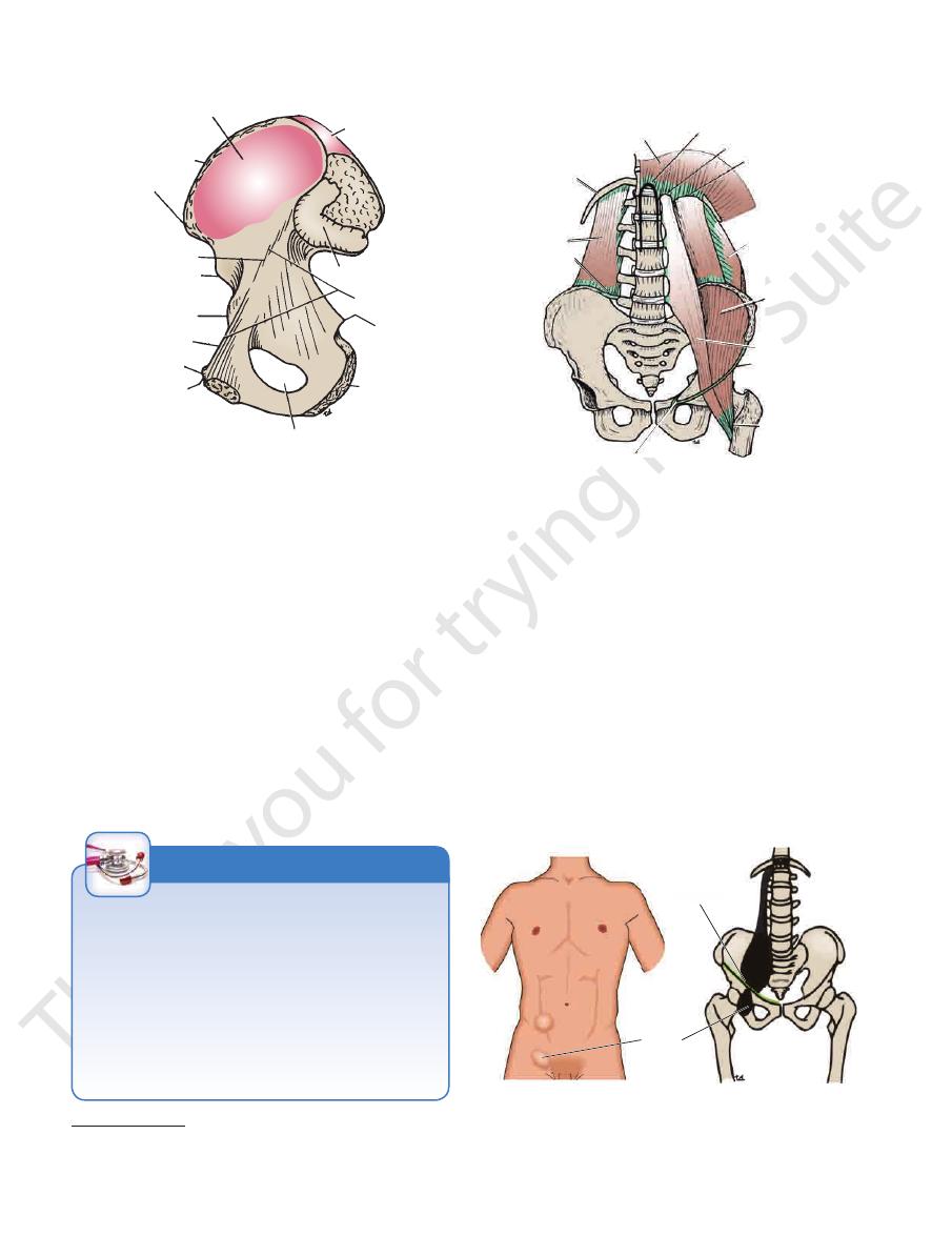
diaphragm
inguinal ligament
iliacus
transversus
lateral arcuate
ligament
medial arcuate
ligament
median arcuate
ligament
12th rib
quadratus
lumborum
iliolumbar
ligament
lacunar ligament
psoas
lesser trochanter
of femur
FIGURE 4.33
Muscles and bones forming the posterior
abdominal wall.
inguinal
ligament
psoas
abscess
inguinal
ligament
psoas
abscess
FIGURE 4.34
Case of advanced tuberculous disease of the
above and below the right inguinal ligament.
abscess is present, and swellings occur in the right groin
thoracolumbar region of the vertebral column. A psoas
the psoas major. It is unimportant and is absent in 40% of patients.
The psoas minor is a small muscle with a long tendon that lies anterior to
†
Psoas Fascia and Tuberculosis
bral bodies, with possible extension of pus laterally under the
The psoas fascia covers the anterior surface of the psoas
muscle and can influence the direction taken by a tuberculous
abscess. Tuberculous disease of the thoracolumbar region of
the vertebral column results in the destruction of the verte-
psoas fascia (Fig. 4.34). From there, the pus tracks downward,
following the course of the psoas muscle, and appears as a
swelling in the upper part of the thigh below the inguinal liga-
ment. It may be mistaken for a femoral hernia.
C L I N I C A L N O T E S

Basic Anatomy
diaphragmatic fascia
structure it overlies. For example, the
vic walls. It is customary to name the fascia according to the
continuous below with a similar fascial layer lining the pel
the parietal peritoneum and the muscles (Fig. 4.35). It is
one continuous layer of connective tissue that lies between
As mentioned previously, the abdominal walls are lined by
Fascial Lining of the Abdominal Walls
in Table 4.2.
abdominal wall, their nerve supply, and their action is given
on page 44. A summary of the muscles of the posterior
forms part of the posterior abdominal wall. It is described
(see Fig. 4.33) also
diaphragm
The posterior part of the
on the thigh.
the hip joint, or if the thigh is fixed, it flexes the trunk
The iliopsoas flexes the thigh on the trunk at
Action:
nerve, a branch of the lumbar plexus.
This muscle is supplied by the femoral
Nerve supply:
iliopsoas.
often referred to as the
lesser trochanter of the femur. The combined muscles are
the lateral side of the psoas tendon to be inserted into the
part of the iliac fossa (see Figs. 4.32 and 4.33). Its fibers join
The iliacus muscle is fan shaped and arises from the upper
page 120.
The transversus abdominis muscle is fully described on
Transversus Abdominis
137
Iliacus
■
■
■
■
-
covers the undersurface of the diaphragm, the
also supply the overlying muscles and skin.
plied segmentally by intercostal and lumbar nerves, which
lining the anterior and posterior abdominal walls is sup
supplied by the lower intercostal nerves. The peritoneum
plied by the phrenic nerves, and the peripheral part is
The central part of the diaphragmatic peritoneum is sup
(see Fig. 4.35). For further details, see pages 278 and 296.
ous below with the parietal peritoneum lining the pelvis
of mesothelium resting on connective tissue. It is continu
neum. This is a thin serous membrane consisting of a layer
The walls of the abdomen are lined with parietal perito
Walls
matic fascia (see Fig. 4.4).
tinued over the cord as a tubular sheath, the internal sper
Fig. 4.8). From the margins of the ring, the fascia is con
the fascia transversalis to form the deep inguinal ring (see
spine and the symphysis pubis, the spermatic cord pierces
At the midpoint between the anterior superior iliac
and peritoneum only (see page 122).
Figs. 4.10 and 4.13) and is formed by the fascia transversalis
of the rectus sheath is devoid of muscular aponeuroses (see
level of the anterior superior iliac spines, the posterior wall
performs particularly important functions. Inferior to the
In certain areas of the abdominal wall, the fascial lining
(see page 463).
nerve lies outside the fascial envelope, it has no sheath
thigh, behind the inguinal ligament. Because the femoral
vessels and lymphatics, for about 1.5 in. (4 cm) into the
prolongation of the fascial lining around the femoral
femoral sheath (see Fig. 4.35). This is simply a downward
fascia. This fact is important in the understanding of the
fascial lining, whereas the principal nerves lie outside the
The abdominal blood and lymph vessels lie within this
ers the iliacus muscle.
cov
covers the quadratus lumborum, and the
quadratus lumborum fascia
covers the psoas muscle, the
psoas fascia
lines the transversus abdominis, the
transversalis
fascia
iliaca fascia
-
-
-
Peritoneal Lining of the Abdominal
-
-
Nerve Supply
-
-
Muscles of the Posterior Abdominal Wall
T A B L E 4 . 2
Transverse processes,
Name of Muscle
Origin
Insertion
Nerve Supply
Action
Psoas
bodies, and interver-
tebral discs of 12th
thoracic and five
lumbar vertebrae
With iliacus into lesser
trochanter of femur
Lumbar plexus
Flexes thigh on trunk; if thigh
is fixed, it flexes trunk on
thigh, as in sitting up from
lying position
Quadratus lumborum
Iliolumbar ligament,
iliac crest, tips of
transverse pro-
cesses of lower
lumbar vertebrae
12th rib
Lumbar plexus
Fixes 12th rib during inspira-
tion; depresses 12th rib
during forced expiration;
laterally flexes vertebral
column same side
Iliacus
Iliac fossa
With psoas into lesser
trochanter of femur
Femoral nerve
Flexes thigh on trunk; if thigh
is fixed, it flexes the trunk
on the thigh, as in sitting up
from lying position