138
CHAPTeR 4
The Abdomen: Part I—The Abdominal Wall
Development of the Abdominal Wall
Wharton’s
vessels to form the tubular umbilical cord. The mesenchymal core
amnion encloses the body stalk and the yolk sac with their blood
yolk sac (Fig. 4.37). The amnion and chorion now fuse, so that the
lie on the anterior surface of the embryo, close to the remains of the
of the body stalk to the caudal end of the embryonic disc comes to
As the tail fold of the embryo develops, the embryonic attachment
chyme forms the linea alba, and on either side of this, the rectus
meet in the midline and fuse. The line of fusion of the mesen
closes in the midline at 3 months, when the right and left sides
transversus abdominis muscles. The anterior body wall finally
layers, which form the external oblique, internal oblique, and
somatopleuric mesoderm becomes split tangentially into three
gin, as seen by the presence of the tendinous intersections. The
ribs, and the mesenchyme fuses to form large sheets of muscle.
the segmental arrangement becomes lost due to the absence of
from the anterior rami of the spinal nerves. Unlike the thorax,
somatopleuric mesoderm and retain their segmental innervation
muscles of the anterior abdominal wall are derived from the
ciated with ectoderm and entoderm, respectively (Fig. 4.36). The
Following segmentation of the mesoderm, the lateral mesoderm
(see page 33) splits into a somatic and a splanchnic layer asso-
The rectus abdominis retains indications of its segmental ori-
-
muscles come to lie within their rectus sheaths.
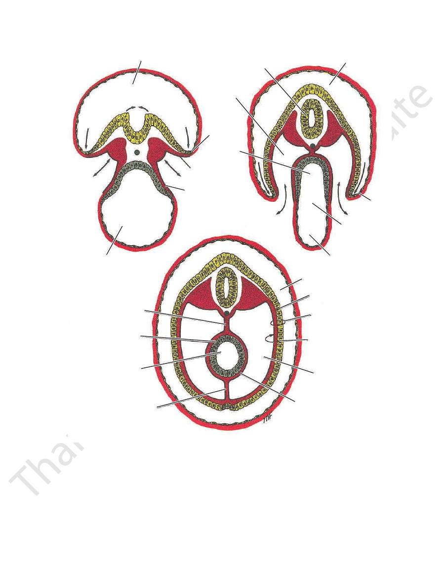
Development of the Umbilical Cord and the Umbilicus
of the cord forms the loose connective tissue called
jelly.
embedded in this are the remains of the yolk sac, the vitelline
at the end of pregnancy, it is about 20 in. (50 cm) long—that is,
duct, the remains of the allantois, and the umbilical blood vessels.
The umbilical vessels consist of two arteries that carry deox-
ygenated blood from the fetus to the chorion (later the placenta).
The two umbilical veins convey oxygenated blood from the pla-
centa to the fetus. The right vein soon disappears (see Fig. 4.37).
The umbilical cord is a twisted tortuous structure that mea-
sures about 0.75 in. (2 cm) in diameter. It increases in length until,
about the same length as the child.
E M B R Y O L O G I C N O T E S
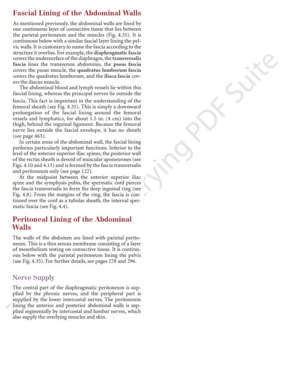
diaphragmatic fascia
peritoneum
quadratus lumborum fascia
pelvic fascia
fascia transversalis
ileum
inguinal ligament
femoral sheath
fascia iliaca
femoral nerve
peritoneum
fascia
femoral sheath
femoral artery femoral vein
femoral canal
lymph node
femoral ring
lymph vessel
femoral sheath
femoral nerve
anterior view
lateral view
transverse colon
FIGURE 4.35
Sagittal section of the abdomen showing arrangement of the fascial and peritoneal linings of walls. The femoral
sheath with its contained vessels is also shown. Note that the femoral nerve is devoid of a fascial sheath.
Basic Anatomy
139
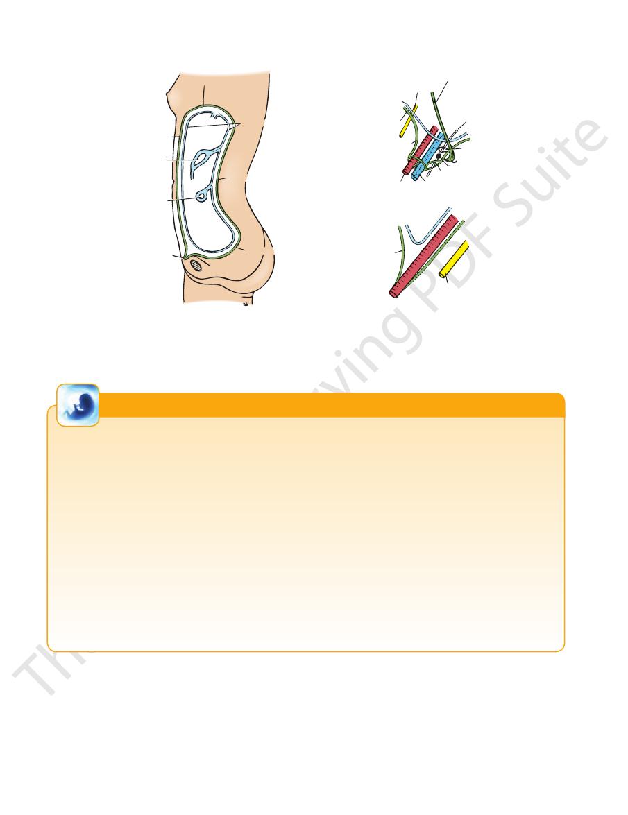
amniotic cavity
B
A
C
extra-
embryonic
coelom
yolk sac
lateral fold
somatic
mesenchyme
splanchnic
mesenchyme
developing
gut
intraembryonic
coelom
neural tube
amniotic cavity
lateral fold
beginning of
development of
vitelline duct
yolk sac
extraembryonic coelom
amniotic cavity
ectoderm
body wall
somatic mesenchyme
closed-off
intraembryonic coelom
splanchnic mesenchyme
dorsal mesentery
entoderm
foregut
ventral mesentery
FIGURE 4.36
Transverse sections through the embryo at different stages of development showing the formation of the
lom or future peritoneal cavity. Most of the ventral mesentery will break down and disappear.
The lateral folds of the embryo finally fused in the midline and closing off the intraembryonic coe
intraembryonic coelom.
The development of the lateral folds of the embryo and the beginning of the closing off of the
double-headed arrows
The intraembryonic coelom in free communication with the extraembryonic coelom
abdominal wall and peritoneal cavity. A.
(
). B.
C.
-
140
CHAPTeR 4
The Abdomen: Part I—The Abdominal Wall
amnion
amniotic cavity
chorion laeve
stomodeum
extraembryonic
coelom
yolk sac
chorion frondosum
ectoderm
proctodeum
allantois
body stalk
placenta
amnion
chorion
vitelline duct
yolk sac
foregut
midgut
hindgut
allantois
umbilical vein
vitelline duct
umbilical arteries
remains of
allantois
placenta
amniotic cavity
filled with
amniotic fluid
FIGURE 4.37
The formation of the umbilical cord. Note the expansion of the amniotic cavity (
becomes covered with amnion. Note also that the umbilical vessels have been reduced to one vein and two arteries.
arrows) so that the cord