Basic Anatomy
263
Contents of the Pelvic Cavity
The rectum is about 5 in. (13 cm) long and begins in front
Location and Description
inferior hypogastric plexuses.
The sympathetic and parasympathetic nerves from the
Nerve Supply
inferior mesenteric nodes.
moid arteries; from these nodes, the lymph travels to the
The lymph drains into nodes along the course of the sig
Lymph Drainage
joins the portal venous system.
The veins drain into the inferior mesenteric vein, which
Veins
Sigmoid branches of the inferior mesenteric artery.
Arteries
part of the ileum.
colon is also related to the lower coils of the terminal
The rectum and the sacrum. The sigmoid
Posteriorly:
part of the vagina
female, the posterior surface of the uterus and the upper
In the male, the urinary bladder; in the
Anteriorly:
Relations
sigmoid mesocolon.
wall by the fan-shaped
The sigmoid colon is attached to the posterior pelvic
the form of a loop.
colon is mobile and hangs down into the pelvic cavity in
rectum in front of the 3rd sacral vertebra. The sigmoid
of the pelvic brim. Below, it becomes continuous with the
begins as a continuation of the descending colon in front
The sigmoid colon is 10 to 15 in. (25 to 38 cm) long and
Location and Description
Sigmoid Colon
■
■
■
■
Blood Supply
-
Rectum
of the third sacral vertebra as a continuation of the sigmoid
natomy
asic
B
a
The pelvic cavity, or cavity of the true pelvis, can be defined
into the main pelvic cavity above and the perineum below
It is customary to subdivide it by the pelvic diaphragm
as the area between the pelvic inlet and the pelvic outlet.
(Fig. 7.1). This chapter is concerned with the contents of
neum is given in Chapter 8.
the main pelvic cavity. A detailed description of the peri-
■
ectopic pregnancy, spontaneous abortion, and acute pelvic
Emergency situations involving the bladder, the pregnant uterus,
The organs project up into the peritoneal cavity, causing the
their nerve supply, blood supply, and lymphatic drainage.
■
The pelvic cavity contains the lower ends of the intestinal and
urinary tracts and the internal organs of reproduction as well as
■
■
peritoneum to be draped over them in folds, producing impor-
tant fossae that are the sites for the accumulation of blood and
pus in different types of pelvic disease.
■
■
The physician is often confronted with problems involving infec-
tions, injuries, and prolapses of the rectum, uterus, and vagina.
■
■
inflammatory disease are examples of problems found in the
female.
■
■
The urinary bladder and the prostate in the male are frequent
sites of disease.
■
■
The purpose of this chapter is to consider the important
anatomy relative to common clinical conditions involving the
pelvic organs.
thoracic cavity
perineum
pelvic outlet
main
pelvic cavity
abdominal cavity
diaphragm
costal
margin
iliac crest
pelvic inlet
pelvic diaphragm
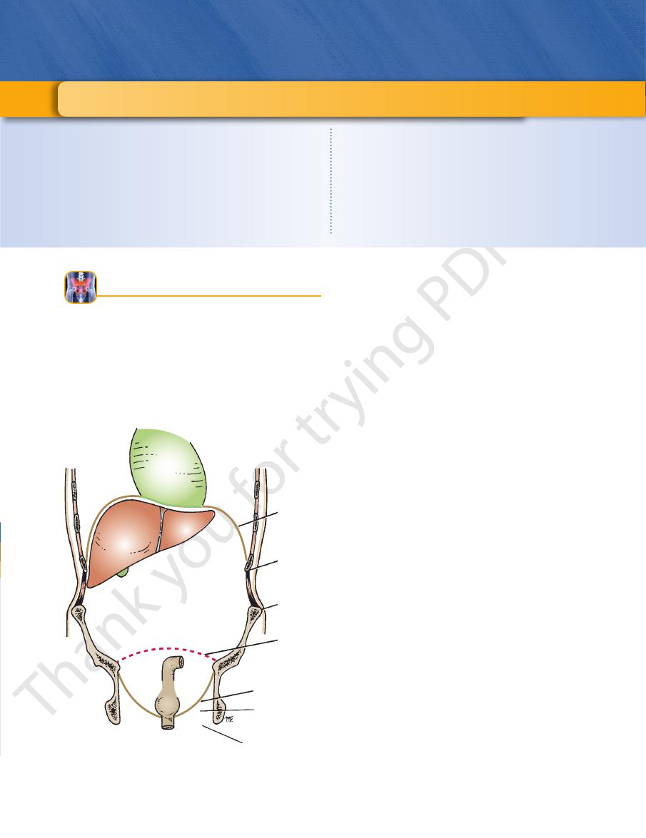
FIGURE 7.1
Coronal section through the thorax, abdomen,
and pelvis showing the thoracic, abdominal, and pelvic
cavities and the perineum.
C H A P T E R O B J E C T I V E S
Basic Anatomy
265
A
B
S1
S2
S3
sigmoid
colon
umbilicus
area of
referred
discomfort or
pain as instrument
enters sigmoid
colon
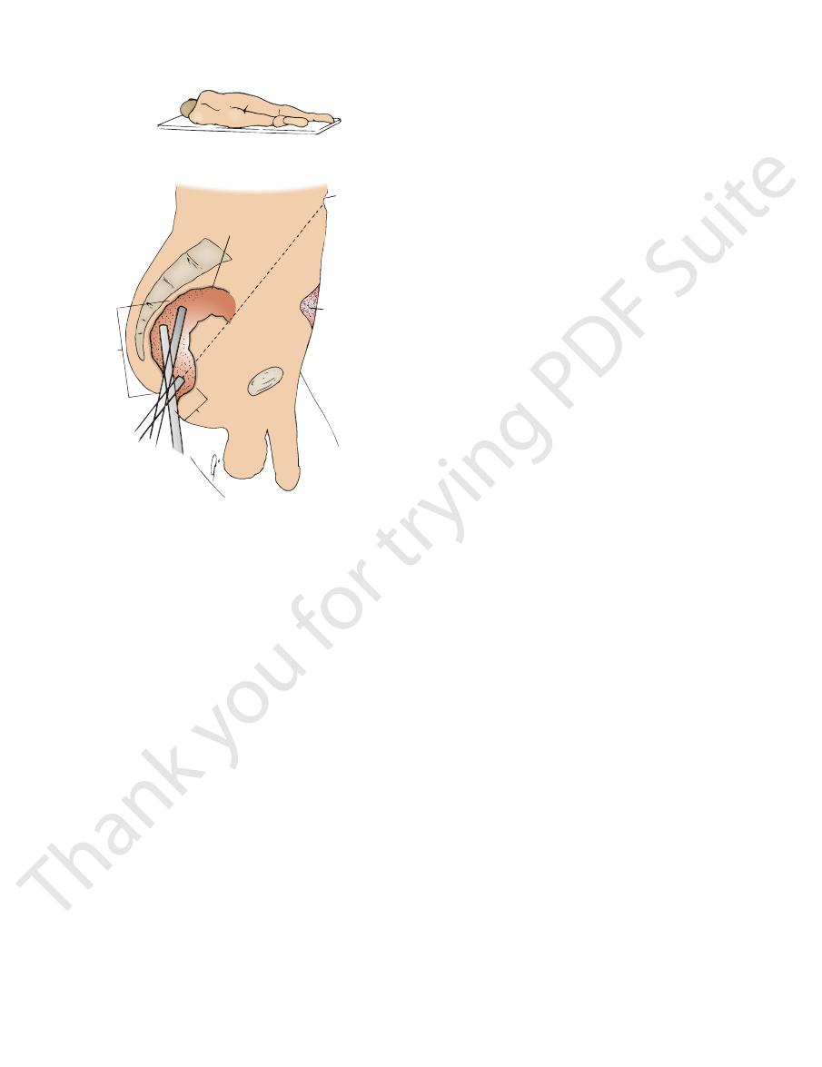
6.5 in.
1 1/2 in.
1
2
3
FIGURE 7.2
Sigmoidoscopy.
thetic nerves from the inferior hypogastric plexuses. The
The nerve supply is from the sympathetic and parasympa
Nerve Supply
middle rectal artery to the internal iliac nodes.
Lymph vessels from the lower part of the rectum follow the
and then into inferior mesenteric nodes.
arectal nodes
The lymph vessels of the rectum drain first into the
Lymph Drainage
tomosis (see Chapter 5).
the rectal veins forms an important portal–systemic anas
internal pudendal veins, respectively. The union between
drain into the internal iliac and
inferior rectal veins
middle
and drains into the inferior mesenteric vein. The
is a tributary of the portal circulation
superior rectal vein
The veins of the rectum correspond to the arteries. The
Veins
middle rectal artery at the anorectal junction.
pudendal artery in the perineum. It anastomoses with the
is a branch of the internal
inferior rectal artery
The
iliac artery and is distributed mainly to the muscular coat.
is a small branch of the internal
middle rectal artery
The
another and with the middle and inferior rectal arteries.
supply the mucous membrane. They anastomose with one
right and left branches, which pierce the muscular coat and
ing in the root of the sigmoid mesocolon and divides into
ing the mucous membrane. It enters the pelvis by descend
inferior mesenteric artery and is the chief artery supply
is a direct continuation of the
superior rectal artery
The
supply the rectum.
The superior, middle, and inferior rectal arteries (Fig. 7.6)
Arteries
vagina (see Fig. 7.5).
of peritoneum, is related to the posterior surface of the
of Douglas). The lower third of the rectum, which is devoid
coils of ileum that occupy the rectouterine pouch (pouch
covered by peritoneum, is related to the sigmoid colon and
the upper two thirds of the rectum, which is
In the female,
vesicles on each side, and to the prostate (see Fig. 7.4).
der, to the termination of the vas deferens and the seminal
peritoneum, is related to the posterior surface of the blad
pouch. The lower third of the rectum, which is devoid of
moid colon and coils of ileum that occupy the rectovesical
tum, which is covered by peritoneum, is related to the sig
the upper two thirds of the rec
Anteriorly: In the male,
(see Fig. 6.18).
muscles; the sacral plexus; and the sympathetic trunks
and coccyx; the piriformis, coccygeus, and levatores ani
The rectum is in contact with the sacrum
Posteriorly:
Relations
(see Fig. 7.3); they vary in position.
transverse folds of the rectum
permanent folds called the
the circular muscle layer, forms two or three semicircular
of the rectum, together with
mucous membrane
The
band on the anterior and posterior surfaces of the rectum.
come together so that the longitudinal fibers form a broad
muscle. The three teniae coli of the sigmoid colon, however,
usual outer longitudinal and inner circular layers of smooth
of the rectum is arranged in the
muscular coat
The
toneum (Figs. 7.4 and 7.5).
of the middle third, leaving the lower third devoid of peri
of the first third of the rectum and only the anterior surface
covers the anterior and lateral surfaces
peritoneum
The
producing the anorectal angle.
with the anal canal and pulls this part of the bowel forward,
forms a sling (see page 247) at the junction of the rectum
The puborectalis portion of the levator ani muscles
canal (Fig. 7.4).
ing downward and backward at its junction with the anal
follows the anterior concavity of the sacrum before bend
to the median plane (Fig. 7.3). On lateral view, the rectum
The rectum deviates to the left, but it quickly returns
rectal ampulla.
dilated to form the
tinuous with the anal canal. The lower part of the rectum is
cyx by piercing the pelvic diaphragm and becoming con
sacrum and coccyx, and ends in front of the tip of the coc
colon. It passes downward, following the curve of the
referred to the skin of the anterior abdominal wall below the
discomfort or pain experienced by the patient as the tube
patient as it ascends the anal canal and rectum. The area of
) of the tube of the sigmoidoscope relative to the
Sagittal section of the male pelvis showing the positions
tion with the left knee flexed and the right knee extended.
Patient in the left lateral posi
A.
-
B.
(1, 2, and 3
is negotiated around the bend into the sigmoid colon is
umbilicus.
-
-
-
-
■
■
■
■
-
-
-
Blood Supply
-
-
and
-
par-
-
to stretch.
only
rectum is sensitive
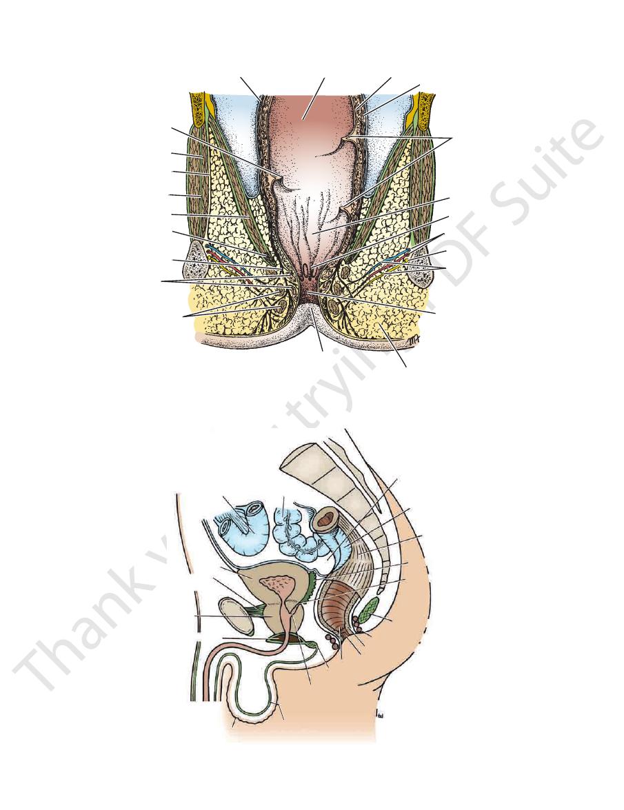
266
CHAPTER 7
The Pelvis: Part II—The Pelvic Cavity
peritoneum
middle transverse
fold of rectum
obturator internus
obturator internus
fascia
obturator membrane
levator ani
puborectalis
outer longitudinal
muscle
internal anal
sphincter
external anal sphincter
anus
fat in ischiorectal fossa
anal canal
inferior rectal
vessels and
nerve
pudendal nerve
internal pudendal
vessels
anal column
ampulla of rectum
upper and lower
transverse folds
of rectum
outer longitudinal muscle
mucous membrane of rectum
inner circular muscle
FIGURE 7.3
Coronal section through the pelvis showing the rectum and the pelvic floor.
sigmoid colon
coil of ileum
bladder
puboprostatic
ligaments
prostate
urogenital diaphragm
scrotum
membranous layer of superficial fascia
opening of ejaculatory
duct into prostatic urethra
perineal body
anal canal
anus
internal sphincter
external sphincter
anococcygeal body
ejaculatory duct
seminal vesicle
rectum
peritoneum
rectovesical pouch
S3
FIGURE 7.4
Sagittal section of the male pelvis.
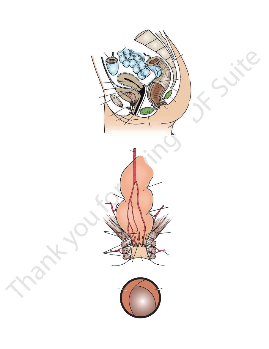
Basic Anatomy
267
sigmoid colon
coil of ileum
cavity of uterus
uterovesical pouch
bladder
cervix
urogenital diaphragm
urethra
vagina
perineal body
anus
anal canal
anococcygeal body
rectum
rectouterine pouch
peritoneum
S3
FIGURE 7.5
Sagittal section of the female pelvis.
superior rectal
artery
right
transverse
fold of
rectum
middle rectal
artery
RIGHT
anal
columns
external
anal
sphincter
anal
valves
anus
inferior rectal
artery
puborectalis
muscle
upper left
transverse
fold of rectum
LEFT
A
B
lower left
transverse fold
of rectum
right transverse
fold of rectum
lower left
transverse
fold of rectum
FIGURE 7.6
The transverse folds of the rectum as seen through a sigmoidoscope.
Blood supply to the rectum.
A.
B.