
12
Lecture
- Cortical Areas
- Internal Structure of
the Cerebral
Hemispheres

Functional areas of Brain (Cortical Areas)
Frontal Lobe:
area:
precentral
-
which is divided into ant and post regions
- posterior region (motor area, primary motor area, or Brodmann area 4), the function of
the primary motor area is to carry out the individual movements of different parts of
the body
-
- anterior region (premotor area, secondary motor area, or Brodmann area 6 and
parts of areas 8, 44, and 45) the premotor area programs the activity of the primary
motor area
-
The supplementary motor area
-
-
Removal of the supplementary motor area produces no permanent loss of
movement
-
The frontal eye field
-
-
(parts of Brodmann areas 6, 8, and 9) Responsible for conjugate movements of the
eyes
-
Broca
The motor speech area of
-
-
Brodmann area 45, The ablation of this region in the nondominant hemisphere has
no effect on speech.
-
The prefrontal cortex
-
-
Brodmann areas 9, 10, 11, and 12, concerned with the makeup of the individual's
personality and emotion

Parietal Lobe
,
2
, and
1
,
3
areas
Brodmann
:
area ( primary sensory)
somesthetic
The primary
contralateral sensation
area
somesthetic
The secondary
). This area has many
7
and
5
areas
Brodmann
(
association area:
somesthetic
The
connections with other sensory areas of the cortex. It is believed that its main
function is to receive and integrate different sensory modalities.
Occipital Lobe
)
17
area
Brodmann
(
primary visual area
)
19
and
18
areas
Brodmann
(
secondary visual area
Temporal Lobe
)
41
area
Brodmann
(
primary auditory area
22
area
Brodmann
(auditory association cortex),
secondary auditory area
is localized in the left dominant hemisphere, The
Wernicke
The sensory speech area of
Wernicke area is connected to the Broca area by a bundle of nerve fibers called the
arcuate fasciculus, It receives fibers from the visual cortex in the occipital lobe and
the auditory cortex in the superior temporal gyrus.
The Wernicke area permits the understanding of the written and spoken language and
enables a person to read a sentence, understand it, and say it out loud
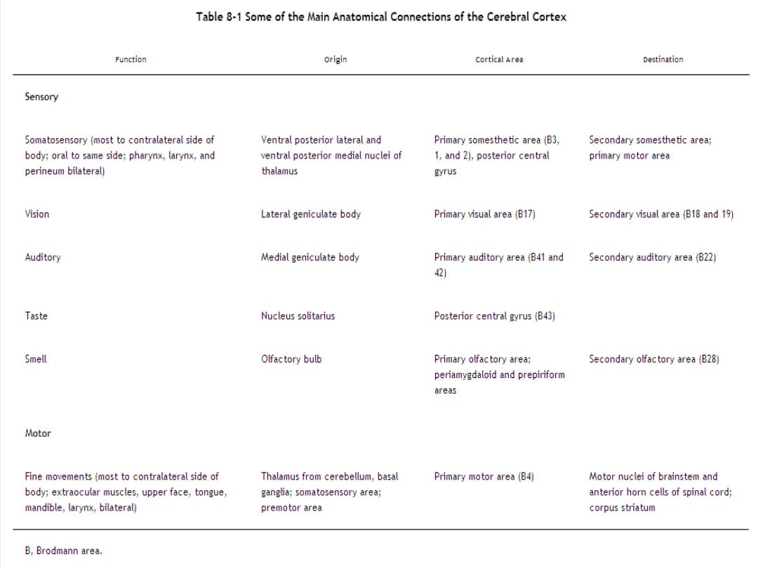
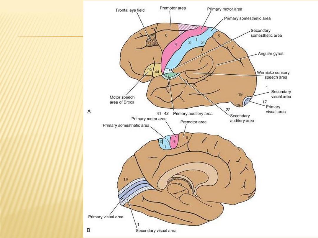
Functional
localization of
the cerebral
cortex.
A: Lateral view of
the left
cerebral
hemisphere.
B: Medial view of
the left
cerebral
hemisphere
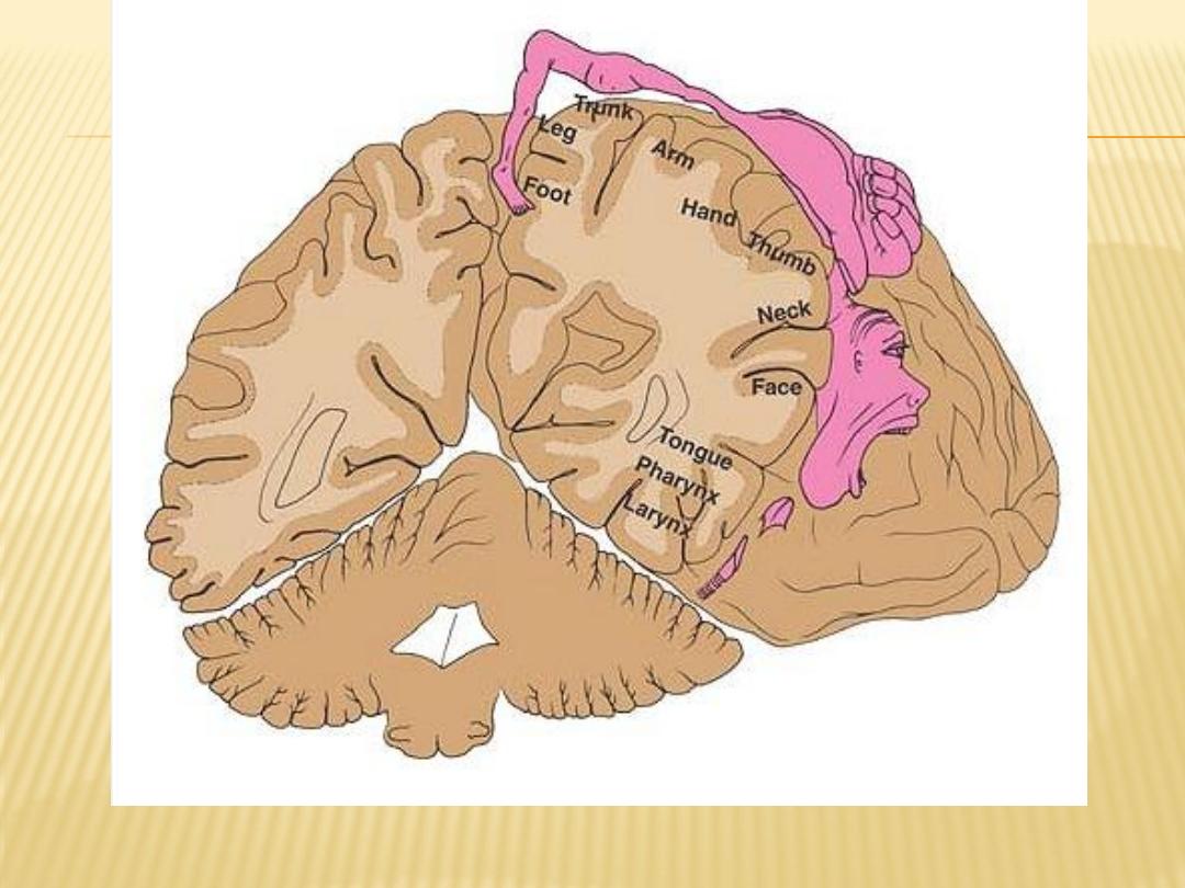
Motor homunculus on the precentral gyrus.
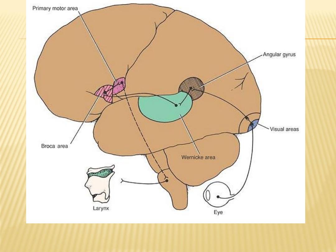
Probable nerve pathways involved in reading a sentence and repeating it out loud
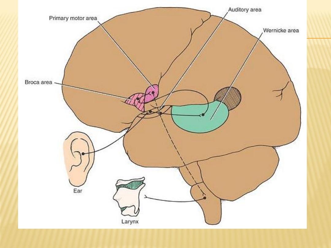
Probable nerve pathways involved with hearing a question and answering it

Internal Structure of the Cerebral
Hemispheres
Basal Nuclei
The term basal nuclei (basal ganglia) is applied to a collection of masses of
gray matter situated within each cerebral hemisphere.
They are:
- the corpus striatum,
- the amygdaloid nucleus
- claustrum

Corpus Striatum
The corpus striatum is situated lateral to the thalamus.
It is almost completely divided by a band of nerve fibers, the internal capsule,
into:
-
shaped mass of
-
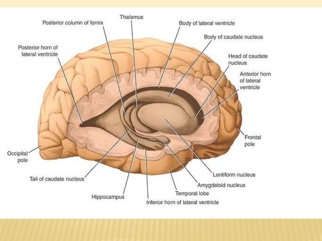
The caudate nucleus is a large C
The caudate nucleus:
-
gray matter that is closely related to the lateral ventricle and lies lateral to
the thalamus
-
it can be divided into a head, a body, and a tail
-
shaped mass of gray matter whose
-
is a wedge
nucleus:
lentiform
The
-
broad convex base is directed laterally and whose blade is directed medially
A vertical plate of white matter divides the nucleus into: the putamen larger,
darker lateral portion,, and the globus pallidus an inner lighter portion,

Functions of corpus striatum
1- regulation of muscle tone
2- contral autonomic movement
3- contral voluntary movement
Clinical application
Lesion of corpus striatum lead to tremor due to loss of dopamine
(See functions of Basal n.)
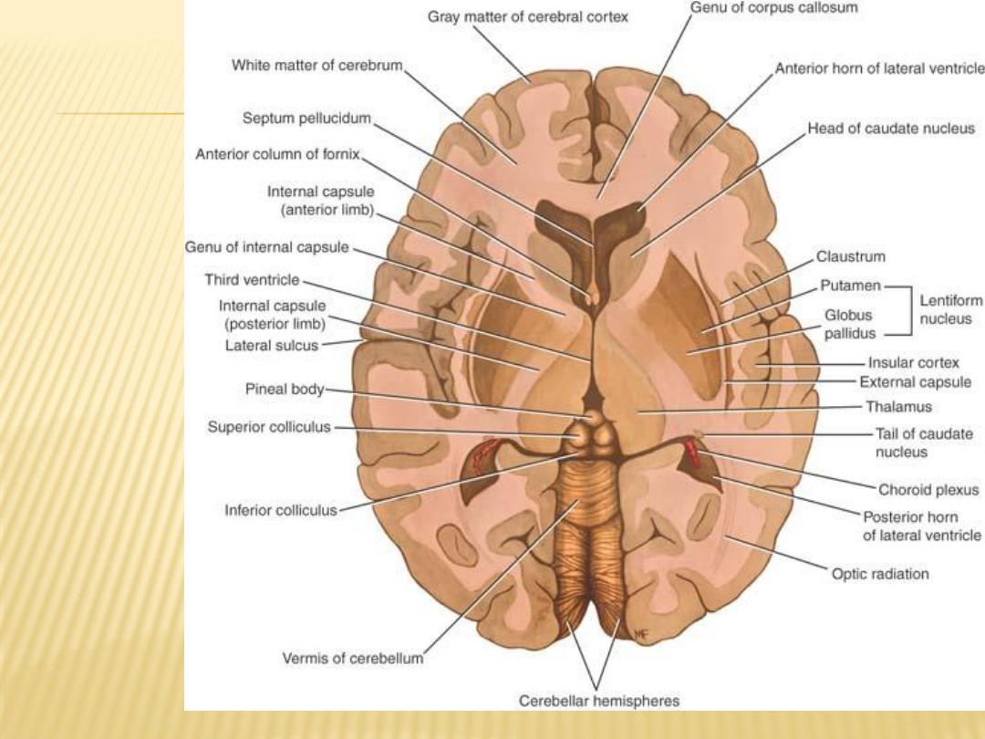
Horizontal
section of
the
cerebrum,
as seen
from
above,
showing
the
relationshi
p between
the
lentiform
nucleus,
the
caudate
nucleus,
the
thalamus,
and the
internal
capsule

Amygdaloid Nucleus
The amygdaloid nucleus is situated in the temporal lobe close to the uncus
it can influence the body's response to environmental changes. In the sense of
fear, for example, it can change the heart rate, blood pressure, skin color,
and rate of respiration.
Claustrum
The claustrum is a thin sheet of gray matter that is separated from the lateral
surface of the lentiform nucleus by the external capsule

Connections of the Corpus Striatum
Afferent Fibers
-
- Thalamostriate Fibers
-
- Nigrostriate Fibers
-
- Brainstem Striatal Fibers
-
Efferent Fibers
-
- Striatopallidal Fibers
-
- Striatonigral Fibers
-
Pallidus
Globus
Connections of the
-
Afferent Fibers
-
Striatopallidal Fibers
-
Efferent Fibers
Pallidofugal Fibers can be divided into groups: (1) the ansa lenticularis, (2)
the fasciculus lenticularis, (3) the pallidotegmental fibers and (4) the
pallidosubthalamic fibers

Functions of the Basal Nuclei
the basal nuclei control muscular movements by influencing the cerebral cortex
and have no direct control through descending pathways to the brainstem
and spinal cord. In this way, the basal nuclei assist in the regulation of
voluntary movement and the learning of motor skills.
Writing the letters of the alphabet, drawing a diagram, passing a football, using
the vocal cords in talking and singing, and using the eye muscles when
looking at an object are a few examples where the basal nuclei influence
the skilled cortical motor activities.

White Matter of the Cerebral Hemispheres
The white matter is composed of myelinated nerve fibers of different diameters
supported by neuroglia. The nerve fibers may be classified into three groups
according to their connections:
(1) commissural fibers: Commissure fibers essentially connect corresponding
regions of the two hemispheres. They are as follows: the corpus callosum,
the anterior commissure, the posterior commissure, the fornix, optic
chiasma and the habenular commissure
(2) association fibers: Association fibers are nerve fibers that essentially
connect various cortical regions within the same hemisphere and may be
divided into short and long groups ex: cingulate gyrus.
(3) projection fibers: Afferent and efferent nerve fibers passing to and from the
brainstem to the entire cerebral cortex must travel between large nuclear
masses of gray matter within the cerebral hemisphere ex: corona radiata,
internal capsule, external capsule and corticospinal tract.
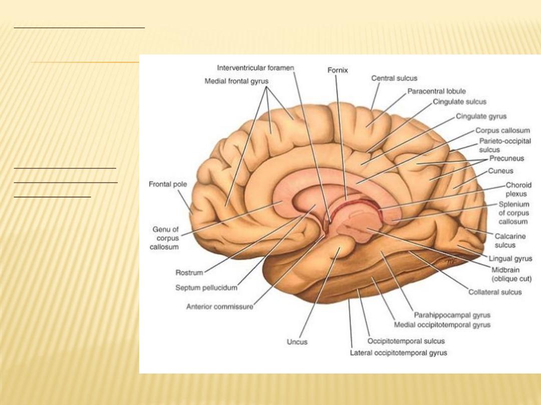
callosum
The corpus
the largest
commissure of the
brain, connects the
two cerebral
hemispheres.It lies
at the bottom of the
longitudinal fissure.
For purposes of
description, it is
divided into:
the rostrum (connect
both temporal
lobes),
the genu (connect both
frontal lobes),
the body (connect part
of frontal parietal
and occipital lobes)
the splenium (connect
the occipital lobes).

The septum pellucidum
The septum pellucidum is a thin vertical sheet of nervous tissue consisting of
white and gray matter covered on either side by ependyma
The septum pellucidum forms a partition between the anterior horns of the
lateral ventricles.
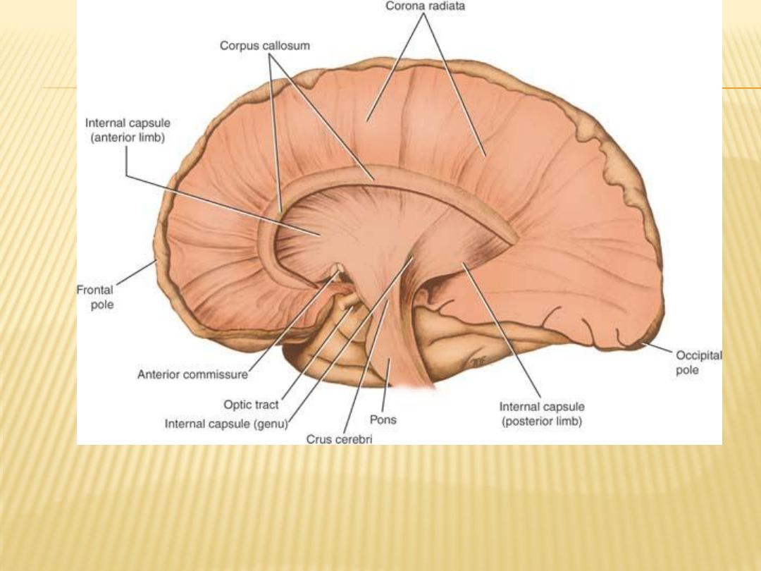
Medial view of the right cerebral hemisphere, which has been dissected to
show the internal capsule and the corona radiata. The thalamus has been
removed

Structure of the Cerebral Cortex
The cerebral cortex forms a complete covering of the cerebral hemisphere. It is
composed of gray matter and has been estimated to contain approximately
10 billion neurons. The surface area of the cortex has been increased by
throwing it into convolutions, or gyri, which are separated by fissures or
sulci. The thickness of the cortex varies from 1.5 to 4.5 mm. The cortex is
thickest over the crest of a gyrus and thinnest in the depth of a sulcus. The
cerebral cortex, like gray matter elsewhere in the central nervous system,
consists of a mixture of nerve cells, nerve fibers, neuroglia, and blood
vessels.
The following types of nerve cells are present in the cerebral cortex:
(1) pyramidal cells, (2) stellate cells, (3) fusiform cells, (4) horizontal cells of
Cajal, and (5) cells of Martinotti
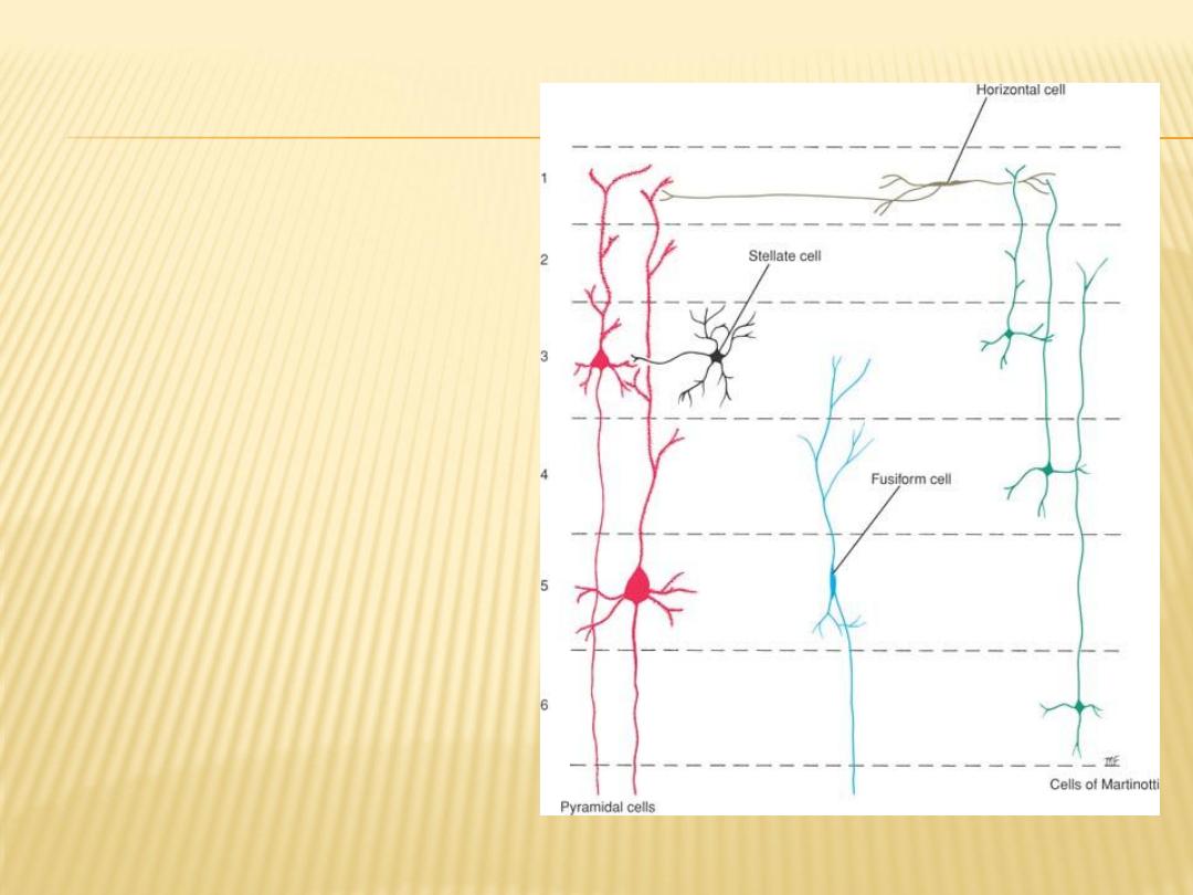
Main types of neurons found
in the cerebral cortex
Layers of the Cerebral Cortex
1- Molecular layer (plexiform layer).
2- External granular layer.
3- External pyramidal layer.
4- Internal granular layer.
5- Ganglionic layer (internal
pyramidal layer).

The insula
is an area of the cortex that is buried within the lateral sulcus and forms its
floor
It can be examined only when the lips of the lateral sulcus are separated
widely.
Histologically, the posterior part is granular and the anterior part is agranular,
thus resembling the adjoining cortical areas.
Its fiber connections are incompletely known. It is believed that this area is
important for planning or coordinating the articulatory movements
necessary for speech.