phonuclear cells, all have a granular appearance, as shown in cell numbers 7,
The first three types of cells, the polymor-
megakaryocyte.
bone marrow, the
platelets,
In addition, there are large numbers of
plasma cells.
sionally,
and, occa-
eosinophils, polymorphonuclear basophils, monocytes, lymphocytes,
polymorphonuclear neutrophils, polymorphonuclear
the blood. They are
Types of White Blood Cells.
invader.
locytes and monocytes have a special ability to “seek out and destroy” a foreign
rapid and potent defense against infectious agents. As we see later, the granu-
transported to areas of serious infection and inflammation, thereby providing a
The real value of the white blood cells is that most of them are specifically
). After formation, they are transported in the blood
protective system. They are formed partially in the bone marrow (
of the body’s
white blood cells,
The leukocytes, also called
of these methods, and Chapter 34 with the second.
may destroy or inactivate the invader. This chapter is concerned with the first
sensitized lymphocytes,
phagocytosis,
disease: (1) by actually destroying invading bacteria or viruses by
cells derived from leukocytes. These cells work together in two ways to prevent
toxic agents. This is comprised of blood leukocytes (white blood cells) and tissue
fever.
acute lethal diseases such as pneumonia, streptococcal infection, and typhoid
teria and viruses besides those that are normally present, and these can cause
tissues. In addition, we are exposed intermittently to other highly infectious bac-
urinary tract. Many of these infectious agents are
tract, the lining membranes of the eyes, and even the
mouth, the respiratory passageways, the intestinal
normally and to varying degrees in the skin, the
viruses, fungi, and parasites, all of which occur
Monocyte-Macrophage System,
Granulocytes, the
to Infection: I. Leukocytes,
C
H
A
P
T
E
R
3
3
429
Resistance of the Body
and Inflammation
Our bodies are exposed continually to bacteria,
capable of causing serious abnormal physiologic
function or even death if they invade the deeper
Our bodies have a special system for combating the different infectious and
and (2) by forming antibodies and
one or both of which
Leukocytes (White Blood Cells)
are the mobile units
granulocytes
and monocytes and a few lymphocytes) and partially in the lymph tissue (lym-
phocytes and plasma cells
to different parts of the body where they are needed.
General Characteristics of Leukocytes
Six types of white blood cells are normally present in
which are
fragments of another type of cell similar to the white blood cells found in the
million red blood cells). Of the total white blood cells,
The adult human being has about 7000 white blood
blood clotting mechanism, which is discussed in
immune system; this is discussed in Chapter 34. Finally,
The lymphocytes and
phagocytosis.
them—that is, by
The granulocytes and monocytes protect the body
or, in clinical terminology, “polys,”
granulocytes,
10, and 12 in Figure 33–1, for which reason they are
Blood Cells, Immunity, and Blood Clotting
430
Unit VI
called
because of the multiple nuclei.
against invading organisms mainly by ingesting
plasma cells function mainly in connection with the
the function of platelets is specifically to activate the
Chapter 36.
Concentrations of the Different White Blood Cells in the Blood.
cells per microliter of blood (in comparison with 5
the normal percentages of the different types are
approximately the following:
Monocytes 5.3%
Polymorphonuclear basophils
0.4%
Polymorphonuclear eosinophils
2.3%
Polymorphonuclear neutrophils
62.0%
tonsils, and various pockets of lymphoid tissue
tissues—especially the lymph glands, spleen, thymus,
in the bone marrow. Lymphocytes and plasma cells
The granulocytes and monocytes are formed only
lymphocytic lineage,
lineage,
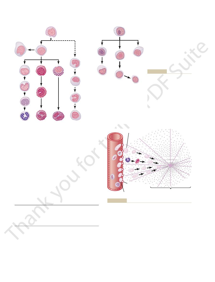
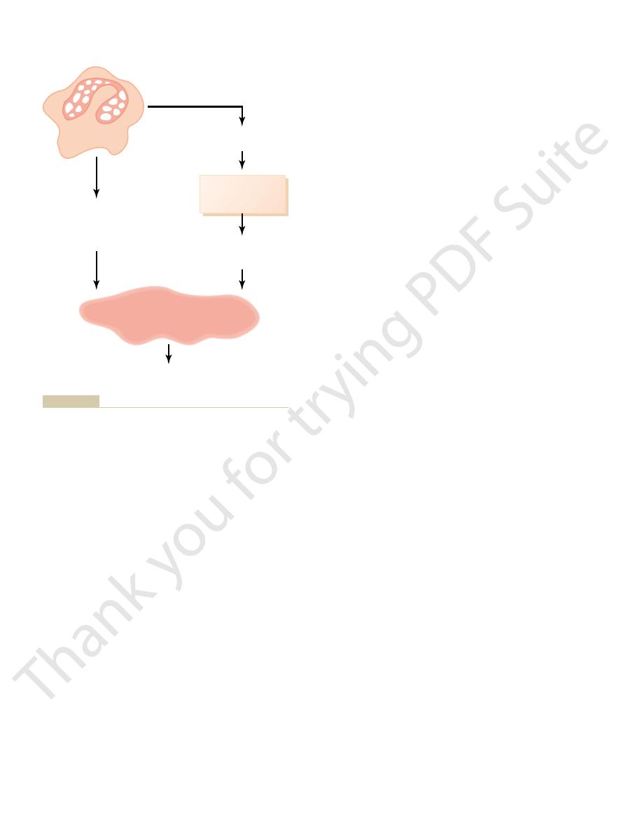
The left side of Figure 33–1 shows the
formed, the myelocytic and the lymphocytic lineages.
blood cells, two major lineages of
chapter. Aside from those cells committed to form red
stem cells is shown in Figure 32–2 in the previous
Genesis of the White Blood Cells
ments, in each microliter of blood is normally about
The number of platelets, which are only cell frag-
Lymphocytes
30.0%
300,000.
Early differentiation of the pluripotential hematopoi-
etic stem cell into the different types of committed
white blood cells are
myelocytic
beginning with the myeloblast; the right
shows the
beginning with the
lymphoblast.
are produced mainly in the various lymphogenous
Genesis of Myelocytes
Genesis of Lymphocytes
8
11
10
12
1
2
3
4
5
6
7
16
9
13
14
15
stages of monocyte formation.
7, polymorphonuclear neutrophil;
“band” neutrophil metamyelocyte;
neutrophil metamyelocyte; 6,
4, neutrophil myelocyte; 5, young
promyelocyte; 3, megakaryocyte;
series are 1, myeloblast; 2,
different cells of the myelocyte
Figure 33–1
Genesis of white blood cells. The
8, eosinophil myelocyte; 9,
eosinophil metamyelocyte; 10,
polymorphonuclear eosinophil;
11, basophil myelocyte; 12, poly-
morphonuclear basophil; 13–16,
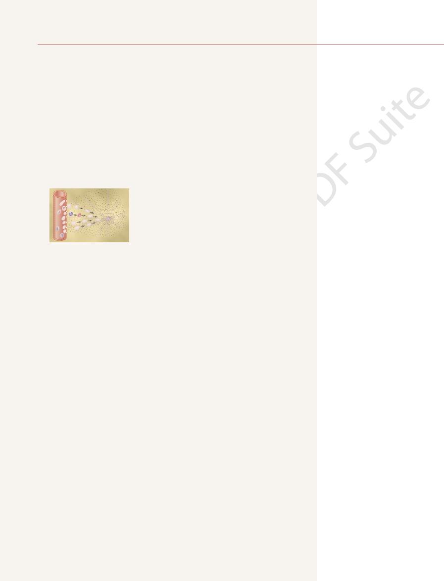
Diapedesis
Chemotaxis
source
Chemotactic
substance
Increased
permeability
Margination
toward an area of tissue damage.
through capillary pores
Movement of neutrophils by
Figure 33–2
diapedesis
and by chemotaxis

wise, normal cells and structures of the body might be
selective of the material that is phagocytized; other-
ingestion of the offending agent. Phagocytes must be
phagocytosis,
The most important function of the neutrophils and
away from a capillary, the chemotactic signal can easily
eters away from an inflamed tissue. Therefore, because
white cells. Chemotaxis is effective up to 100 microm-
stance. The concentration is greatest near the source,
As shown in Figure 33–2, chemotaxis depends on
substances.
clotting in the inflamed area, as well as other
(discussed in Chapter 34) activated in inflamed tissues,
reaction products of the “complement complex”
ucts of the inflamed tissues themselves, (3) several
of the bacterial or viral toxins, (2) degenerative prod-
taxis toward the inflamed area. They include (1) some
When a tissue becomes inflamed, at least a dozen
taxis.
chemo-
nomenon, shown in Figure 33–2, is known as
move toward the source of the chemical. This phe-
White Blood Cells Are Attracted to Inflamed Tissue Areas by
each minute.
m/min, a distance as great as their own length
Chapter 2. Some cells move at velocities as great as
through the tissues by ameboid motion, described in
White Blood Cells Move Through Tissue Spaces by Ameboid
to the size of the pore, as shown in Figure 33–2.
even though a pore is much smaller than a cell, a small
That is,
diapedesis.
Neutrophils and monocytes can squeeze through the
White Blood Cells Enter the Tissue Spaces by Diapedesis.
bating intratissue disease agents.
macrophages,
seen with the naked eye. These cells are now called
great as 60 to 80 micrometers, a size that can barely be
enter the tissues, they begin to swell—sometimes
fight infectious agents at that time. However, once they
begin life as blood monocytes, which are immature
circulating blood. Conversely, the tissue macrophages
other injurious agents. The neutrophils are mature
that attack and destroy invading bacteria, viruses, and
Neutrophils and Macrophages
every 10 days; in other words, about 30,000 platelets
The platelets in the blood are replaced about once
cells.
this life span depends on the body’s need for these
The lymphocytes have life spans of weeks or months;
tinual circulation of lymphocytes through the body.
return to the blood again and again; thus, there is con-
pedesis. Then, still later, they re-enter the lymph and
nodes and other lymphoid tissue. After a few hours,
ally, along with drainage of lymph from the lymph
Lymphocytes enter the circulatory system continu-
later, which provides continuing defense against
tions. These tissue macrophages are the basis of the
unless destroyed while performing phagocytic func-
and, in this form, can live for months
macrophages,
tissues, they swell to much larger sizes to become
capillary membranes into the tissues. Once in the
20 hours in the blood, before wandering through the
The monocytes also have a short transit time, 10 to
and, in the process, are themselves destroyed.
rapidly to the infected area, perform their functions,
hours because the granulocytes proceed even more
they are needed. In times of serious tissue infection,
The life of the granulocytes after being released from
Life Span of the White Blood Cells
tion of blood clotting.
into the blood. They are very important in the initia-
), then pass
ments, known as
ocytes fragment in the bone marrow; the small frag-
are also formed in the bone marrow. These megakary-
As shown in Figure 33–1, megakaryocytes (cell 3)
lymphoid tissues, except for a small number that are
The lymphocytes are mostly stored in the various
about a 6-day supply of these cells.
marrow as circulate in the entire blood.This represents
factors are discussed later). Normally, about three
the circulatory system. Then, when the need arises,
The white blood cells formed in the bone marrow
in so-called Peyer’s patches underneath the epithelium
elsewhere in the body, such as in the bone marrow and
Resistance of the Body to Infection: I. Leukocytes, Granulocytes, the Monocyte-Macrophage System
Chapter 33
431
in the gut wall.
are stored within the marrow until they are needed in
various factors cause them to be released (these
times as many white blood cells are stored in the
temporarily being transported in the blood.
platelets (or thrombocytes
the bone marrow is normally 4 to 8 hours circulating
in the blood and another 4 to 5 days in tissues where
this total life span is often shortened to only a few
tissue
tissue macrophage system, discussed in greater detail
infection.
they pass out of the blood back into the tissues by dia-
are formed each day for each microliter of blood.
Defend Against Infections
It is mainly the neutrophils and tissue macrophages
cells that can attack and destroy bacteria even in the
cells while still in the blood and have little ability to
increasing their diameters as much as fivefold—to as
and they are extremely capable of com-
pores of the blood capillaries by
portion of the cell slides through the pore at a time;
the portion sliding through is momentarily constricted
Motion.
Both neutrophils and macrophages can move
40
m
Chemotaxis.
Many different chemical substances in the
tissues cause both neutrophils and macrophages to
different products are formed that can cause chemo-
and (4) several reaction products caused by plasma
the concentration gradient of the chemotactic sub-
which directs the unidirectional movement of the
almost no tissue area is more than 50 micrometers
move hordes of white cells from the capillaries into the
inflamed area.
Phagocytosis
macrophages is
which means cellular

macrophages, fixed tissue macrophages, and a few
The total combination of monocytes, mobile
tually all tissue areas.
a widespread “monocyte-macrophage system” in vir-
related to the inflammatory process. Thus, the body has
stimulated, they can break away from their attach-
foreign particles in the tissue. And, when appropriately
quantities of bacteria, viruses, necrotic tissue, or other
as the mobile macrophages to phagocytize large
protective functions. They have the same capabilities
another large portion of monocytes becomes attached
entering the tissues and becoming macrophages,
of wandering through the tissues. However, after
In the preceding paragraphs, we described the
Monocyte-Macrophage Cell
tuberculosis.
many of the chronic diseases, an example of which is
macrophages. These bacteria are responsible for
bacillus, have coats that are resistant to lysosomal
Some bacteria, however, notably the tuberculosis
hypochlorite, which is exceedingly bactericidal.
lysosomal enzymes, myeloperoxidase, catalyzes the
bacteria, even in small quantities. Also, one of the
), all of which are lethal to most
hydroxyl ions
), and
hydrogen peroxide
superoxide
These oxidizing agents include large quantities of
peroxisome.
oxidizing agents
destruction by digestive enzymes. Much of the killing
cially important, because some bacteria have protec-
the lysosomal enzymes fail to digest them. This is espe-
phagosomes, neutrophils and macrophages contain
tuberculosis bacillus.
lipases,
and other foreign protein matter. The lysosomes of
immediately.
and digestion of the phagocytized particle begins
digestive vesicle,
phagocytic vesicle now becomes a
and bactericidal agents into the vesicle. Thus, the
branes fuse, thereby dumping many digestive enzymes
contact with the phagocytic vesicle, and their mem-
tized, lysosomes and other cytoplasmic granules in
Once a foreign particle has been phagocy-
more months.
ing particles, macrophages can extrude the residual
particles much larger than bacteria. Also, after digest-
whereas neutrophils are not capable of phagocytizing
red blood cells or, occasionally, malarial parasites,
the ability to engulf much larger particles, even whole
phagocytizing as many as 100 bacteria. They also have
ful phagocytes than neutrophils, often capable of
described in Chapter 34, they are much more power-
the blood. When activated by the immune system as
stage product of monocytes that enter the tissues from
and dies.
single neutrophil can usually phagocytize 3 to 20 bac-
) inside the cytoplasm. A
cytoplasmic cavity and breaks away from the outer cell
ticle. Then the chamber invaginates to the inside of the
enclosed chamber that contains the phagocytized par-
another on the opposite side and fuse. This creates an
tions around the particle. The pseudopodia meet one
be phagocytized, the neutrophil first attaches itself to
ately begin phagocytosis. On approaching a particle to
The neutrophils entering
phagocytosis. This selection and phagocytosis process
receptors on the phagocyte membrane, thus initiating
next chapter. The C3 molecules, in turn, attach to
complement cascade,
do this, the antibody molecule also combines with the
the bacteria especially susceptible to phagocytosis. To
tious agents such as bacteria. The antibodies then
Third, the immune system of the body (described in
phagocytosis.
have no protective coats, which makes them subject to
Conversely, most dead tissues and foreign particles
protective protein coats that repel the phagocytes.
Second, most natural substances of the body have
surface is rough, the likelihood of phagocytosis is
smooth surfaces, which resist phagocytosis. But if the
First, most natural structures in the tissues have
especially on three selective procedures.
ingested. Whether phagocytosis will occur depends
Blood Cells, Immunity, and Blood Clotting
432
Unit VI
increased.
detail in Chapter 34) develops antibodies against infec-
adhere to the bacterial membranes and thereby make
C3 product of the
which is an
additional part of the immune system discussed in the
is called opsonization.
Phagocytosis by Neutrophils.
the tissues are already mature cells that can immedi-
the particle and then projects pseudopodia in all direc-
membrane to form a free-floating phagocytic vesicle
(also called a phagosome
teria before the neutrophil itself becomes inactivated
Phagocytosis by Macrophages.
Macrophages are the end-
products and often survive and function for many
Once Phagocytized, Most Particles Are Digested by Intracellu-
lar Enzymes.
the neutrophil or macrophage immediately come in
Both neutrophils and macrophages contain an
abundance of lysosomes filled with proteolytic
enzymes especially geared for digesting bacteria
macrophages (but not of neutrophils) also contain
large amounts of
which digest the thick lipid
membranes possessed by some bacteria such as the
Both Neutrophils and Macrophages Can Kill Bacteria.
In
addition to the digestion of ingested bacteria in
bactericidal agents that kill most bacteria even when
tive coats or other factors that prevent their
effect results from several powerful
formed by enzymes in the membrane of the phago-
some or by a special organelle called the
(O
2
–
),
(H
2
O
2
(–OH
–
reaction between H
2
O
2
and chloride ions to form
digestion and also secrete substances that partially
resist the killing effects of the neutrophils and
System (Reticuloendothelial
System)
macrophages mainly as mobile cells that are capable
to the tissues and remains attached for months or even
years until they are called on to perform specific local
ments and once again become mobile macrophages
that respond to chemotaxis and all the other stimuli
phagocytosis of a single bacterium in less than
of phagocytosis by Kupffer cells have demonstrated
general systemic circulation. Indeed, motion pictures
shown in Figure 33–4. These cells
Kupffer cells,
culation, it passes through the sinusoids of the liver;
portal blood. Before this blood enters the general cir-
body is through the gastrointestinal tract. Large
bacilli, silica dust particles, and even carbon particles.
time—if ever—that it can be slowly dissolved. Such
a “giant cell” capsule around the particle until such
particle is not digestible, the macrophages often form
release the digestive products into the lymph. If the
digestible, the macrophages can also digest them and
become entrapped in the alveoli. If the particles are
the alveolar walls. They can phagocytize particles that
is through the lungs. Large numbers of tissue
Alveolar Macrophages in the Lungs.
prevent general dissemination throughout the body.
of the lymph, the macrophages phagocytize them and
sinuses, and if any particles enter the sinuses by way
nodal medullary sinuses,
afferent lymphatics,
the lymph node, showing lymph entering through the
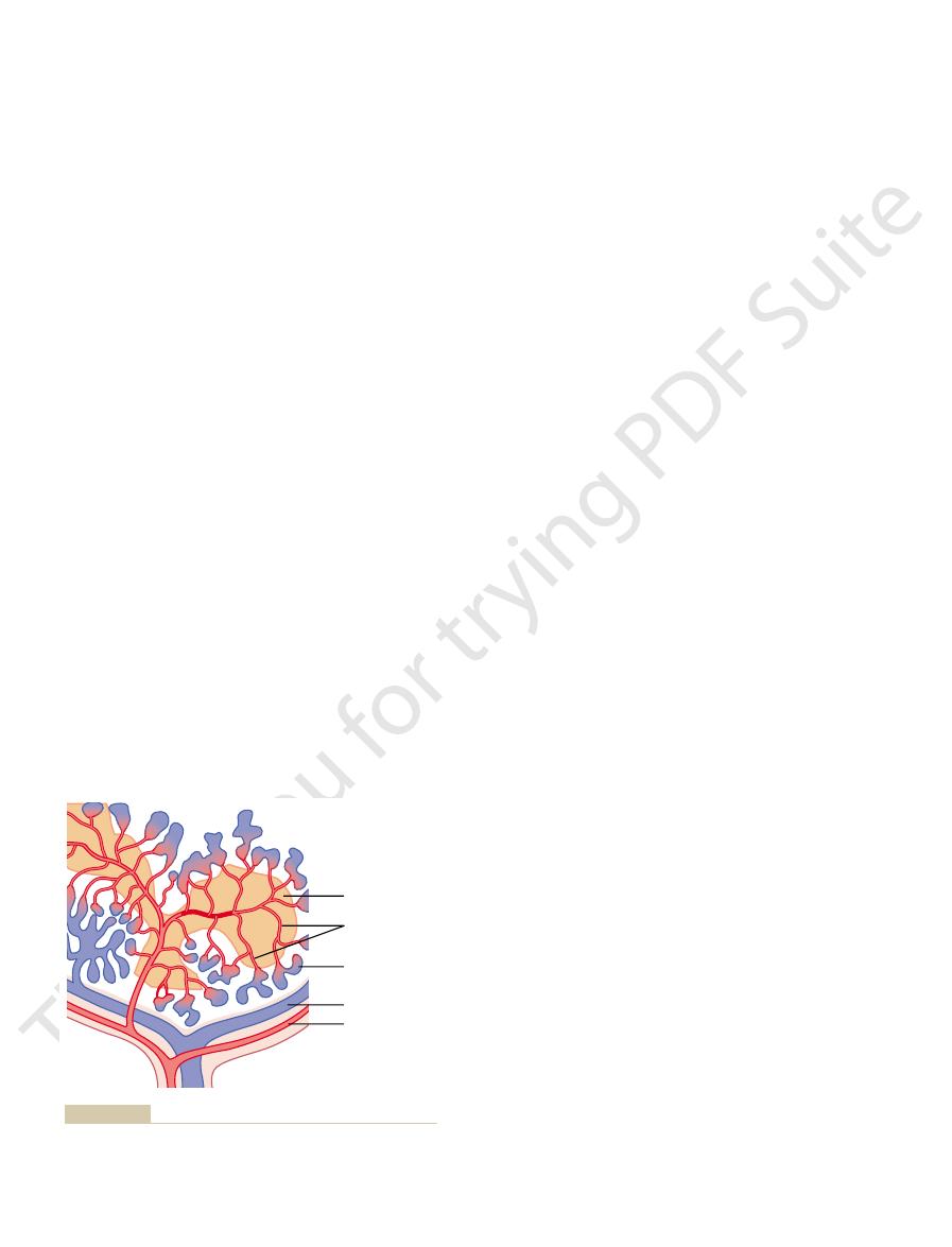
Figure 33–3 illustrates the general organization of
tissue macrophages.
along the course of the lymph flow. The foreign parti-
destroyed locally in the tissues, they enter the lymph
into the blood. Instead, if the particles are not
late matter that enters the tissues, such as bacteria, can
Macrophages in the Lymph Nodes.
described earlier.
of attacking and destroying the infectious agents, as
macrophages. Then they perform the usual functions
tissue and local inflammation ensues, local tissue
is broken. When infection begins in a subcutaneous
infectious agents, this is no longer true when the skin
Tissue Macrophages in the Skin and Subcutaneous Tissues (His-
of particles, toxins, and other unwanted substances
generalized phagocytic system located in all tissues,
the monocyte-macrophage system. Because the term
inate from monocytic stem cells; therefore, the
However, all or almost all these cells orig-
spleen, and lymph nodes is called the
specialized endothelial cells in the bone marrow,
Resistance of the Body to Infection: I. Leukocytes, Granulocytes, the Monocyte-Macrophage System
Chapter 33
433
reticuloendothe-
lial system.
reticuloendothelial system is almost synonymous with
reticuloendothelial system is much better known
in medical literature than the term monocyte-
macrophage system, it should be remembered as a
especially in those tissue areas where large quantities
must be destroyed.
tiocytes).
Although the skin is mainly impregnable to
macrophages can divide in situ and form still more
Essentially no particu-
be absorbed directly through the capillary membranes
and flow to the lymph nodes located intermittently
cles are then trapped in these nodes in a meshwork of
sinuses lined by
lymph node capsule by way of
then flowing through the
and
finally passing out the hilus into efferent lymphatics
that eventually empty into the venous blood.
Large numbers of macrophages line the lymph
Another route by
which invading organisms frequently enter the body
macrophages are present as integral components of
capsules are frequently formed around tuberculosis
Macrophages (Kupffer Cells) in the Liver Sinusoids.
Still
another favorite route by which bacteria invade the
numbers of bacteria from ingested food constantly
pass through the gastrointestinal mucosa into the
these sinusoids are lined with tissue macrophages
called
form such an effective particulate filtration system that
almost none of the bacteria from the gastrointestinal
tract succeeds in passing from the portal blood into the
1
/
100
of a
second.
Afferent lymphatics
Capsule
Subcapsular
sinus
Germinal
center
Medullary cord
Hilus
Lymph in
medullary
sinuses
Valve
Primary
nodule
Efferent lymphatics
from Gartner LP, Hiatt JL: Color Textbook of Histology, 2nd ed.
Histology, 6th ed. Philadelphia: JB Lippincott, 1969.) (Modified
Functional diagram of a lymph node. (Redrawn from Ham AW:
Figure 33–3
Philadelphia, WB Saunders, 2001.)
Kupffer cells
tology, 10th ed. Baltimore: Williams & Wilkins, 1971.)
(Redrawn from Copenhaver WM, et al: Bailey’s Textbook of His-
India ink particles into the cytoplasm of the Kupffer cells.
Kupffer cells lining the liver sinusoids, showing phagocytosis of
Figure 33–4
others, immediately begin their phagocytic actions.
macrophages in the lungs, microglia in the brain, or
histiocytes in the subcutaneous tissues, alveolar
macrophages already present in the tissues, whether
Within minutes after inflammation begins, the
Tissue Macrophage Is a First Line of Defense Against Infection.
Macrophage and Neutrophil
tissues.
body and cause death than do staphylococci, even
have a far greater tendency to spread through the
reproduce and migrate. As a result, streptococci often
ops slowly over many hours, while many streptococci
destruction. Therefore, the walling-off process devel-
cocci, in contrast, do not cause such intense local tissue
prevented from spreading through the body. Strepto-
ply and spread. Therefore, local staphylococcal in-
inflammation develops rapidly—indeed, much more
release extremely lethal cellular toxins. As a result,
invade tissues, they
instance, when
proportional to the degree of tissue injury. For
The intensity of the inflammatory process is usually
delays the spread of bacteria or toxic products.
flows through the spaces. This walling-off process
by fibrinogen clots so that after a while, fluid barely
injury from the remaining tissues. The tissue spaces
results of inflammation is to “wall off ” the area of
“Walling-Off” Effect of Inflammation.
cells.
devour the destroyed tissues. But at times, the
and within a few hours, the macrophages begin to
system; also discussed in Chapter 34). Several of these
released by sensitized T cells (part of the immune
tonin, prostaglandins,
histamine, bradykinin, sero-
tissue cells. Some of the many tissue products that
and monocytes into the tissue; and (5) swelling of the
laries; (4) migration of large numbers of granulocytes
the interstitial spaces; (3) often clotting of the fluid in
ies, allowing leakage of large quantities of fluid into
blood flow; (2) increased permeability of the capillar-
the local blood vessels, with consequent excess local
rounding uninjured tissues. This entire complex of
trauma, chemicals, heat, or any other phenomenon,
When tissue injury occurs, whether caused by bacteria,
Neutrophils and Macrophages
blood cells.
in the blood, including especially old and abnormal red
exceptional means of phagocytizing unwanted debris
also lined with macrophages. This peculiar passage of
numbers of macrophages, and the venous sinuses are
The trabeculae of the red pulp are lined with vast
venous sinuses.
squeezes
The blood then
cords of red pulp.
ies are highly porous, allowing whole blood to pass out
and terminates in small capillaries. The capillar-
eral segment of spleen tissue. Note that a small artery
spaces of the spleen. Figure 33–5 shows a small periph-
blood, instead of lymph, flows through the tissue
The spleen is similar to the lymph nodes, except that
phagocytized.
cles come in contact with these macrophages, they are
meshwork of the two organs, and when foreign parti-
spleen and bone marrow. In both these tissues,
macrophage system, especially by macrophages of the
tion, there are other lines of defense by the tissue
Macrophages of the Spleen and Bone Marrow.
Blood Cells, Immunity, and Blood Clotting
434
Unit VI
If an invad-
ing organism succeeds in entering the general circula-
macrophages have become entrapped by the reticular
penetrates from the splenic capsule into the splenic
pulp
of the capillaries into
gradually
through the trabecular meshwork
of these cords and eventually returns to the circulation
through the endothelial walls of the
blood through the cords of the red pulp provides an
Inflammation: Role of
Inflammation
multiple substances are released by the injured tissues
and cause dramatic secondary changes in the sur-
tissue changes is called inflammation.
Inflammation is characterized by (1) vasodilation of
the interstitial spaces because of excessive amounts of
fibrinogen and other proteins leaking from the capil-
cause these reactions are
several different reaction prod-
ucts of the complement system (described in Chapter
34), reaction products of the blood clotting system, and
multiple substances called lymphokines that are
substances strongly activate the macrophage system,
macrophages also further injure the still-living tissue
One of the first
and the lymphatics in the inflamed area are blocked
staphylococci
rapidly than the staphylococci themselves can multi-
fection is characteristically walled off rapidly and
though staphylococci are far more destructive to the
Responses During Inflammation
Pulp
Vein
Artery
Capillaries
Venous sinuses
Fawcett DW: A Textbook of Histology, 10th ed. Philadelphia: WB
Functional structures of the spleen. (Modified from Bloom W,
Figure 33–5
Saunders, 1975.)

days, and the end products are eventually absorbed
tion has been suppressed, the dead cells and necrotic
pus.
trophils, dead macrophages, and tissue fluid. This
contains varying portions of necrotic tissue, dead neu-
macrophages eventually die. After several days, a
all the neutrophils and many, if not most, of the
numbers of bacteria and necrotic tissue, essentially
When neutrophils and macrophages engulf large
Formation of Pus
tively. This combination of TNF, IL-1, and colony-stim-
late granulocyte and monocyte production, respec-
production; the other two, G-CSF and M-CSF, stimu-
GM-CSF, stimulates both granulocyte and monocyte
the three colony-stimulating factors, one of which,
cytes and monocytes by the bone marrow is mainly
The cause of the increased production of granulo-
quantities by other inflamed tissue cells.
(M-CSF). These factors are formed by activated
(G-CSF), and (5)
(GM-CSF), (4)
(IL-1), (3)
(TNF), (2)
nant roles. They are shown in Figure 33–6 and consist
inflammation, five of these are believed to play domi-
Neutrophil Responses
Feedback Control of the Macrophage and
years, sometimes at a rate 20 to 50 times normal.
tinues, the bone marrow can continue to produce these
marrow. If the stimulus from the inflamed tissue con-
and monocytes reach the stage of leaving the bone
takes 3 to 4 days before newly formed granulocytes
cytic progenitor cells of the marrow. However, it
results from stimulation of the granulocytic and mono-
ulocytes and monocytes by the bone marrow. This
The fourth line of
Bone Marrow Is a Fourth Line of Defense.
role in initiating the development of antibodies, as we
neutrophils. Also, the macrophages play an important
selves and large quantities of necrotic tissue, than can
far larger particles, including even neutrophils them-
As already pointed out, macrophages can phagocy-
duction of new monocytes, as explained later.
come to dominate the phagocytic cells of the inflamed
several days to several weeks, the macrophages finally
for phagocytosis. Yet, after
of lysosomes; only then do they acquire the full capac-
immature cells, requiring 8 hours or more to swell to
after invading the inflamed tissue, monocytes are still
several days to become effective. Furthermore, even
area is much slower than that of neutrophils, requiring
fore, the buildup of macrophages in the inflamed tissue
marrow is much less than that of neutrophils. There-
also, the storage pool of monocytes in the bone
number of monocytes in the circulating blood is low:
and enlarge to become macrophages. However, the
monocytes from the blood enter the inflamed tissue
Along with the invasion of neutrophils,
Second Macrophage Invasion into the Inflamed Tissue Is a Third
into the circulating blood. This makes even more neu-
are transported to the bone marrow, and there act on
neutrophils in the blood. Neutrophilia is caused by
25,000 neutrophils per microliter. This is called
acute, severe inflammation, the number of neutrophils
Acute Increase in Number of Neutrophils in the Blood—“Neu-
foreign matter.
mature cells, they are ready to immediately begin their
trophils. Because the blood neutrophils are already
begins, the area becomes well supplied with neu-
Thus, within several hours after tissue damage
toward the injured tissues, as explained earlier.
chemotaxis
into the tissue spaces. (3) Other products of inflam-
to loosen, allowing openings large enough for neu-
They cause the intercellular attachments between the
and is shown in Figure 33–2. (2)
to the capillary walls in the inflamed area. This effect
the capillary endothelium, causing neutrophils to stick
lowing reactions: (1) They alter the inside surface of
the inflamed area from the blood. This is caused by
begins, large numbers of neutrophils begin to invade
Within the first hour or so after inflammation
are not great, but they are lifesaving.
against infection during the first hour or so. The
and become mobile, forming the first line of defense
each of these cells. Next, many of the previously sessile
inflammation, the first effect is rapid enlargement of
When activated by the products of infection and
Resistance of the Body to Infection: I. Leukocytes, Granulocytes, the Monocyte-Macrophage System
Chapter 33
435
macrophages break loose from their attachments
numbers of these early mobilized macrophages often
Neutrophil Invasion of the Inflamed Area Is a Second Line of
Defense.
products from the inflamed tissues that initiate the fol-
is called margination
endothelial cells of the capillaries and small venules
trophils to pass by diapedesis directly from the blood
mation then cause
of the neutrophils
scavenger functions for killing bacteria and removing
trophilia.”
Also within a few hours after the onset of
in the blood sometimes increases fourfold to five-
fold—from a normal of 4000 to 5000 to 15,000 to
neu-
trophilia, which means an increase in the number of
products of inflammation that enter the blood stream,
the stored neutrophils of the marrow to mobilize these
trophils available to the inflamed tissue area.
Line of Defense.
much larger sizes and develop tremendous quantities
ity of tissue macrophages
area because of greatly increased bone marrow pro-
tize far more bacteria (about five times as many) and
discuss in Chapter 34.
Increased Production of Granulocytes and Monocytes by the
defense is greatly increased production of both gran-
cells in tremendous quantities for months and even
Although more than two dozen factors have been
implicated in control of the macrophage response to
of (1) tumor necrosis factor
interleukin-1
granulocyte-monocyte colony-stimulating
factor
granulocyte colony-stimulating
factor
monocyte colony-stimulating
factor
macrophage cells in the inflamed tissues and in smaller
ulating factors provides a powerful feedback mecha-
nism that begins with tissue inflammation and
proceeds to formation of large numbers of defensive
white blood cells that help remove the cause of the
inflammation.
cavity is often excavated in the inflamed tissues that
mixture is commonly known as
After the infec-
tissue in the pus gradually autolyze over a period of
ifestations. These reactions are discussed in greater
tions that cause many, if not most, of the allergic man-
These cause local vascular and tissue reac-
enzymes.
substance of anaphylaxis,
histamine, bradykinin, serotonin, heparin, slow-reacting
reacts with the antibody, the resulting attachment of
mast cells and basophils. Then, when the specific
34), has a special propensity to become attached to
tions, the immunoglobulin E (IgE) type (see Chapter
The mast cells and basophils play an exceedingly
Indeed, it is mainly the mast cells in
histamine,
The mast cells and basophils also release
into the blood, a substance
many of the capillaries in the body. Both mast cells and
mast cells
The basophils in the circulating blood are similar to
inflammatory process.
complexes, thus preventing excess spread of the local
also to phagocytize and destroy allergen-antibody
allergic tissue. The eosinophils are believed to detox-
eosinophil chemotactic factor
we discuss in the next paragraph. The mast cells and
cells and basophils participate in allergic reactions, as
This is caused at least partly by the fact that many mast
asthma and in the skin after allergic skin reactions.
in tissues in which allergic reactions occur, such as in
Trichinella
invasion of the body’s muscles by the
This results from
trichinosis.
In a few areas of the world, another parasitic disease
especially lethal to parasites; and (3) by releasing from
which are modified lysosomes; (2) probably by also
by releasing hydrolytic enzymes from their granules,
and kill many of them. They do so in several ways: (1)
parasite can invade any part of the body. Eosinophils
of the population of some Third World countries; the
asis,
schistosomi-
stances that kill many of the parasites. For instance,
cells, eosinophils attach themselves to the parasites by
phagocytized by eosinophils or any other phagocytic
asites. Although most parasites are too large to be
numbers in people with parasitic infections, and they
Eosinophils, however, are often produced in large
parison with the neutrophils, it is doubtful that the
phagocytes, and they exhibit chemotaxis, but in com-
of all the blood leukocytes. Eosinophils are weak
The eosinophils normally constitute about 2 per cent
the evidence of tissue damage is gone.
Blood Cells, Immunity, and Blood Clotting
436
Unit VI
into the surrounding tissues and lymph until most of
Eosinophils
eosinophils are significant in protecting against the
usual types of infection.
migrate in large numbers into tissues diseased by par-
way of special surface molecules and release sub-
one of the most widespread infections is
a parasitic infection found in as many as one third
attach themselves to the juvenile forms of the parasite
releasing highly reactive forms of oxygen that are
the granules a highly larvacidal polypeptide called
major basic protein.
that causes eosinophilia is
par-
asite (“pork worm”) after a person eats undercooked
infested pork.
Eosinophils also have a special propensity to collect
the peribronchial tissues of the lungs in people with
basophils release an
that
causes eosinophils to migrate toward the inflamed
ify some of the inflammation-inducing substances
released by the mast cells and basophils and probably
Basophils
the large tissue
located immediately outside
basophils liberate heparin
that can prevent blood coagulation.
as well as smaller quantities of bradykinin and
serotonin.
inflamed tissues that release these substances during
inflammation.
important role in some types of allergic reactions
because the type of antibody that causes allergic reac-
antigen for the specific IgE antibody subsequently
antigen to antibody causes the mast cell or basophil
to rupture and release exceedingly large quantities of
and a number of lysosomal
detail in Chapter 34.
Leukopenia
A clinical condition known as leukopenia occasionally
occurs in which the bone marrow produces very few
Activated
macrophage
INFLAMMATION
TNF
IL-1
GM-CSF
G-CSF
M-CSF
GM-CSF
G-CSF
M-CSF
Granulocytes
Monocytes/macrophages
Bone marrow
TNF
IL-1
Endothelial cells,
fibroblasts,
lymphocytes
colony-stimulating factor; TNF, tumor necrosis factor.
colony-stimulating factor; IL-1, interleukin-1; M-CSF, monocyte
ulocyte colony-stimulating factor; GM-CSF, granulocyte-monocyte
from activated macrophages in an inflamed tissue. G-CSF, gran-
cyte-macrophages in response to multiple growth factors released
Control of bone marrow production of granulocytes and mono-
Figure 33–6

graft-versus-leukaemia effect. Nat Rev Cancer 4:371, 2004.
Bleakley M, Riddell SR: Molecules and mechanisms of the
ration by signals from toll-like receptors. Science 304:1014,
Blander JM, Medzhitov R: Regulation of phagosome matu-
86:1190, 2000.
immune targeting and cardiovascular disease. Circ Res
ated by vascular adhesion protein-1: implications for
Alexander JS, Granger DN: Lymphocyte trafficking medi-
enough, this alone is sufficient to cause death.
tated. After metabolic starvation has continued long
leukemic tissues grow, other tissues become debili-
the normal protein tissues of the body. Thus, while the
sequently, the energy of the patient is greatly depleted,
for foodstuffs, specific amino acids, and vitamins. Con-
substrates by the growing cancerous cells. The
Finally, perhaps the most important effect of
cells.
cytopenia (lack of platelets). These effects result
anemia, and a bleeding tendency caused by thrombo-
in leukemia are the development of infection, severe
the bone marrow or the lymph nodes. Common effects
spleen, lymph nodes, liver, and other vascular regions,
fracture easily.
causing pain and, eventually, a tendency for bones to
so greatly that they invade the surrounding bone,
leukemic cells in abnormal areas of the body.
The first effect of leukemia is metastatic growth of
Effects of Leukemia on the Body
cells, especially the very undifferentiated cells, are
times developing slowly over 10 to 20 years. Leukemic
chronic,
ferentiated cells, the process can be
few months if untreated. With some of the more dif-
is the leukemia, often leading to death within a
Usually, the more undifferentiated the cell, the more
not identical to any of the normal white blood cells.
More frequently, however, the
leukemia, eosinophilic leukemia, basophilic leukemia,
occasionally produces partially differentiated cells,
In myelogenous leukemia, the cancerous process
spleen, and liver.
extramedullary tissues—especially in the lymph nodes,
type of leukemia, myelogenous leukemia, begins by
and spreading to other areas of the body. The second
beginning in a lymph node or other lymphocytic tissue
by cancerous production of lymphoid cells, usually
The lymphocytic leukemias are caused
nous leukemias.
general types:
Types of Leukemia.
lymphogenous cell. This causes
ward off infection, usually develops enough new bone
with transfusions, plus antibiotics and other drugs to
sufficient time is available. A patient properly treated
capable of regenerating the bone marrow, provided
marrow, some stem cells, myeloblasts, and hemocyto-
malady.
hypnotics, on very rare occasions cause leukopenia,
treat thyrotoxicosis), and even various barbiturate
chloramphenicol (an antibiotic), thiouracil (used to
bone marrow. Indeed, some common drugs, such as
or anthracene nuclei, is likely to cause aplasia of the
Irradiation of the body by x-rays or gamma rays, or
week after acute total leukopenia begins.
Without treatment, death often ensues in less than a
severe respiratory infection. Bacteria from the ulcers
and colon, or the person might develop some form of
ing white blood cells, ulcers may appear in the mouth
Within 2 days after the bone marrow stops produc-
of the eyes, urethra, and vagina. Any decrease in the
thermore, one can always find bacteria on the surfaces
nal tract is especially loaded with colon bacilli. Fur-
in the entire respiratory tract. The distal gastrointesti-
spirochetal, pneumococcal, and streptococcal bacteria,
bacteria. The mouth almost always contains various
many bacteria, because all the mucous membranes of
Normally, the human body lives in symbiosis with
tissues.
white blood cells, leaving the body unprotected against
Resistance of the Body to Infection: I. Leukocytes, Granulocytes, the Monocyte-Macrophage System
Chapter 33
437
many bacteria and other agents that might invade the
the body are constantly exposed to large numbers of
and these same bacteria are present to a lesser extent
number of white blood cells immediately allows inva-
sion of adjacent tissues by bacteria that are already
present.
rapidly invade surrounding tissues and the blood.
exposure to drugs and chemicals that contain benzene
thus setting off the entire infectious sequence of this
After moderate irradiation injury to the bone
blasts may remain undestroyed in the marrow and are
marrow within weeks to months for blood cell con-
centrations to return to normal.
The Leukemias
Uncontrolled production of white blood cells can be
caused by cancerous mutation of a myelogenous or
leukemia, which is
usually characterized by greatly increased numbers
of abnormal white blood cells in the circulating
blood.
Leukemias are divided into two
lymphocytic leukemias and myeloge-
cancerous production of young myelogenous cells in
the bone marrow and then spreads throughout the
body so that white blood cells are produced in many
resulting in what might be called neutrophilic
or monocytic leukemia.
leukemia cells are bizarre and undifferentiated and
acute
some-
usually nonfunctional for providing the normal pro-
tection against infection.
Leukemic cells from the bone marrow may reproduce
Almost all leukemias eventually spread to the
regardless of whether the origin of the leukemia is in
mainly from displacement of the normal bone marrow
and lymphoid cells by the nonfunctional leukemic
leukemia on the body is excessive use of metabolic
leukemic tissues reproduce new cells so rapidly that
tremendous demands are made on the body reserves
and excessive utilization of amino acids by the
leukemic cells causes especially rapid deterioration of
References
2004.

phages, a serious business. Science 304:1123, 2004.
Zullig S, Hengartner MO: Cell biology: tickling macro-
factors and cytokines. Physiol Rev 83:835, 2003.
Werner S, Grose R: Regulation of wound healing by growth
Rev Immunol 3:223, 2003.
antigen-induced regulatory T cells in autoimmunity. Nat
von Herrath MG, Harrison LC: Regulatory lymphocytes:
syndrome revisited. Annu Rev Med 54:169, 2003.
Roufosse F, Cogan E, Goldman M: The hypereosinophilic
leukemia. N Engl J Med 350:1535, 2004.
Pui CH, Relling MV, Downing JR: Acute lymphoblastic
ulation in wound healing. Am J Surg 187:11S, 2004.
Park JE, Barbul A: Understanding the role of immune reg-
84:579, 2004.
contribution to physiology and disease. Physiol Rev
Ossovskaya VS, Bunnett NW: Protease-activated receptors:
Trends Immunol 24:327, 2003.
leukocyte transmigration and the inflammatory response.
Muller WA: Leukocyte-endothelial-cell interactions in
25:75, 2004.
ple levels of leukocyte migration control. Trends Immunol
Moser B, Wolf M, Walz A, Loetscher P: Chemokines: multi-
Immunol 4:325, 2004.
lymphoid tissues and sites of inflammation. Nat Rev
Ley K, Kansas GS: Selectins in T-cell recruitment to non-
transcellular? News Physiol Sci 16:15, 2001.
Kvietys PR, Sandig M: Neutrophil diapedesis: paracellular or
Immunol 3:822, 2003.
Kunkel EJ, Butcher EC: Plasma-cell homing. Nat Rev
for the resolution of inflammation. J Endocrinol 178:29,
macrophage phagocytosis of apoptotic cells: implications
mediated regulation of granulocyte apoptosis and
Heasman SJ, Giles KM, Ward C, et al: Glucocorticoid-
leukemia. Semin Oncol 31(Suppl 4):60, 2004.
Ferrajoli A, O’Brien SM: Treatment of chronic lymphocytic
Blood Cells, Immunity, and Blood Clotting
438
Unit VI
2003.