onds. These originate from large neuronal cell bodies that lie throughout the
The signals passing through the thalamus are of two types. One type is rapidly
as to multiple subcortical areas.
tion. Most of these go first to the thalamus, where they excite a different set of
downward signals, this area also sends a profusion of signals in the upward direc-
and to control levels of activity of the spinal cord reflexes. In addition to these
downward to the spinal cord
. We also discuss this area in Chapter
. This area is also known
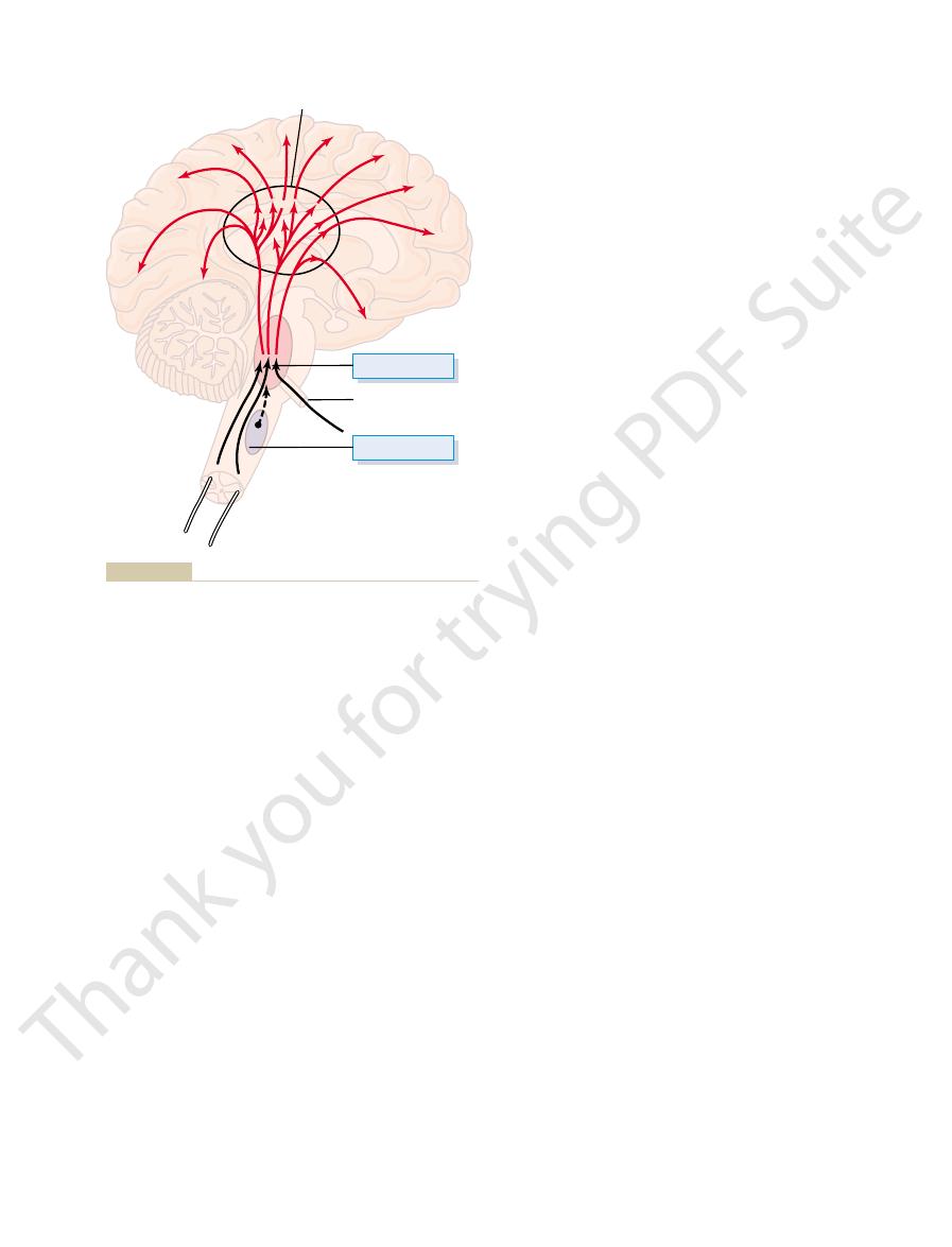
brain. The central driving component of this system is an excitatory area located
Figure 58–1 shows a general system for controlling the level of activity of the
Reticular Excitatory Area of the Brain Stem
Signals from the Brain Stem
Control of Cerebral Activity by Continuous Excitatory
ways: (1) by directly stimulating a background level of neuronal activity in wide
ting coma lasting for the remainder of his or her life.
times results from a pineal tumor, often causes the person to go into unremit-
brain stem at the juncture between the mesencephalon and cerebrum, as some-
cerebrum, the cerebrum becomes useless. In fact, severe compression of the
Without continuous transmission of nerve signals from the lower brain into the
, meaning the “border”
brain, which together are loosely called the
of the learning process and feelings of pleasure and punishment. These func-
motivational drives, especially motivational control
parts of the brain. Then we discuss the causes of
In this chapter, we deal first with those mecha-
important behavioral patterns.
cycle discussed in Chapter 59 is one of our most
nervous system. Even the wakefulness and sleep
Mechanisms of the Brain—The
C
H
A
P
T
E
R
5
8
728
Behavioral and Motivational
Limbic System and the
Hypothalamus
Control of behavior is a function of the entire
nisms that control levels of activity in the different
tions of the nervous system are performed mainly by the basal regions of the
limbic system
system.
Activating-Driving Systems of the Brain
Nerve signals in the brain stem activate the cerebral part of the brain in two
areas of the brain and (2) by activating neurohormonal systems that release
specific facilitory or inhibitory hormone-like neurotransmitter substances into
selected areas of the brain.
in the reticular substance of the pons and mesencephalon
by the name bulboreticular facilitory area
55 because it is the same brain stem reticular area that transmits facilitory
signals
to maintain tone in the antigravity muscles
neurons that transmit nerve signals to all regions of the cerebral cortex as well
transmitted action potentials that excite the cerebrum for only a few millisec-
detail later.
crucial points in the brain; we will discuss this in more
; these in turn
tions of the brain as well. One of the mechanisms for
medulla. In Chapter 55, we learned that this area can
, located medially and ventrally in the
in controlling brain activity. This is the reticular
Figure 58–1 shows still another area that is important
A Reticular Inhibitory Area Located in the
these purposes.
thought processes? Proof of this is still lacking, but the
reverberation of signals.
exciting the thalamus by way of return fibers. It has
between the thalamus and the cerebral cortex, the
more, signals regularly reverberate back and forth
its own specific small region of the cortex. Further-
area in the thalamus. Therefore, electrical stimulation
and shown in Figure 57–2, almost every area of the
support still more activity, thus leading to an “awake”
positive feedback
This is a general mechanism of
to the cortex. This helps to maintain the level of
area, which in turn sends still more excitatory signals
brain thought processes or motor processes, signals
cortex back to this same area. Therefore, any time
Increased Activity of the Excitatory Area Caused by Feedback
sensory signals from the facial and oral regions, the
the fifth nerves, which leaves much input of
below
state of coma. But when the brain stem is transected
of greatly reduced activity, approaching a permanent
abruptly, and the brain proceeds instantly to a state
When all these input sensory signals are gone, the level
nerves enter the pons. These nerves are the highest
The importance of sensory signals in activating the
the periphery. Pain signals in particular increase activ-
brain, is determined to a great extent by the number
stem, and therefore the level of activity of the entire
The level of activity of the excitatory area in the brain
Excitation of the Excitatory Area by Peripheral Sensory Signals.
longer-term background excitability level of the brain.
many seconds to a minute or more, which suggests that
the cerebral cortex. The excitatory effect caused by
lar nuclei over the surface of the thalamus. From here,
these pass to the thalamus, but this time through small,
brain stem reticular excitatory area. Again, most of
The second type of excitatory signal originates from
serves as an excitatory agent, lasting for only a few mil-
, which
acetylcholine
brain stem reticular area. Their nerve endings release
Behavioral and Motivational Mechanisms of the Brain—The Limbic System and the Hypothalamus
Chapter 58
729
the neurotransmitter substance
liseconds before it is destroyed.
large numbers of small neurons spread throughout the
slowly conducting fibers that synapse mainly in the
intralaminar nuclei of the thalamus and in the reticu-
additional small fibers are distributed everywhere in
this system of fibers can build up progressively for
its signals are especially important for controlling
and type of sensory signals that enter the brain from
ity in this excitatory area and therefore strongly excite
the brain to attention.
excitatory area is demonstrated by the effect of cutting
the brain stem above the point where the fifth cerebral
nerves entering the brain that transmit significant
numbers of somatosensory signals into the brain.
of activity in the brain excitatory area diminishes
coma is averted.
Signals Returning from the Cerebral Cortex.
Not only do
excitatory signals pass to the cerebral cortex from
the bulboreticular excitatory area of the brain stem,
but feedback signals also return from the cerebral
the cerebral cortex becomes activated by either
are sent from the cortex to the brain stem excitatory
excitation of the cerebral cortex or even to enhance it.
that
allows any beginning activity in the cerebral cortex to
mind.
Thalamus Is a Distribution Center That Controls Activity in Spe-
cific Regions of the Cortex.
As pointed out in Chapter 57
cerebral cortex connects with its own highly specific
of a specific point in the thalamus generally activates
thalamus exciting the cortex and the cortex then re-
been suggested that the thinking process establishes
long-term memories by activating such back-and-forth
Can the thalamus also function to call forth specific
memories from the cortex or to activate specific
thalamus does have appropriate neuronal circuitry for
Lower Brain Stem
inhibitory area
inhibit the reticular facilitory area of the upper brain
stem and thereby decrease activity in the superior por-
this is to excite serotonergic neurons
secrete the inhibitory neurohormone serotonin at
Thalamus
Inhibitory area
Excitatory area
5th Cranial nerve
in the medulla that can inhibit or depress the acti-
inhibitory area
Excitatory-activating system
Figure 58–1
of the brain. Also shown is an
vating system.
The substantia nigra is discussed in Chapter 56 in
important role in causing dreaming, thus leading
59, we will see that this system probably plays an
receptors at certain neuronal synapses. In Chapter
increased activity. However, it has inhibitory
. The
Figure 58–2, and they secrete
from this area spread throughout the brain, the
the pons and mesencephalon. Nerve fibers
The locus ceruleus is a small area located
. Some of the specific functions of these are as
acetylcholine
discussed for the rat and one other, the
vating four neurohormonal systems, the same three
Figure 58–3
midline structures.
brain, whereas the serotonin and dopamine systems
excitability in different parts of the brain. The norepi-
but inhibitory in others. As would be expected, these
inhibitory, and dopamine is excitatory in some areas
as an excitatory hormone, whereas serotonin usually is
. Norepinephrine usually functions
, and (3)
, (2) a
that have been mapped in detail in the rat brain: (1) a
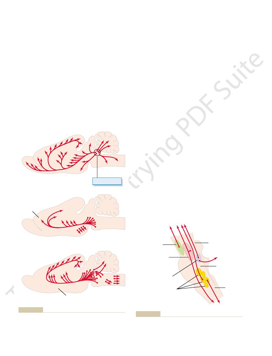
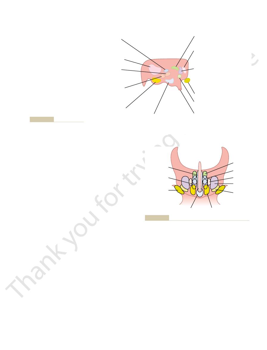
Figure 58–2 shows three neurohormonal systems
control, rather than just instantaneous activation or
of the brain. These neurohormones often persist for
brain activity. This is to secrete
areas to the cortical regions of the brain, still another
Brain Activity
Neurohormonal Control of
The Nervous System: C. Motor and Integrative Neurophysiology
730
Unit XI
Aside from direct control of brain activity by specific
transmission of nerve signals from the lower brain
physiologic mechanism is very often used to control
excitatory or inhibitory
neurotransmitter hormonal agents into the substance
minutes or hours and thereby provide long periods of
inhibition.
norepinephrine system
dopamine system
a serotonin system
three systems have different effects on levels of
nephrine system spreads to virtually every area of the
are directed much more to specific brain regions—
the dopamine system mainly into the basal ganglial
regions and the serotonin system more into the
Neurohormonal Systems in the Human Brain.
shows the brain stem areas in the human brain for acti-
system
follows:
1. The locus ceruleus and the norepinephrine system.
bilaterally and posteriorly at the juncture between
same as shown for the rat in the top frame of
norepinephrine
norepinephrine generally excites the brain to
effects in a few brain areas because of inhibitory
to a type of sleep called rapid eye movement
sleep (REM sleep).
2. The substantia nigra and the dopamine system.
Locus cerulus
Basal brain areas
Brain stem
Cerebellum
Caudate nucleus
Olfactory
region
Cerebral cor
tex
Cerebral cor
tex
NOREPINEPHRINE
Frontal
cortex
Cingulate
cortex
DOPAMINE
Midline nuclei
SEROTONIN
New York: Elsevier, 1985.)
in Kandel ER, Schwartz JH: Principles of Neural Science, 2nd ed.
. (Adapted from Kelly, after Cooper, Bloom, and Roth,
sero-
norepinephrine system
Three neurohormonal systems that have been mapped in the rat
Figure 58–2
brain: a
, a dopamine system, and a
tonin system
To diencephalon
and cerebrum
To cord
Medulla
Pons
To cerebellum
Mesencephalon
Substantia nigra
(dopamine)
Gigantocellular
neurons of
reticular formation
(acetylcholine)
Locus ceruleus
(norepinephrine)
Nuclei of the raphe
(serotonin)
cerebrum and downward into the spinal cord.
neurons send control signals upward into the diencephalon and
different transmitter substances (specified in parentheses). These
Multiple centers in the brain stem, the neurons of which secrete
Figure 58–3

spinal cord synapses. In Chapter 60, we will see that
and their associated nuclei. It was pointed out in
and the lower limbic structures.
associated with overall behavior and emotions. In turn,
Thus, on the medial and ventral surfaces of each
, and finally (4) passing behind the corpus callo-
, (3) then
on the ventral surface of the frontal lobes, (2)
, composed of a ring of cerebral cortex in
, and the
, the
, the
system, including the
of the central elements of the limbic system. Figure
, which from a physiologic point of view is one
connected complex of basal brain elements. Located
limbic system, demonstrating that they are an inter-
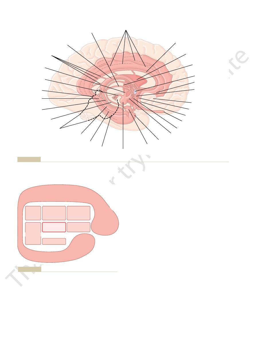
Figure 58–4 shows the anatomical structures of the
to behavior.
of the brain, and their control is closely related
eat and drink and to control body weight. These inter-
ature, osmolality of the body fluids, and the drives to
internal conditions of the body, such as body temper-
roles in behavioral control, these areas control many
, with its related structures. In addition to their
controls emotional behavior and motivational drives.
limbic system, the term
tures around the basal regions of the cerebrum, but as
term “limbic” was used to describe the border struc-
The word “limbic” means “border.” Originally, the
brain, the activation of each of which plays its own role
endorphins, angiotensin II, and neurotensin. Thus,
adrenocorticotropic hormone, epinephrine, histamine,
gamma-aminobutyric acid, glutamate, vasopressin,
by release into the fluids of the brain: enkephalins,
Without describing their function,
Other Neurotransmitters and Neurohormonal Substances
neurotransmitter. Activation of these
. In most places,
acetylcholine
cord. The neurohormone secreted at their
branches, one passing upward to the higher levels
the pons and mesencephalon. The fibers from
excitatory area and the acetylcholine system.
cause normal sleep, as we discuss in Chapter 59.
which was discussed in Chapter 48. The serotonin
the spinal cord. The serotonin secreted at the cord
to the cerebral cortex; still other fibers descend to
. They
thin nuclei called the raphe nuclei. Many of the
disease.
substantia nigra is the basic cause of Parkinson’s
excitatory. Also, remember from Chapter 56 that
inhibitory transmitter in the basal ganglia, but
system. The dopamine is believed to act as an
regions also secrete dopamine, but they send their
. Other neurons located in adjacent
putamen of the cerebrum, where they secrete
the superior mesencephalon, and its neurons send
relation to the basal ganglia. It lies anteriorly in
Behavioral and Motivational Mechanisms of the Brain—The Limbic System and the Hypothalamus
Chapter 58
731
nerve endings mainly to the caudate nucleus and
dopamine
endings into more ventral areas of the brain,
especially to the hypothalamus and the limbic
in some other areas of the brain it is possibly
destruction of the dopaminergic neurons in the
3. The raphe nuclei and the serotonin system. In
the midline of the pons and medulla are several
neurons in these nuclei secrete serotonin
send fibers into the diencephalon and a few fibers
fiber endings has the ability to suppress pain,
released in the diencephalon and cerebrum almost
certainly plays an essential inhibitory role to help
4. The gigantocellular neurons of the reticular
Earlier we discussed the gigantocellular neurons
(the giant cells) in the reticular excitatory area of
these large cells divide immediately into two
of the brain and the other passing downward
through the reticulospinal tracts into the spinal
terminals is
the acetylcholine functions as an excitatory
acetylcholine neurons leads to an acutely awake
and excited nervous system.
Secreted in the Brain.
the following is a list of still other neurohormonal
substances that function either at specific synapses or
there are multiple neurohormonal systems in the
in controlling a different quality of brain function.
Limbic System
we have learned more about the functions of the
limbic system has been
expanded to mean the entire neuronal circuitry that
A major part of the limbic system is the hypothala-
mus
nal functions are collectively called vegetative func-
tions
Functional Anatomy of the
Limbic System; Key Position
of the Hypothalamus
in the middle of all these is the extremely small hypo-
thalamus
58–5 illustrates schematically this key position of the
hypothalamus in the limbic system and shows sur-
rounding it other subcortical structures of the limbic
septum
paraolfactory area,
the anterior nucleus of the thalamus, portions of the
basal ganglia
hippocampus
amygdala.
And surrounding the subcortical limbic areas is the
limbic cortex
each side of the brain (1) beginning in the orbitofrontal
area
extending upward into the subcallosal gyrus
over the top of the corpus callosum onto the medial
aspect of the cerebral hemisphere in the cingulate
gyrus
sum and downward onto the ventromedial surface of
the temporal lobe to the parahippocampal gyrus and
uncus.
cerebral hemisphere is a ring of mostly paleocortex
that surrounds a group of deep structures intimately
this ring of limbic cortex functions as a two-way com-
munication and association linkage between the neo-
cortex
Many of the behavioral functions elicited from the
hypothalamus and other limbic structures are also
mediated through the reticular nuclei in the brain stem
Chapter 55 as well as earlier in this chapter that stim-
ulation of the excitatory portion of this reticular for-
mation can cause high degrees of cerebral excitability
while also increasing the excitability of much of the
most of the hypothalamic signals for controlling the
autonomic nervous system are also transmitted
through synaptic nuclei located in the brain stem.
An important route of communication between
the limbic system and the brain stem is the medial
tant of the control pathways of the limbic system. It
1 per cent of the brain mass, is one of the most impor-
Thus, the hypothalamus, which represents less than
both the posterior and the anterior pituitary glands.
diencephalon and cerebrum, especially to the anterior
system; (2) upward toward many higher areas of the
encephalon, pons, and medulla and from these areas
brain stem, mainly into the reticular areas of the mes-
in three directions: (1) backward and downward to the
pathways with all levels of the limbic system. In turn,
few cubic centimeters, has two-way communicating
The hypothalamus, despite its very small size of only a
Control Headquarters for the
brain stem, thalamus, hypothalamus, and most other
system. A second route of communication is through
both directions, forming a trunk line communication
stem reticular formation. This bundle carries fibers in
, which extends from the septal and
The Nervous System: C. Motor and Integrative Neurophysiology
732
Unit XI
forebrain bundle
orbitofrontal regions of the cerebral cortex downward
through the middle of the hypothalamus to the brain
short pathways among the reticular formation of the
contiguous areas of the basal brain.
Hypothalamus, a Major
Limbic System
it and its closely allied structures send output signals
into the peripheral nerves of the autonomic nervous
thalamus and limbic portions of the cerebral cortex;
and (3) into the hypothalamic infundibulum to control
or partially control most of the secretory functions of
controls most of the vegetative and endocrine func-
tions of the body as well as many aspects of emotional
Connecting spinal cord
Stria terminalis
Fimbria
of fornix
Gyrus
fasciolaris
Isthmus
Mamillotegmental
tract
Mamillothalamic
tract
Dorsal fornix
Body of fornix
Stria medullaris
thalami
Cingulate gyrus and cingulum
Indusium griseum
and longitudinal striae
Septum pellucidum
(supracommissural septum)
Anterior nuclear
group of thalamus
Anterior commissure
Subcallosal gyrus
Paraterminal gyrus
(precommissural
septum)
Orbitofrontal cortex
Prehippocampal rudiment
Paraolfactory area
Olfactory bulb
HYPOTHALAMUS
Column of fornix
(postcommissural fornix)
Uncus
Amygdaloid body
Mamillary body
Parahippocampal gyrus
Dentate gyrus
Hippocampus
Longman Group Ltd, 1973.)
Anatomy of the limbic system, shown in the dark pink area. (Redrawn from Warwick R, Williams PL: Gray’s Anatomy, 35th Br. ed. L
Figure 58–4
ondon:
Cingulate gyrus
Subcallosal
gyrus
Orbitofrontal
cortex
Uncus
Paraolfactory
area
Septum
area
Anterior
nuclei of
thalamus
Hypothalamus
Hippocampus
Amygdala
Parahippocampal gyrus
Portions
of basal
ganglia
Figure 58–5
Limbic system, showing the key position of the hypothalamus.
, is concerned
hypothalamus, especially the
The anterior portion of the
Regulation of Body Temperature.
arterial pressure. These effects are transmitted mainly
site effects, causing a decrease in both heart rate and
increases the arterial pressure and heart rate,
In general, stimulation in the
pressure, increased heart rate, and decreased heart rate.
including increased arterial pressure, decreased arterial
etative and control functions of the hypothalamus.
nuclei located elsewhere. With this caution in mind, we
in the figures. Also, it is not known whether the effects
thirst, hunger, and many of the emotional drives.
58–7) is present on each side of the hypothalamus. The
area (shown in Figure
lated. In addition to the centers shown in Figure 58–6,
to study these diagrams, especially to see in Figure
only a small area in Figure 58–4. Take a few minutes
coronal views of the hypothalamus, which represents
Figures 58–6 and 58–7 show enlarged sagittal and
endocrine control in Chapter 75. To illustrate the
29, temperature regulation in Chapter 73, and
Chapter 18, thirst and water conservation in Chapter
out this text. For instance, the role of the hypothala-
The different hypothalamic mechanisms for control-
Functions of the Hypothalamus
Vegetative and Endocrine Control
these operate together.
behavior. Let us discuss first the vegetative and
Behavioral and Motivational Mechanisms of the Brain—The Limbic System and the Hypothalamus
Chapter 58
733
endocrine control functions and then return to the
behavioral functions of the hypothalamus to see how
ling multiple functions of the body are so important
that they are discussed in multiple chapters through-
mus to help regulate arterial pressure is discussed in
organization of the hypothalamus as a functional unit,
let us summarize the more important of its vegetative
and endocrine functions here as well.
58–6 the multiple activities that are excited or inhib-
ited when respective hypothalamic nuclei are stimu-
a large lateral hypothalamic
lateral areas are especially important in controlling
A word of caution must be issued for studying these
diagrams because the areas that cause specific activi-
ties are not nearly as accurately localized as suggested
noted in the figures result from stimulation of specific
control nuclei or whether they result merely from
activation of fiber tracts leading from or to control
can give the following general description of the veg-
Cardiovascular Regulation.
Stimulation of different areas
throughout the hypothalamus can cause every known
type of neurogenic effect on the cardiovascular system,
posterior and lateral hypo-
thalamus
whereas stimulation in the preoptic area often has oppo-
through specific cardiovascular control centers in the
reticular regions of the pons and medulla.
preoptic area
HYPOTHALAMUS
POSTERIOR
Posterior hypothalamus
(Increased blood pressure)
(Pupillary dilation)
(Shivering)
Perifornical nucleus
(Hunger)
(Increased blood pressure)
(Rage)
Ventromedial nucleus
(Satiety)
(Neuroendocrine control)
Dorsomedial nucleus
(GI stimulation)
Mamillary body
(Feeding reflexes)
Arcuate nucleus and periventricular zone
(Neuroendocrine control)
Lateral hypothalamic area (not shown)
(Thirst and hunger)
ANTERIOR
Paraventricular nucleus
(Oxytocin release)
(Water conservation)
Supraoptic nucleus
(Vasopressin release)
Optic chiasm (Optic nerve)
Medial preoptic area
(Bladder contraction)
(Decreased heart rate)
(Decreased blood pressure)
Posterior preoptic and
anterior hypothalamic areas
(Body temperature regulation)
(Panting)
(Sweating)
(Thyrotropin inhibition)
Infundibulum
Control centers of the hypothala-
Figure 58–6
mus (sagittal view).
Paraventricular
Dorsomedial
Fornix
Lateral
hypothalamic
Supraoptic
Ventromedial
Arcuate
Thalamus
Optic
tract
Anterior
hypothalamic
Periventricular
tions of the respective hypothalamic nuclei.
Coronal view of the hypothalamus, showing the mediolateral posi-
Figure 58–7

hypothalamus.
areas of the hypothalamus, especially the most
this portion of the hypothalamus), usually leads to
, located immediately adjacent to the third
thin zone of periventricular
3. Stimulation of a
, and
stimulation—that is, a sense of
2. Stimulation in the
fighting, as discussed subsequently.
animal, sometimes leading to overt rage and
causes thirst and eating, as discussed above, but
1. Stimulation in the
In animals, some of the behavioral effects of stimu-
animals and human beings.
tive and endocrine functions of the hypothalamus,
Hypothalamus and Associated
Behavioral Functions of the
ferent hypothalamic functions is partially tentative.
These areas are still poorly delimited, so much so that
control specific vegetative and endocrine functions.
Summary.
pituitary hormones.
to the anterior pituitary gland, where they act on the
These hormones are then transported via the blood
itary, specific
pituitary vascular sinuses. As the blood courses through
The anterior pituitary gland receives its blood supply
the following.
the endocrine glands. Briefly, the basic mechanisms are
secrete its endocrine hormones. This subject is discussed
Anterior Pituitary Gland.
reflexes, such as licking the lips and swallowing.
; these
eventually in tremendous obesity. Another area of the
overactive, so that it has a voracious appetite, resulting
instead, its hypothalamic hunger centers become
destroyed bilaterally, the animal cannot be satiated;
complete indifference to food. However, if this area is
When this center is stimulated electrically, an animal
, is located in the
A center that opposes the desire for food, called the
versely, damage to this area on both sides of the hypo-
intense desire to search for food. One area associated
experience extreme hunger, a voracious appetite, and an
Stimulation of
so that the baby can nourish itself. These functions are
of the breast, thereby expelling milk through the nipples
also causes oxytocin release, and the oxytocin now per-
whenever the baby suckles the mother’s breast, a reflex
promote labor contractions that expel the baby. Then,
oxytocin are secreted, and this secretion helps to
At the end of pregnancy, especially large quantities of
the alveoli to empty their milk through the nipples.
rounding the alveoli of the breasts, which then causes
This in turn causes increased contractility of the uterus
oxytocin
back toward normal. These functions are presented in
the urine but allows continuing excretion of electrolytes,
reabsorption of water. This decreases loss of water into
). This hormone is then absorbed into
mus into the posterior pituitary gland, where the nerve
stimulated. Nerve fibers from these neurons project
too concentrated, the neurons of these areas become
nuclei. When the body fluids become
drink water; it will search out the nearest source of
concentrated, the animal develops an intense desire to
lateral hypothalamus. When the fluid electrolytes in
urine. An area called the
thirst, which makes the animal or person drink water,
body water in two ways: (1) by creating the sensation of
The hypothalamus regulates
Regulation of Body Water.
increasing or decreasing body temperature, as discussed
ity. In turn, these neurons control mechanisms for
increases the activity of temperature-sensitive neurons,
with regulation of body temperature. An increase in the
The Nervous System: C. Motor and Integrative Neurophysiology
734
Unit XI
temperature of the blood flowing through this area
while a decrease in temperature decreases their activ-
in Chapter 73.
and (2) by controlling the excretion of water into the
thirst center is located in the
either this center or closely allied areas become too
water and drink enough to return the electrolyte con-
centration of the thirst center to normal.
Control of renal excretion of water is vested mainly
in the supraoptic
downward through the infundibulum of the hypothala-
endings secrete the hormone antidiuretic hormone (also
called vasopressin
the blood and transported to the kidneys where it acts
on the collecting ducts of the kidneys to cause increased
thus decreasing the concentration of the body fluids
Chapter 28.
Regulation of Uterine Contractility and of Milk Ejection from the
Breasts.
Stimulation of the paraventricular nuclei causes
their neuronal cells to secrete the hormone
.
as well as contraction of the myoepithelial cells sur-
signal from the nipple to the posterior hypothalamus
forms the necessary function of contracting the ductules
discussed in Chapter 82.
Gastrointestinal and Feeding Regulation.
several areas of the hypothalamus causes an animal to
with hunger is the lateral hypothalamic area. Con-
thalamus causes the animal to lose desire for food,
sometimes causing lethal starvation.
satiety center
ventromedial nuclei.
that is eating food suddenly stops eating and shows
hypothalamus that enters into overall control of gas-
trointestinal activity is the mamillary bodies
control at least partially the patterns of many feeding
Hypothalamic Control of Endocrine Hormone Secretion by the
Stimulation of certain areas of the
hypothalamus also causes the anterior pituitary gland to
in detail in Chapter 74 in relation to neural control of
mainly from blood that flows first through the lower
part of the hypothalamus and then through the anterior
the hypothalamus before reaching the anterior pitu-
releasing and inhibitory hormones are
secreted into the blood by various hypothalamic nuclei.
glandular cells to control release of specific anterior
A number of areas of the hypothalamus
the specification given above of different areas for dif-
Limbic Structures
Effects Caused by Stimulation.
In addition to the vegeta-
stimulation of or lesions in the hypothalamus often
have profound effects on emotional behavior of
lation are the following:
lateral hypothalamus not only
also increases the general level of activity of the
ventromedial nucleus
and surrounding areas mainly causes effects
opposite to those caused by lateral hypothalamic
satiety, decreased
eating
tranquility.
nuclei
ventricle (or also stimulation of the central gray
area of the mesencephalon that is continuous with
fear and punishment reactions.
4. Sexual drive can be stimulated from several
anterior and most posterior portions of the
pleasure and reward
punishment and fear can take precedence over
reward and pleasure centers completely, demonstrat-
pocampus. It is particularly interesting that stimulation
thalamus and thalamus. Less potent punishment areas
By means of this technique, the most potent areas
sickness.
displeasure, fear, terror, pain, punishment, and even
immediately learns to turn it off. Stimulation in these
areas; but when it is in certain other areas, the animal
case, the animal will not press the lever to turn the
when the lever is pressed. In this
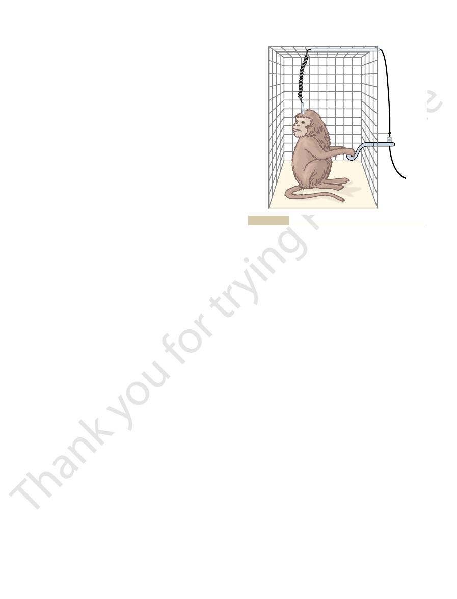
The apparatus shown in Figure 58–8 can also be
certain areas of the thalamus and basal ganglia, and
hypothalamus, are found in the septum, the amygdala,
sense of punishment. Less potent reward centers,
cause rage. But this is true in many areas, with weaker
reward areas—indeed, it is one of the most potent of
. It is strange
, especially in the
By using this procedure, the major reward centers
center, the animal often chooses the electrical
even thousands of times per hour. Furthermore, when
again and again, sometimes as much as hundreds or
animal a sense of reward, then it will press the lever
the lever. If stimulating the particular area gives the
makes electrical contact with a stimulator. Electrodes
brain. In this figure, a lever is placed at the side of
Figure 58–8 shows a technique that has been used for
Reward Centers
and all the other elements of punishment. The degrees
causes terror, pain, fear, defense, escape reactions,
animal, whereas electrical stimulation of other regions
. Electrical stimula-
, or
reward
These affective qualities are also called
nature of sensory sensations—that is,
From the discussion thus far, it is already clear that
of the Limbic System
will discuss some of these in more detail later.
similar to those elicited from the hypothalamus. We
and areas in the mesencephalon, often cause effects
system, especially in the amygdala, the septal area,
hypothalamus: excessive drinking and eating as
2. Bilateral lesions of the ventromedial areas of the
of most of its overt drives.
of the animal as well, with loss
leading to lethal starvation. These lesions cause
decrease drinking and eating almost to zero, often
1. Bilateral lesions in the lateral hypothalamus will
those caused by stimulation. For instance:
hypothalamus, in general, cause effects opposite to
Behavioral and Motivational Mechanisms of the Brain—The Limbic System and the Hypothalamus
Chapter 58
735
Effects Caused by Hypothalamic Lesions.
Lesions in the
extreme passivity
hypothalamus cause effects that are mainly
opposite to those caused by lesions of the lateral
well as hyperactivity and often continuous
savagery along with frequent bouts of extreme
rage on the slightest provocation.
Stimulation or lesions in other regions of the limbic
“Reward” and “Punishment” Function
several limbic structures are particularly concerned
with the affective
whether the sensations are pleasant or unpleasant.
or pun-
ishment
satisfaction or aversion
tion of certain limbic areas pleases or satisfies the
of stimulation of these two oppositely responding
systems greatly affect the behavior of the animal.
localizing specific reward and punishment areas of the
the cage and is arranged so that depressing the lever
are placed successively at different areas in the brain
so that the animal can stimulate the area by pressing
offered the choice of eating some delectable food as
opposed to the opportunity to stimulate the reward
stimulation.
have been found to be located along the course of the
medial forebrain bundle
lateral and
ventromedial nuclei of the hypothalamus
that the lateral nucleus should be included among the
all—because even stronger stimuli in this area can
stimuli giving a sense of reward and stronger ones a
which are perhaps secondary to the major ones in the
extending downward into the basal tegmentum of the
mesencephalon.
Punishment Centers
connected so that the stimulus to the brain continues
all the time except
stimulus off when the electrode is in one of the reward
areas causes the animal to show all the signs of
for punishment and escape tendencies have been
found in the central gray area surrounding the aque-
duct of Sylvius in the mesencephalon and extending
upward into the periventricular zones of the hypo-
are found in some locations in the amygdala and hip-
in the punishment centers can frequently inhibit the
ing that
.
Rage—Its Association with
Punishment Centers
An emotional pattern that involves the punish-
ment centers of the hypothalamus and other limbic
brain of a monkey.
Technique for localizing reward and punishment centers in the
Figure 58–8

visual, auditory, tactile, and other types of hallucina-
various psychomotor effects, including olfactory,
During hippocampal seizures, the person experiences
signals even under normal functioning conditions.
seconds after the stimulation is over, suggesting that
of the hippocampi. These often persist for many
become hyperexcitable. For instance, weak electrical
rage, passivity, or excess sex drive.
of the different behavioral patterns such as pleasure,
poses. As in other limbic structures, stimulation of dif-
nicating pathway. Thus, the hippocampus is an addi-
, a major commu-
system, especially through the
mus, hypothalamus, and other parts of the limbic
the hippocampus, and the hippocampus in turn dis-
and the mamillary bodies. Almost any type of sensory
system—the amygdala, the hypothalamus, the septum,
parietal lobe structures, all together called the
The hippocampus (and its adjacent temporal and
lobe.
parahippocampal gyrus, which is the cerebral cortex
nuclei, and along its lateral border it fuses with the
surface of much of the inside of the lateral ventricle.
The hippocampus is the elongated portion of the cere-
Functions of the Hippocampus
Parts of the Limbic System
the information that we learn, usually throwing away
that are either rewarding or punishing but, conversely,
. An
away, and the response is said to be
ment rather than indifference, the cerebral cortical
is, the animal becomes
plete extinction of the cerebral cortical response. That
elicit a sense of either reward or punishment, repeti-
bral cortex. But, if the sensory experience does not
hardly remembered at all. Electrical recordings from
Versus Reinforcement
in Learning and Memory—Habituation
Importance of Reward or Punishment
reactivity of the animal. Therefore, it is presumed that
ishment centers, thereby decreasing the affective
mazine, usually inhibits both the reward and the pun-
Administration of a tranquilizer, such as chlorpro-
Effect of Tranquilizers on the Reward or Punishment Centers.
activities, our drives, our aversions, our motivations.
ing, we cease to do it. Therefore, the reward and pun-
that is rewarding, we continue to do it; if it is punish-
to reward and punishment. If we are doing something
stimulated: placidity and tameness.
Placidity and Tameness.
gyri and subcallosal gyri, help suppress the rage
limbic cortex, especially in the anterior cingulate
addition, portions of the hippocampi and anterior
from the ventromedial nuclei of the hypothalamus. In
Fortunately, in the normal animal, the rage phe-
animal being severely punished, and it is a pattern of
causes an immediate savage attack. This is approxi-
pupils. Furthermore, even the slightest provocation
(7) develop piloerection, wide-open eyes, and dilated
its claws, (3) lift its tail, (4) hiss, (5) spit, (6) growl, and
the animal to (1) develop a defense posture, (2) extend
, causes
periventricular zone of the
the brain, especially in the
, described as follows.
structures, and has also been well characterized, is the
The Nervous System: C. Motor and Integrative Neurophysiology
736
Unit XI
rage pattern
Strong stimulation of the punishment centers of
hypothalamus and in the lateral hypothalamus
mately the behavior that one would expect from an
behavior that is called rage.
nomenon is held in check mainly by inhibitory signals
phenomenon.
Exactly the opposite emotional
behavior patterns occur when the reward centers are
Importance of Reward or Punishment
in Behavior
Almost everything that we do is related in some way
ishment centers undoubtedly constitute one of the
most important of all the controllers of our bodily
tranquilizers function in psychotic states by suppress-
ing many of the important behavioral areas of the
hypothalamus and its associated regions of the limbic
brain.
Animal experiments have shown that a sensory expe-
rience that causes neither reward nor punishment is
the brain show that a newly experienced sensory stim-
ulus almost always excites multiple areas in the cere-
tion of the stimulus over and over leads to almost com-
habituated to that specific
sensory stimulus and thereafter ignores it.
If the stimulus does cause either reward or punish-
response becomes progressively more and more
intense during repeated stimulation instead of fading
reinforced
animal builds up strong memory traces for sensations
develops complete habituation to indifferent sensory
stimuli.
It is evident that the reward and punishment centers
of the limbic system have much to do with selecting
more than 99 per cent of it and selecting less than 1
per cent for retention.
Specific Functions of Other
bral cortex that folds inward to form the ventral
One end of the hippocampus abuts the amygdaloid
on the ventromedial outside surface of the temporal
hip-
pocampal formation) has numerous but mainly indi-
rect connections with many portions of the cerebral
cortex as well as with the basal structures of the limbic
experience causes activation of at least some part of
tributes many outgoing signals to the anterior thala-
fornix
tional channel through which incoming sensory signals
can initiate behavioral reactions for different pur-
ferent areas in the hippocampus can cause almost any
Another feature of the hippocampus is that it can
stimuli can cause focal epileptic seizures in small areas
the hippocampi can perhaps give off prolonged output
tions that cannot be suppressed as long as the seizure
persists even though the person has not lost con-
sciousness and knows these hallucinations to be

animals, animals of the wrong sex, or even animals of a
to eat solid objects, and (5) often has a sex drive so
everything, (3) forgets rapidly, (4) has a tendency to
not afraid of anything, (2) has extreme curiosity about
drome, which is demonstrated by an animal that (1) is
causes changes in behavior called the Klüver-Bucy syn-
that lie inside these parts of the temporal lobes. This
lobes are destroyed in a monkey, this removes not only
When the anterior parts of both temporal
Effects of Bilateral Ablation of the Amygdala—The Klüver-Bucy
activity, and premature labor.
copulatory movements, ejaculation, ovulation, uterine
Finally, excitation of still other portions of the amyg-
and pleasure.
the hypothalamus, as described earlier. Stimulation of
pain, and fear similar to the rage pattern elicited from
can cause a pattern of rage, escape, punishment, severe
In addition, stimulation of certain amygdaloid nuclei
licking, chewing, and swallowing.
ments associated with olfaction and eating, such as
rhythmical movements; and (4) different types of move-
body; (2) circling movements; (3) occasionally clonic,
movements, such as raising the head or bending the
types of involuntary movement. These include (1) tonic
thalamus, amygdala stimulation can also cause several
adrenocorticotropic hormone.
pituitary hormones, especially the gonadotropins and
(6) piloerection, and (7) secretion of various anterior
micturition, (5) pupillary dilation or, rarely, constriction,
gastrointestinal motility and secretion, (4) defecation or
or decreases in heart rate, (3) increases or decreases in
increases or decreases in arterial pressure, (2) increases
plus other effects. Effects initiated from the amygdala
those elicited by direct stimulation of the hypothalamus,
In general, stimulation
hypothalamus.
septum, (4) into the thalamus, and (5) especially into the
tical areas, (2) into the hippocampus, (3) into the
sees the place of the person in the world. In turn, the
called the “window” through which the limbic system
of these multiple connections, the amygdala has been
from the auditory and visual association areas. Because
of the temporal, parietal, and occipital lobes—especially
tions of the limbic cortex, as well as from the neocortex
The amygdala receives neuronal signals from all por-
, has
portion of the amygdala, the
area of the temporal lobe. In the human being, another
, which lies immediately
with the limbic brain. Indeed, it is pointed out in
In lower animals, the amygdala is concerned to a great
medial anterior pole of each temporal lobe. It has abun-
The amygdala is a complex of multiple small nuclei
Functions of the Amygdala
poor or does not take place.
manent storage takes place. Whatever the mechanism,
rehearse over and over
term memory—that is, the hippocampus transmits
rience that causes either pleasure or pain. But what is
Thus, a person rapidly becomes habituated to indif-
to be committed to memory.
neuronal input is important, the information is likely
making. Therefore, if the hippocampus signals that a
established, presumably the remainder of the brain
ing the importance of the incoming sensory signals.
ical decision-making neuronal mechanism, determin-
the brain, the hippocampus presumably became a crit-
importance.Very early in evolutionary development of
inviting, thus making decisions that are of life-or-death
object suggests danger, or whether the odor is sexually
a particular food, whether the smell of a particular
cortex. In many lower animals, this cortex plays essen-
The
pletely abolished. This is the phenomenon called
memory for seconds up to a minute or two, although
their activities. Thus, they are capable of short-term
in contact every day. Yet they can remember for a
based on verbal symbolism. In fact, they often cannot
ously learned memories satisfactorily. However, they
ment of epilepsy. These people can recall most previ-
Portions of the hippocampi have been surgically
Role of the Hippocampus in Learning
of its areas instead of the six layers found elsewhere.
cerebrum, having only three nerve cell layers in some
unreal. Probably one of the reasons for this hyper-
Behavioral and Motivational Mechanisms of the Brain—The Limbic System and the Hypothalamus
Chapter 58
737
excitability of the hippocampi is that they have a dif-
ferent type of cortex from that elsewhere in the
Effect of Bilateral Removal of the Hippocampi—Inability to
Learn.
removed bilaterally in a few human beings for treat-
often can learn essentially no new information that is
even learn the names of people with whom they come
moment or so what transpires during the course of
their ability to establish memories lasting longer than
a few minutes is either completely or almost com-
anterograde amnesia that was discussed in Chapter 57.
Theoretical Function of the Hippocampus in Learning.
hippocampus originated as part of the olfactory
tial roles in determining whether the animal will eat
Once this critical decision-making capability had been
also began to call on the hippocampus for decision
ferent stimuli but learns assiduously any sensory expe-
the mechanism by which this occurs? It has been sug-
gested that the hippocampus provides the drive that
causes translation of short-term memory into long-
some signal or signals that seem to make the mind
the new information until per-
without the hippocampi, consolidation of long-term
memories of the verbal or symbolic thinking type is
located immediately beneath the cerebral cortex of the
dant bidirectional connections with the hypothalamus
as well as with other areas of the limbic system.
extent with olfactory stimuli and their interrelations
Chapter 53 that one of the major divisions of the olfac-
tory tract terminates in a portion of the amygdala
called the corticomedial nuclei
beneath the cerebral cortex in the olfactory pyriform
basolateral nuclei
become much more highly developed than the olfactory
portion and plays important roles in many behavioral
activities not generally associated with olfactory stimuli.
amygdala transmits signals (1) back into these same cor-
Effects of Stimulating the Amygdala.
in the amygdala can cause almost all the same effects as
and then sent through the hypothalamus include (1)
Aside from these effects mediated through the hypo-
other amygdaloid nuclei can give reactions of reward
dala can cause sexual activities that include erection,
Syndrome.
portions of temporal cortex but also of the amygdalas
place everything in its mouth and sometimes even tries
strong that it attempts to copulate with immature

laughter and humour. Brain 126:2121, 2003.
Wild B, Rodden FA, Grodd W, Ruch W: Neural correlates of
systems in one? Nat Rev Neurosci 5:35, 2004.
Vann SD, Aggleton JP: The mammillary bodies: two memory
neurotransmitters. Neuron 40:1059, 2003.
van den Pol AN: Weighing the role of hypothalamic feeding
83:803, 2003.
daloid complex: anatomy and physiology. Physiol Rev
Sah P, Faber ES, Lopez De Armentia M, Power J: The amyg-
biol 14:198, 2004.
amygdala and hippocampal complex. Curr Opin Neuro-
Phelps EA: Human emotion and memory: interactions of the
receiver/transmitters. J Neuroendocrinol 16:403, 2004.
Morris JF, Ludwig M: Magnocellular dendrites: prototypic
Physiol Sci 19:22, 2004.
tinguishes between escapable and inescapable pain. News
Lumb BM: Hypothalamic and midbrain circuitry that dis-
rosci 23:155, 2000.
LeDoux JE: Emotion circuits in the brain. Annu Rev Neu-
Neurosci Biobehav Rev 27:765, 2004.
role in ingestive behavior and reward-related learning.
Kelley AE: Ventral striatal control of appetitive motivation:
Science, 4th ed. New York: McGraw-Hill, 2000.
Kandel ER, Schwartz JH, Jessell TM: Principles of Neural
1007:367, 2003.
thalamic function after chronic stress. Ann N Y Acad Sci
Joels M, Verkuyl JM, Van Riel E: Hippocampal and hypo-
and reward expectancy. Curr Opin Neurobiol 14:148, 2004.
Holland PC, Gallagher M: Amygdala—frontal interactions
Churchill Livingstone, 1997.
Haines DE: Fundamental Neuroscience. New York:
perception. J Neurophysiol 90:539, 2003.
Guillery RW: Branching thalamic afferents link action and
and wakefulness. Mol Neurobiol 29:41, 2004.
Gerashchenko D, Shiromani PJ: Different neuronal pheno-
in mood disorders. Ann N Y Acad Sci 985:420, 2003
Drevets WC: Neuroimaging abnormalities in the amygdala
U S A 93:7397, 1996.
integration of body fluid regulation. Proc Natl Acad Sci
Denton DA, McKinley MJ, Weisinger RS: Hypothalamic
New York: John Wiley, 1999.
Conlon R, Hobson JA: Understanding the Human Mind.
thalamic feeding circuits. Endocrinology 145:2621, 2004.
Bouret SG, Simerly RB: Leptin and development of hypo-
synaptic plasticity and learning. Mol Neurobiol 29:131,
Blank T, Nijholt I, Spiess J: Molecular determinants mediat-
in decision-making. Ann N Y Acad Sci 985:356, 2003.
Bechara A, Damasio H, Damasio AR: Role of the amygdala
raphe nuclei. Brain Res Brain Res Rev 39:154, 2002.
Adell A, Celada P, Abellan MT, Artigas F: Origin and func-
ciations occur.
lobe. In the middle and posterior cingulate cortex, there
derived from Wernicke’s area of the posterior temporal
pocampal gyri, there is a tendency for complex auditory
and olfactory behavioral associations. In the parahip-
anterior temporal cortex, one especially finds gustatory
tures for control of behavioral patterns. Thus, in the
Until further information is available, it is
Summary.
rage than normally.
prefrontal inhibitory influence. Therefore, the animal
structures. Destruction of these gyri bilaterally releases
The
to sit still and moving about continuously.
ciated with intense motor restlessness, becoming unable
priate animals or even inanimate objects, and loses all
and all objects, has intense sex drives toward inappro-
develops consummatory behavior: it investigates any
Klüver-Bucy syndrome occurs. The animal especially
earlier in this chapter; it was pointed out that the
almost invariably damaged as well. This was discussed
temporal cortex is ablated bilaterally, the amygdalas are
When the anterior
Ablation of the Anterior Temporal Cortex.
follows.
can cause persistent changes in an animal’s behavior, as
cortex. Likewise, ablation of some limbic cortical areas
limbic system, essentially all behavioral patterns can be
However, as is true of so many other portions of the
cortex has failed to give any real idea of their functions.
behavior.
site direction. Therefore, the limbic cortex in effect
This cortex functions as a transitional zone through
that surrounds the subcortical limbic structures.
The most poorly understood portion of the limbic
Function of the Limbic Cortex
mation, the amygdala is believed to make the person’s
surroundings and thoughts. On the basis of this infor-
limbic system one’s current status in relation to both
conscious level. They also seem to project into the
The amygdalas seem to
too different from that of the monkey.
beings are rare, afflicted people respond in a manner not
different species. Although similar lesions in human
The Nervous System: C. Motor and Integrative Neurophysiology
738
Unit XI
Overall Function of the Amygdalas.
be behavioral awareness areas that operate at a semi-
behavioral response appropriate for each occasion.
system is the ring of cerebral cortex called the limbic
cortex
which signals are transmitted from the remainder of the
brain cortex into the limbic system and also in the oppo-
functions as a cerebral association area for control of
Stimulation of the different regions of the limbic
elicited by stimulation of specific portions of the limbic
fear—and thus develops tameness as well.
Ablation of the Posterior Orbital Frontal Cortex.
Bilateral
removal of the posterior portion of the orbital frontal
cortex often causes an animal to develop insomnia asso-
Ablation of the Anterior Cingulate Gyri and Subcallosal Gyri.
anterior cingulate gyri and the subcallosal gyri are the
portions of the limbic cortex that communicate between
the prefrontal cerebral cortex and the subcortical limbic
the rage centers of the septum and hypothalamus from
can become vicious and much more subject to fits of
perhaps best to state that the cortical regions of the
limbic system occupy intermediate associative positions
between the functions of the specific areas of the cere-
bral cortex and functions of the subcortical limbic struc-
associations as well as complex thought associations
is reason to believe that sensorimotor behavioral asso-
References
tional role of the extracellular serotonin in the midbrain
ing effects of acute stress on hippocampus-dependent
2004.
types in the lateral hypothalamus and their role in sleep