
Chapter 8
Third month to
birth
The fetus and
placenta

Development of the fetus
• Fetal period: 9
th
week to birth
• Length of fetus:
CRL (crown-rump length), sitting height
CHL (crown-heel length), standing height
Correlation of height (cm) and age of fetus
Growth in length during 3
rd
,4
th
,5
th
months
• Growth in weight during 8
th
& 9
th
months
• Length of pregnancy:
280 days (40 weeks) after onset of LNMP
266 days or 38 weeks after fertilization
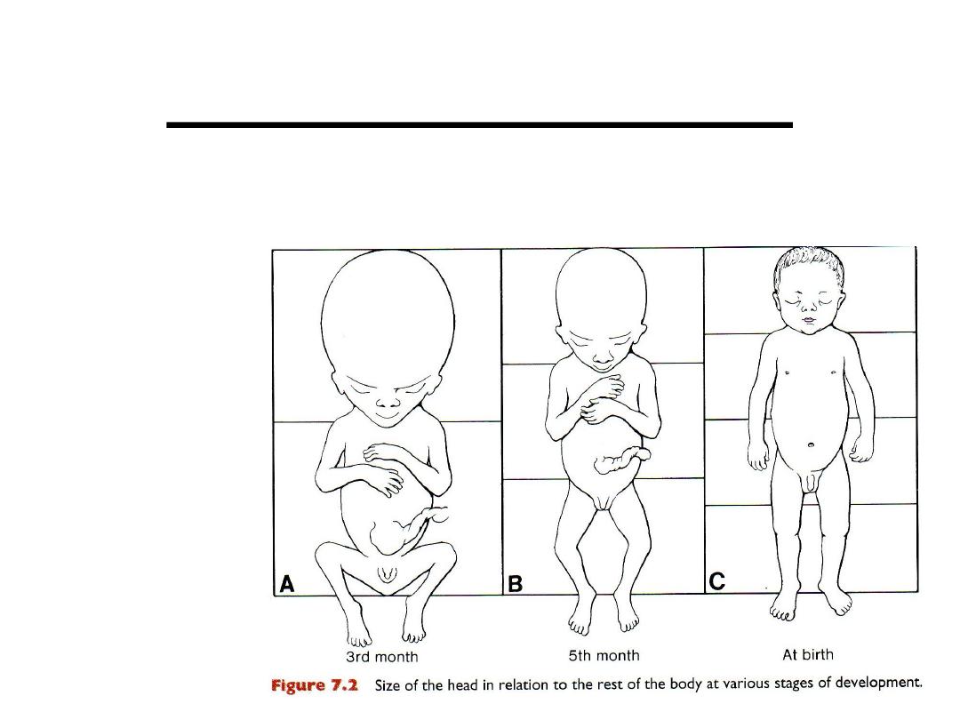
Slowdown of
head growth
Monthly changes

During 3
rd
month
• Face more human
• Limbs lengthen
• Primary ossification centers (at 12
th
week)
• External genitalia (at 12
th
week): sex of fetus determined by ultrasound
• Intestinal loops withdraw into abdominal cavity (by 12
th
week)
During fourth and fifth months
• Increase of length of fetus
• Lanugo hair, eyebrows, head hair appear
• Fetal movements can be felt by mother
During 6
th
month
• Skin red & wrinkled: lack of underlying connective tissue
• Premature birth: difficult to survive because the respiratory system &
nervous system are not differentiated sufficiently.
7
th
month
• Wt.: 1,100g
• If born at 7
th
month: 90% chance of surviving

Last 2 months
• Deposition of subcutaneous fat
• Vernix caseosa : white fatty substance
cover the skin
• End of 9
th
month:
– Skull: widest circumference
– Wt.: 3,000-3,400g
– CRL: 36cm
– CHL: 50cm
• Sexual characteristics: pronounced
• Testes should be in scrotum
Time of birth
• 266 days (38 weeks) after
fertilization: difficulty to
determine date of fertilization
• 280 days (40 weeks) from first day
of LNMP: regular 28-day
menstrual period
• Miscalculations: irregular period,
bleeding at 14 days
• Premature and postmature
Age of embryo
•
Combination of:
– Onset of LNMP,
– Morphological characteristics
•
Ultrasound accurate (1-2 days)
– CRL
– BPD: biparietal diameter
– Head & abdomen circumph.
– Femur length

FETAL MEMBRANES & PLACENTA
Changes in the trophoblast
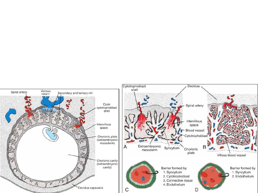
• The
fetal
component of the placenta is derived from the
trophoblast
and
extraembryonic mesoderm
(the chorionic plate);
• The
maternal
component is derived from the uterine endometrium.
Human embryo at the
beginning of
the second month
of development.
Secondary & tertiary villi
Structure of villi at various stages of
development.
4
th
week
4
th
month

Preeclampsia
• Maternal hypertension, proteinuria and edema
• From 20wks to term
• Retardation of fetal growth, death of fetus death of mother
• Failed differentiation of cytotrophoblast cells
• Causes of preeclampsia:
– placental mosiacism: trophoblast cells have genetic defects
– Maternal diabetis
– Smokers

Chorion Frondosum & Decidua Basalis
End of 2
nd
month
End of 3
rd
month
•
In the early weeks of development, villi cover the entire surface of the chorion
•
As pregnancy advances, villi on the embryonic pole continue to grow and expand, giving
rise to the chorion frondosum (bushy chorion).
•
Villi on the abembryonic pole degenerate, and by the third month, this side of the
chorion, is the chorion laeve, is smooth
•
The decidua
: the functional layer of the endometrium, which is shed during parturition.
•
The decidua over the chorion frondosum, the
decidua basalis
,
•
The decidual layer over the abembryonic pole is the
decidua capsularis
.
•
With growth of the chorionic vesicle, this layer becomes stretched and degenerates.
• Subsequently, the chorion laeve
comes into contact with the uterine
wall (
decidua parietalis
) on the
opposite side of the uterus, & the two
fuse obliterating the uterine lumen.
• ONLY the
chorion frondosum
and
the
decidua basalis
, make up
the
placenta
.
• Similarly, fusion of the
amnion
and
chorion
to form the amniochorionic
m. obliterates the chorionic cavity
• It is this membrane that ruptures
during labor (
breaking of the water).
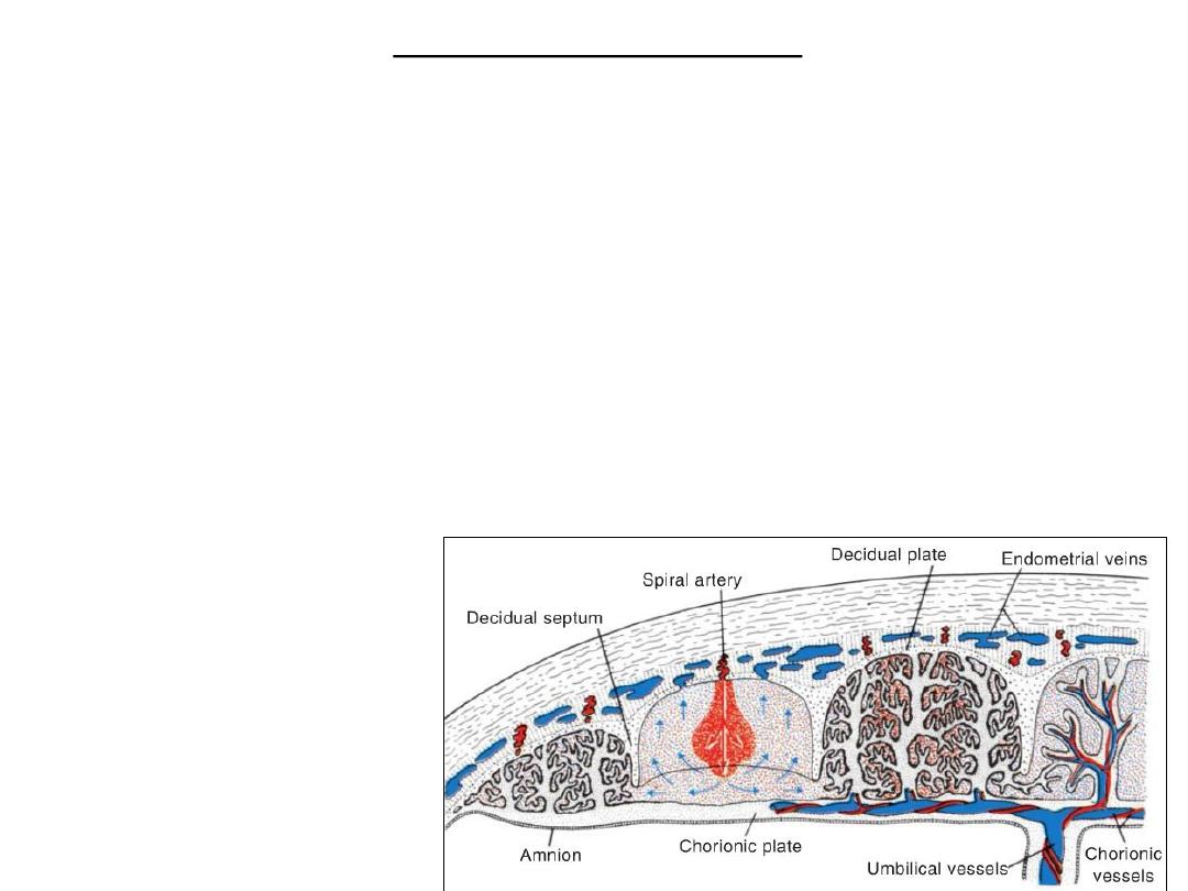
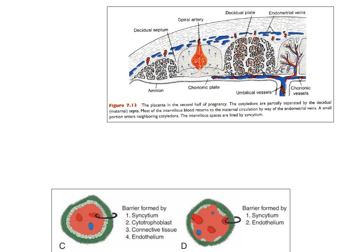
Structure of the placenta
• By the beginning of the fourth month, the placenta has two components:
(1) a fetal portion, formed by the chorion frondosum and
(2) a maternal portion, formed by the decidua basalis
• Between the chorionic and decidual plates are the intervillous spaces, which
are filled with maternal blood. They are derived from lacunae in the
syncytiotrophoblast and are lined with syncytium of fetal origin.
• The villous trees grow into the intervillous blood lakes.
• During the fourth and fifth months, the decidua forms a number of decidual septa,
which project into intervillous spaces but do not reach the chorionic plate. These
septa have a core of maternal tissue, but their surface is covered by a layer of
syncytial cells
• As a result of this septum
formation, the placenta is
divided into a number of
cotyledons.
Full-Term Placenta
• At full term, the placenta is discoid, diameter of 15 to 25 cm, 3 cm thick, and
weighs about 500 to 600 g.
• At birth, it is torn from the uterine wall and, approximately 30 minutes after birth
of the child, is expelled from the uterine cavity as the after birth.
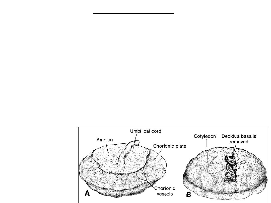
• Maternal side of placenta
: 15 to 20 cotyledons, covered by a thin layer of decidua
basalis, Grooves between the cotyledons are formed by decidual septa.
• The
fetal surface
of the placenta is covered entirely by the chorionic plate. A
number of large arteries and veins, the chorionic vessels, converge toward the
umbilical cord .
• The chorion, in turn, is covered by the amnion.
A- Fetal side
B- Maternal side
Circulation of
placenta
Placental membrane (barrier)
4
th
week
4
th
month
Function of placenta
• Exchange of gases
• Exchange of nutrition & electrolytes
• Transmission of maternal antibodies
• Hormone production:
– hCG…pregnancy test
– Progesterone end of 4
th
month
– estrogens (esteriol)
– somatommamotropin
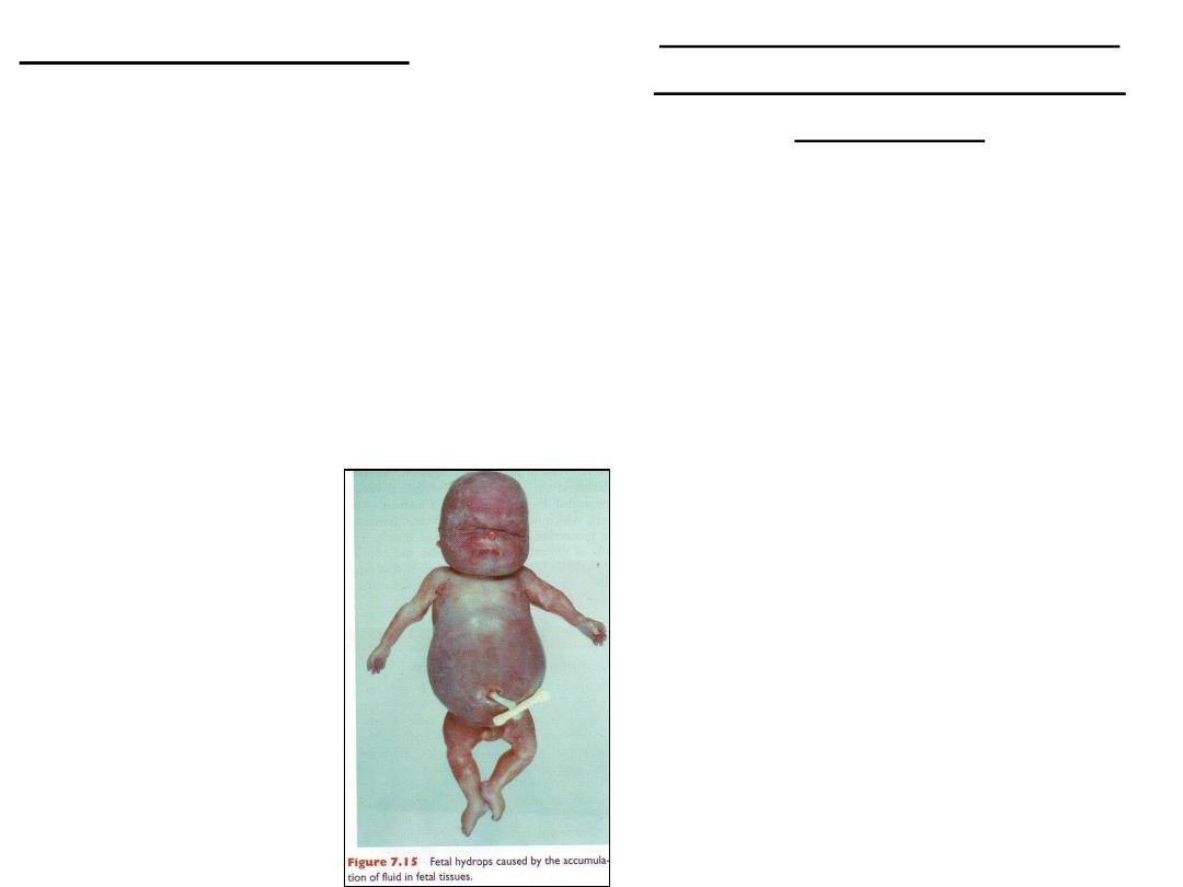
ERYTHROBLASTOSIS FETALIS
(HEMOLYTIC DISEASE OF THE
NEWBORN)
• Fetal hydrops
• Causes: antigens from CDE (Rhesus)
• D most dangerous
• Fetus: D (Rh) =ve
• Mother: D (Rh) –ve
• Treatment: intrauterine or postnatal
transfusion
• Anti-D-immunoglobulin

The placental barrier
• Most maternal hormones do not cross placenta
• Some cross slowly: thyroxine
• Synthetic progestins cross: masculinize female fetuses
• Synthetic estrogen (diethylstilbestrol) cause: vaginal carcinoma & testicular
abnormalities
• Viruses cross placenta:
– Rubella, cytomegalovirus, coxsakie, variola, varicella, measles, poliomyelitis:
cause infections, cell death, or birth defects
• Drugs: damage of embryo
• Heroin & cocain: habituation in infants

Amnion and umbilical cord

• The
amnion
is a large sac containing
amniotic fluid
in which the fetus is suspended
by its umbilical cord.
• The fluid (1) absorbs jolts,(2) allows for fetal movements, and (3) prevents
adherence of the embryo to surrounding tissues.
• The fetus swallows amniotic fluid, which is absorbed through its gut and cleared by
the placenta.
• The fetus adds urine to the amniotic fluid, but this is mostly water.
• An excessive amount of amniotic fluid (
hydramnios
) is associated with
anencephaly
and
esophageal atresia
, whereas an insufficient amount
(
oligohydramnios
) is related to
renal agenesis
.
• The umbilical cord, surrounded by the amnion, contains
1.
two umbilical arteries,
2.
oneumbilical vein, and
3.
Wharton’s jelly, which serves as a protective cushion for the vessels.
Umbilical cord abnormalities
• At birth: diam: 2cm, length: 50-60cm
• 2 art, 1 vein
• Abnormalities: 1 art
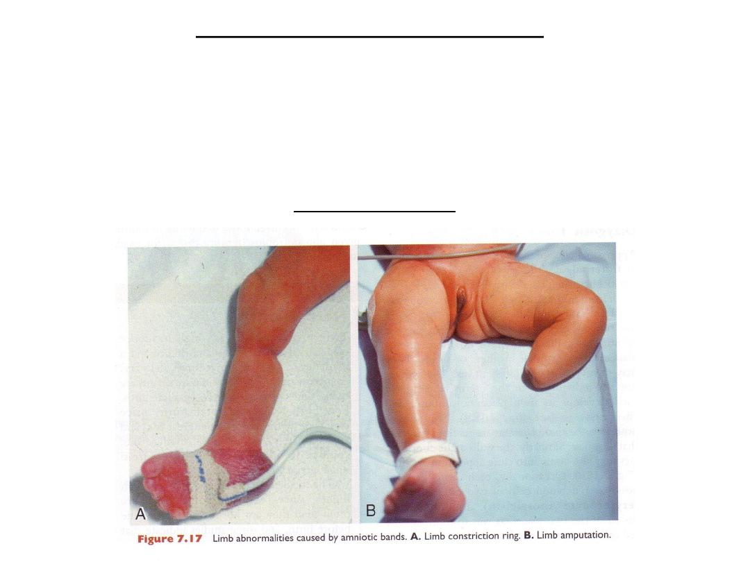
Amniotic bands

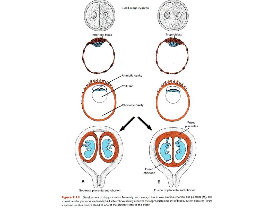
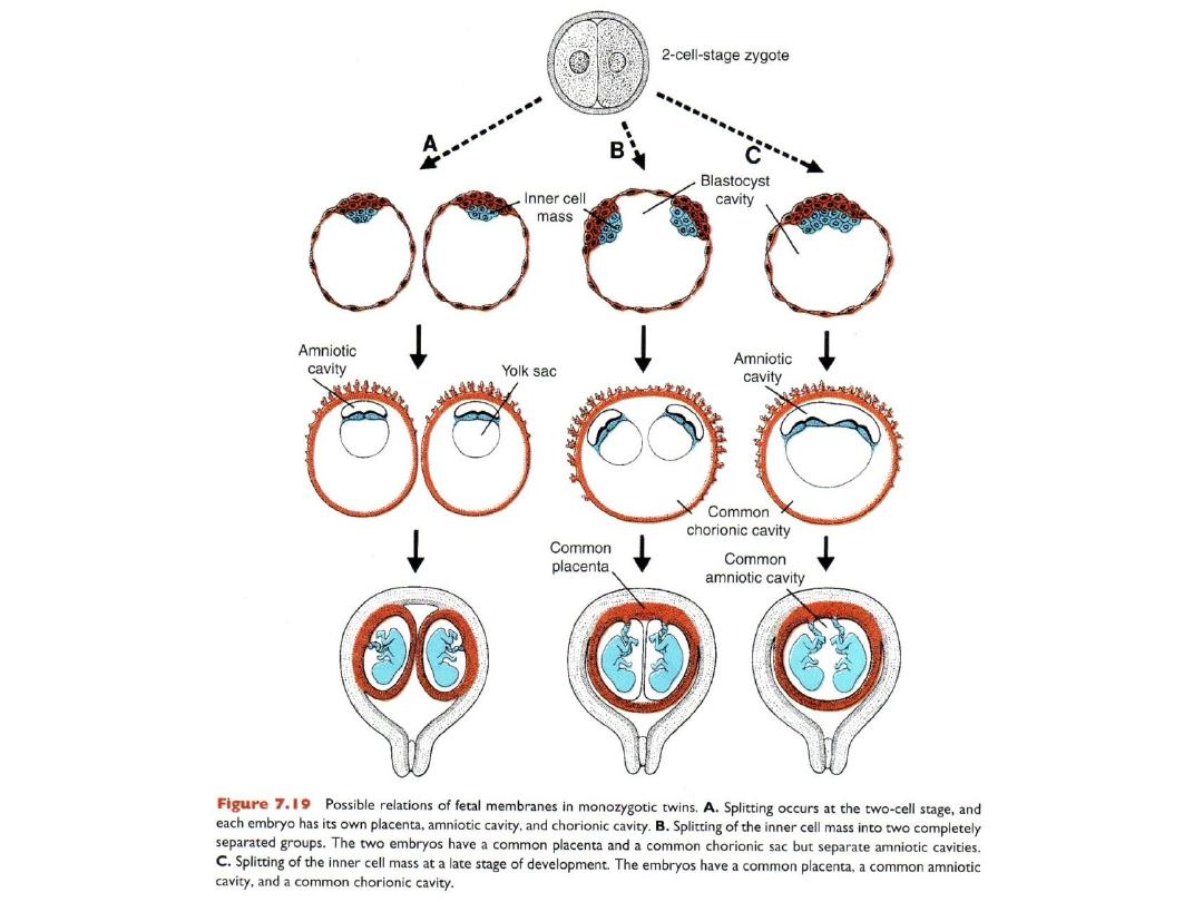
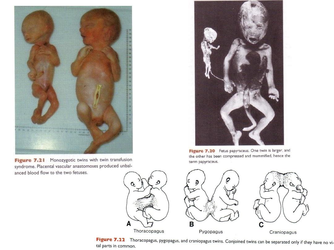
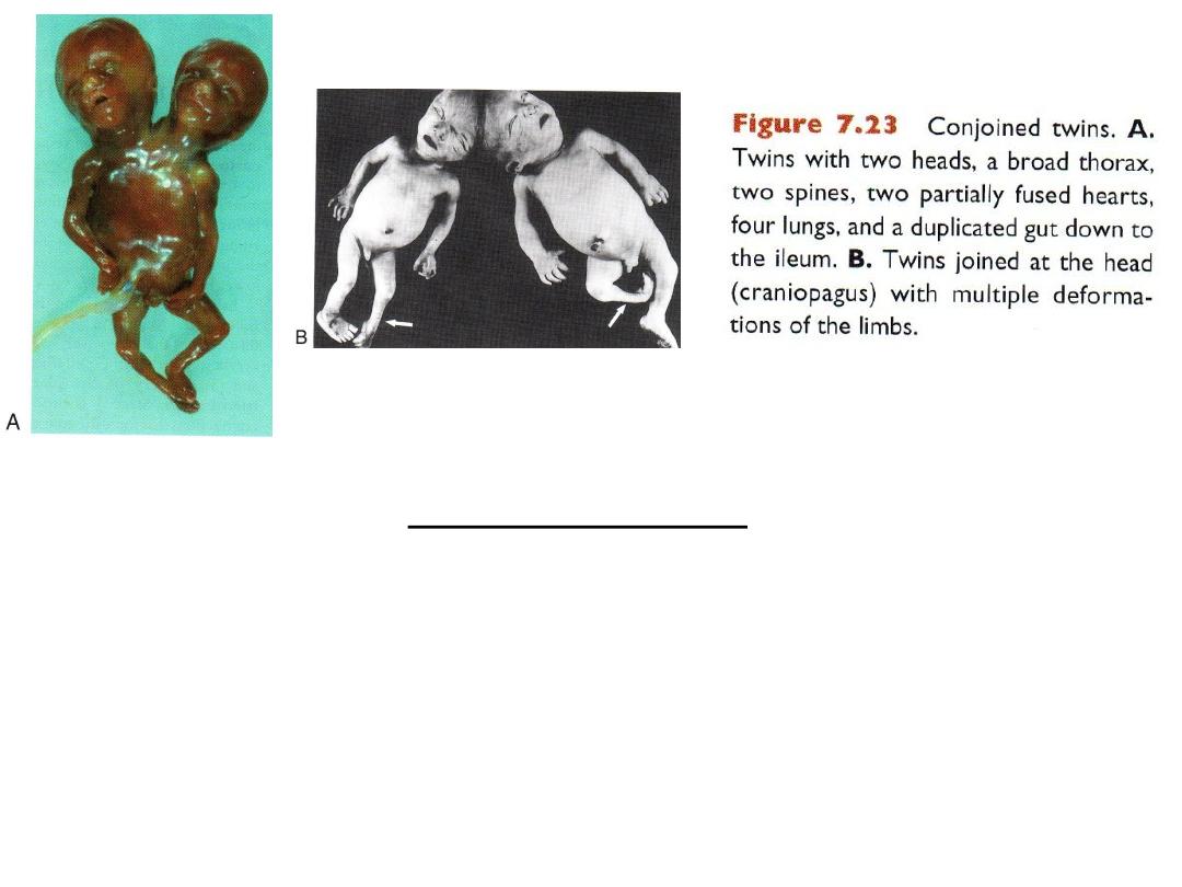
Fetal membranes & twins
Parturition (birth)
• STAGES:
– Effacement
– Delivery of fetus
– Delivery of placenta and fetal membranes