
Basic Anatomy
and passes downward behind the adductor brevis and in
pierces the obturator externus
posterior division
The
the thigh.
rial plexus and supplies the skin on the medial side of
artery. It contributes a variable branch to the subsarto
terminates as a small nerve that supplies the femoral
pectineus. It gives articular branches to the hip joint and
tor brevis, and adductor longus, and occasionally to the
10.30). It gives muscular branches to the gracilis, adduc
the pectineus and adductor longus (Figs. 10.27 and
obturator externus and the adductor brevis and behind
passes downward in front of the
anterior division
The
Branches
anterior and posterior divisions (Fig. 10.27).
obturator foramen (see Fig. 6.12), where it divides into
the lateral wall of the pelvis to reach the upper part of the
cle within the abdomen (see page 222). It runs forward on
and 4) and emerges on the medial border of the psoas mus
The obturator nerve arises from the lumbar plexus (L2, 3,
Compartment of the Thigh
Nerve Supply of the Medial Fascial
vein.
the branches of the artery. It drains into the internal iliac
The obturator vein receives tributaries that correspond to
Obturator Vein
branches and an articular branch to the hip joint.
surface of the obturator membrane. It gives off muscular
eral branches, which pass around the margin of the outer
compartment of the thigh, it divides into medial and lat
tor foramen) (Fig. 10.27). On entering the medial fascial
the obturator canal (i.e., the upper part of the obtura
the pelvis and accompanies the obturator nerve through
465
-
-
Obturator Nerve
-
■
■
-
-
■
■
front of the adductor magnus (Fig. 10.27). It terminates
by descending through the opening in the
or
adduct
tor brevis.
of the adductor magnus, and occasionally to the adduc
branches to the obturator externus, to the adductor part
magnus to supply the knee joint. It gives muscular
-
group of muscles and permits slow recovery of the muscles
crushed. This operation overcomes the spasm of the adductor
severe cases, the posterior division of the obturator nerve is
the anterior division of the obturator nerve. In addition, in some
form a tenotomy of the adductor longus tendon and to divide
In patients with cerebral palsy who have marked spasticity of
Adductor Muscles and Cerebral Palsy
the adductor group of muscles, it is common practice to per-
supplied by the posterior division of the obturator nerve.
C L I N I C A L N O T E S
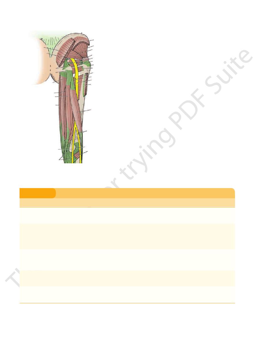
The Back of the Thigh
in Figure 10.31 and are described in Table 10.4.
The muscles of the posterior fascial compartment are seen
Sciatic nerve
Nerve supply:
Branches of the profunda femoris artery
Blood supply:
string muscles)
nosus, and a small part of the adductor magnus (ham
Biceps femoris, semitendinosus, semimembra
Muscles:
group of superficial inguinal lymph nodes (Fig. 10.4).
of the thigh drains upward and forward into the vertical
Lymph from the skin and superficial fascia on the back
Lymph Vessels
vein in the popliteal fossa.
lower part of the back of the thigh join the small saphenous
saphenous vein (Fig. 10.19). Superficial veins from the
aspects of the thigh and ultimately drain into the great
Many small veins curve around the medial and lateral
Superficial Veins
(Fig. 10.1).
skin on the back of the thigh and the upper part of the leg
supplies the skin. It gives off numerous branches to the
and in the popliteal fossa it pierces the deep fascia and
muscle (Fig. 10.1). It descends on the back of the thigh,
from beneath the lower border of the gluteus maximus
the sacral plexus, leaves the gluteal region by emerging
a branch of
posterior cutaneous nerve of the thigh,
The
Cutaneous Nerves
Skin
Contents of the Posterior Fascial
Compartment of the Thigh
■
■
-
-
■
■
■
■
L2 L3 L4
obturator nerve
abdomen
lumbar plexus
pelvis
peritoneum on lateral
wall of pelvis
anterior division
posterior division
adductor
region
of thigh
hip joint
pectineus ?
adductor longus
adductor brevis
gracilis
adductor magnus
(adductor portion)
adductor brevis
knee joint
popliteal artery
femoral artery
subsartorial plexus
with medial cutaneous
nerve of thigh and branch
of saphenous nerve
FIGURE 10.30
Summary of the main branches of the obtura
tor nerve.
-
466
CHAPTER 10
The Lower Limb
gluteus maximus
ischial spine
sacrotuberous ligament
ischial tuberosity
adductor magnus
(hamstring part)
semimembranosus
semitendinosus
gracilis
tibial nerve
semimembranosus
popliteus
oblique popliteal ligament
common peroneal nerve
biceps femoris
(long head)
gluteus maximus
sciatic nerve
nerve to hamstrings
adductor magnus
quadratus femoris
greater trochanter
gemellus inferior
obturator internus
gemellus superior
piriformis
gluteus minimus
gluteus medius
iliac crest
FIGURE 10.31
Structures in the posterior aspect of the right
thigh.
Muscles of the Posterior Fascial Compartment of the Thigh
T A B L E 1 0 . 4
thigh, it ends by dividing into the tibial and common
of the adductor magnus muscle. In the lower third of the
semimembranosus muscles. It lies on the posterior aspect
riorly by the adjacent margins of the biceps femoris and
midline of the thigh (Fig. 10.31). It is overlapped poste
S1, 2, and 3), leaves the gluteal region as it descends in the
The sciatic nerve, a branch of the sacral plexus (L4 and 5;
Sciatic Nerve
of the Thigh
Nerve Supply of the Posterior Compartment
part of the blood from the compartment.
(Fig. 10.27). The profunda femoris vein drains the greater
artery provide a rich blood supply to this compartment
The four perforating branches of the profunda femoris
of the Thigh
Blood Supply of the Posterior Compartment
oblique popliteal ligament.
on the back of the knee joint; the expansion is called the
sion upward and laterally, which reinforces the capsule
The semimembranosus insertion sends a fibrous expan
nerve.
sciatic nerve and the adductor part from the obturator
receives its nerve supply from the tibial portion of the
The hamstring part of the adductor magnus muscle
and the short head from the common peroneal portion.
the sciatic nerve, the long head from the tibial portion,
The biceps femoris muscle receives its nerve supply from
Note the following:
Tibial portion of
Tibial portion of
Tibial portion of
Muscle
Origin
Insertion
Nerve Supply
Nerve Root
a
Action
Biceps femoris
Long head: ischial
tuberosity
Head of fibula
Long head: tibial
portion of
sciatic nerve
L5; S1, 2
Flexes and laterally rotates leg
at knee joint; long head also
extends thigh at hip joint
Short head: linea
aspera, lateral
supracondylar
ridge of shaft of
femur
Short head:
common
peroneal portion
of sciatic nerve
Semitendinosus
Ischial tuberosity
Upper part
of medial
surface of
shaft of tibia
sciatic nerve
L5; S1, 2
Flexes and medially rotates leg
at knee joint; extends thigh at
hip joint
Semimembranosus
Ischial tuberosity
Medial condyle
of tibia
sciatic nerve
L5; S1, 2
Flexes and medially rotates leg
at knee joint; extends thigh at
hip joint
Adductor magnus
(hamstring
portion)
Ischial tuberosity
Adductor
tubercle of
femur
sciatic nerve
L2, 3, 4
Extends thigh at hip joint
a
The predominant nerve root supply is indicated by boldface type.
■
■
■
■
■
■
-
-
Basic Anatomy
467
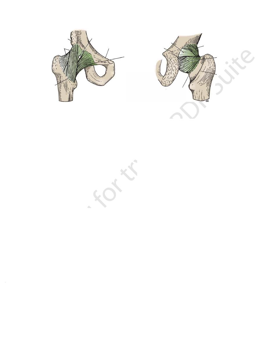
peroneal nerves (Figs. 10.29 and 10.31). Occasionally, the
The synovial membrane lines the capsule and is
membrane (Fig. 10.18).
notch. It lies within the joint and is ensheathed by synovial
the transverse ligament and the margins of the acetabular
on the head of the femur (fovea capitis) and by its base to
angular (Fig. 10.18). It is attached by its apex to the pit
is flat and tri
ligament of the head of the femur
The
through which the blood vessels and nerves enter the joint.
10.18). The ligament converts the notch into a tunnel
acetabular labrum as it bridges the acetabular notch (Fig.
is formed by the
transverse acetabular ligament
The
limits extension.
and are attached to the greater trochanter. This ligament
margin (Fig. 10.32). The fibers pass upward and laterally
attached to the body of the ischium near the acetabular
is spiral shaped and is
ischiofemoral ligament
The
extension and abduction.
part of the intertrochanteric line. This ligament limits
of the pubis, and the apex is attached below to the lower
The base of the ligament is attached to the superior ramus
is triangular (Fig. 10.32).
pubofemoral ligament
The
extension during standing.
teric line of the femur. This strong ligament prevents over
attached to the upper and lower parts of the intertrochan
inferior iliac spine above; below, the two limbs of the Y are
ligament (Fig. 10.32). Its base is attached to the anterior
is a strong, inverted Y-shaped
iliofemoral ligament
The
These blood vessels supply the head and neck of the femur.
retinacula.
reflected upward along the neck as bands called
front, some of its fibers, accompanied by blood vessels, are
behind. At its attachment to the intertrochanteric line in
halfway along the posterior aspect of the neck of the bone
to the intertrochanteric line of the femur in front and
ular labrum medially (Fig. 10.18). Laterally, it is attached
The capsule encloses the joint and is attached to the acetab
The hip joint is a synovial ball-and-socket joint.
Type
The articular surfaces are covered with hyaline cartilage.
(Fig. 10.18).
transverse acetabular ligament
notch and is here called the
The labrum bridges across the acetabular
tabular labrum.
ace
by the presence of a fibrocartilaginous rim called the
The cavity of the acetabulum is deepened
acetabular notch.
lum is horseshoe shaped and is deficient inferiorly at the
hip bone (Fig. 10.18). The articular surface of the acetabu
head of the femur and the cup-shaped acetabulum of the
The hip joint is the articulation between the hemispherical
run medially to supply the muscles (Figs. 10.29 and 10.31).
arise from the tibial component of the sciatic nerve and
hamstring part of the adductor magnus. These branches
ris, the semitendinosus, the semimembranosus, and the
to the long head of the biceps femo
Muscular branches
course is described on page 479.
fossa on the lateral side of the tibial nerve. Its further
sciatic nerve (Figs. 10.29 and 10.31), enters the popliteal
a terminal branch of the
common peroneal nerve,
The
Its further course is described on page 479.
(Figs. 10.17, 10.29, and 10.31), enters the popliteal fossa.
a terminal branch of the sciatic nerve
tibial nerve,
The
Branches
even inside the pelvis.
level—in the upper part of the thigh, the gluteal region, or
sciatic nerve divides into its two terminal parts at a higher
■
■
■
■
■
■
-
Hip Joint
Articulation
-
-
Capsule
-
Ligaments
-
-
-
Synovial Membrane
attached to
the articular surfaces (Fig. 10.18).
the margins of
the joint capsule. It ensheathes the ligament of the head of
It covers the portion of the neck of the femur that lies within
anterior inferior
iliac spine
opening for bursa
superior ramus of pubis
pubofemoral
ligament
intertrochanteric
line
iliofemoral ligament
capsule
A
ischium
iliofemoral ligament
ischiofemoral
ligament
intertrochanteric
crest
area of loose attachment
of capsule
B
FIGURE 10.32
Anterior aspect (A) and posterior aspect (B) of the right hip joint.