508
CHAPTER 10
third, as in the hand.
take place from the midline of the second digit and not the
toes, performed by the interossei muscles, are minimal and
The movements of abduction and adduction of the
verse ligaments connect the joints of the five toes.
resemble those of the hand (see page 412). The deep trans
The metatarsophalangeal and interphalangeal joints closely
sal joint of the big toe has a separate joint cavity.
sal, plantar, and interosseous ligaments. The tarsometatar
joints of the plane variety. The bones are connected by dor
The tarsometatarsal and intermetatarsal joints are synovial
Tarsometatarsal and Intermetatarsal
ligaments.
bones are connected by dorsal, plantar, and interosseous
continuous with that of the cuneonavicular joint. The
vial joints of the plane variety. Their joint cavities are
The intercuneiform and cuneocuboid joints are syno
Intercuneiform and Cuneocuboid Joints
ous ligaments.
the two bones connected by dorsal, plantar, and interosse
The cuboideonavicular joint is usually a fibrous joint, with
Cuboideonavicular Joint
The Lower Limb
-
-
Joints
-
-
Metatarsophalangeal and
Interphalangeal Joints
-
Later, osteoarthritic changes occur in the metatarsophalan
which is a lateral deviation of the great toe
Metatarsophalangeal Joint of the Big Toe
Hallux valgus,
at the metatarsophalangeal joint, is a common condition. Its
incidence is greater in women than in men and is associated
with badly fitting shoes. It is often accompanied by the pres-
ence of a short 1st metatarsal bone. Once the deformity is
established, it is progressively worsened by the pull of the
flexor hallucis longus and extensor hallucis longus muscles.
-
geal joint, which then becomes stiff and painful; the condition
is then known as hallux rigidus.
C L I N I C A L N O T E S
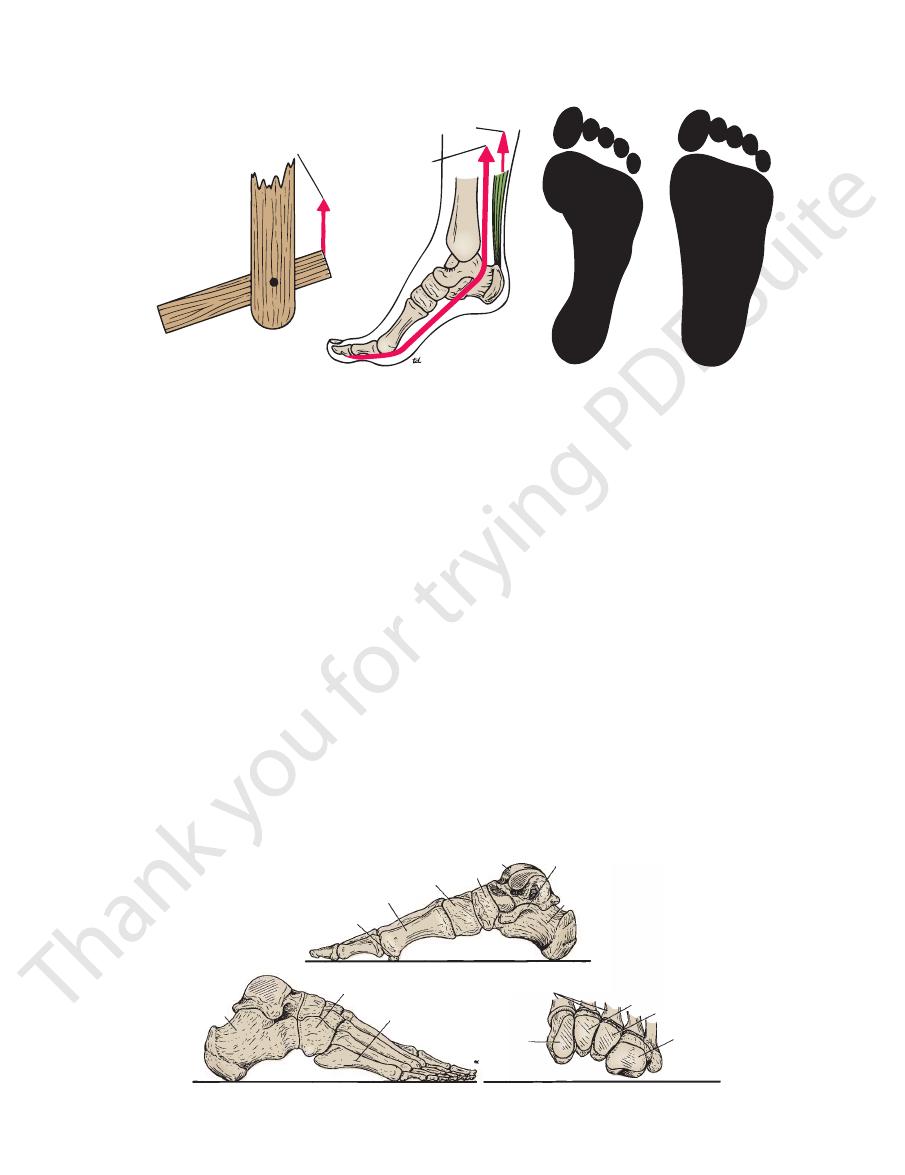
The Foot as a Functional Unit
assist the forward propulsive action of the gastrocnemius
and toes (i.e., the takeoff point of the foot) and greatly
exert their action on the bones of the forepart of the foot
long flexor muscles and the small muscles of the foot can
able and can adapt itself to uneven surfaces. Moreover, the
the lever is segmented with multiple joints, the foot is pli
activities of the gastrocnemius and soleus muscles. Because
forward propulsive action would depend entirely on the
the foot could not adapt itself to uneven surfaces, and the
pulsion (Fig. 10.66). However, with such an arrangement,
body weight and serve well as a rigid lever for forward pro
bone instead of a series of small bones, it could sustain the
walking and running. If the foot possessed a single strong
weight and to serve as a lever to propel the body forward in
The foot has two important functions: to support the body
The Foot as a Weight Bearer
and a Lever
-
-
and soleus muscles (Fig. 10.66).
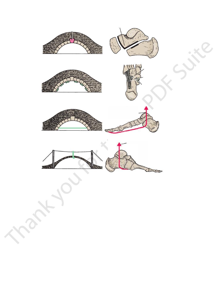
the following engineering methods used for its support
Examination of the design of any stone bridge reveals
Mechanisms of Arch Support
bones (Fig. 10.67).
atarsal bones and the cuboid and the three cuneiform
This consists of the bases of the met
Transverse arch:
(Fig. 10.67).
neum, the cuboid, and the 4th and 5th metatarsal bones
This consists of the calca
Lateral longitudinal arch:
bones, and the first three metatarsal bones (Fig. 10.63).
neum, the talus, the navicular bone, the three cuneiform
This consists of the calca
Medial longitudinal arch:
graph of the foot shows the bones that form the arches.
An examination of an articulated foot or a lateral radio
The Bones of the Arches
metatarsals.
of the first metatarsal and the heads of the remaining four
in front, namely, the two sesamoid bones under the head
the heel behind and six points of contact with the ground
body weight on standing is distributed through a foot via
From this description, it can be understood that the
the two feet are placed together, a complete dome is formed.
likened to a half-dome, so that when the medial borders of
its summit on the foot’s medial border. The foot has been
an arch, with its base on the lateral border of the foot and
the cuboid and cuneiform bones. This is, in fact, only half
transverse arch involves the bases of the five metatarsals and
the presence of the low-lying lateral longitudinal arch. The
5th metatarsal head and least between these areas because of
the lateral margin of the foot is greatest at the heel and the
longitudinal arch. The pressure exerted on the ground by
arched above the ground because of the important medial
gin of the foot, from the heel to the 1st metatarsal head, is
are in contact with the ground (Fig. 10.67). The medial mar
the metatarsal heads, and the pads of the distal phalanges
that the heel, the lateral margin of the foot, the pad under
made with the person in the standing position, one can see
On examination of the imprint of a wet foot on the floor
a large amount of subcutaneous fat on the sole of the foot.
child, the foot appears to be flat because of the presence of
(Fig. 10.67). In the young
transverse arches
and
medial longitudinal, lateral longi
are present at birth: the
in the form of an arch. The foot has three such arches, which
A segmented structure can hold up weight only if it is built
The Arches of the Foot
-
tudinal,
-
-
■
■
-
■
■
-
■
■
-
(Fig. 10.68):
with the thin edge of the wedge lying inferiorly. This
porting the arch is to make the stones wedge shaped,
The most effective way of sup
The shape of the stones:
■
■
-
Basic Anatomy
The cuboid is the keystone.
of the calcaneum and the proximal end of the cuboid.
Minimal shaping of the distal end
Shape of the bones:
Maintenance of the Lateral Longitudinal Arch
and posterior and the medial ligament of the ankle joint.
are the tibialis anterior
Suspending the arch from above
flexor hallucis brevis (Fig. 10.68).
the medial part of the flexor digitorum longus, and the
brevis, the abductor hallucis, the flexor hallucis longus,
aponeurosis, the medial part of the flexor digitorum
are the plantar
Tying the ends of the arch together
terior muscle play an important role in this respect.
tendinous extensions of the insertion of the tibialis pos
the plantar calcaneonavicular ligament (Fig. 10.68). The
the dorsal ligaments. The most important ligament is
plantar ligaments, which are larger and stronger than
by the
The inferior edges of the bones are tied together
the keystone in the center of the arch (Fig. 10.68).
receives the navicular. The rounded head of the talus is
ity of the proximal surface of the medial cuneiform bone
receives the rounded head of the talus; the slight concav
talus; the concave proximal surface of the navicular bone
The sustentaculum tali holds up the
Shape of the bones:
Maintenance of the Medial Longitudinal Arch
ods used to support the arches of the feet (Fig. 10.68).
Using the bridge analogy, one can now examine the meth
a cable above the level of the bridge.
depends on multiple supports suspending the arch from
Here, the maintenance of the arch
A suspension bridge:
ration of the pillars and consequent sagging of the arch.
tie beam connecting the ends effectively prevents sepa
is large and the foundations at either end are insecure, a
When the span of the bridge
The use of the tie beams:
bearing.
edges of the stones to separate when the arch is weight
method effectively counteracts the tendency of the lower
ing their lower edges together with metal staples. This
is accomplished by interlocking the stones or bind
This
The inferior edges of the stones are tied together:
the center of the arch and is referred to as the “keystone.”
applies particularly to the important stone that occupies
509
■
■
-
■
■
-
■
■
-
■
■
-
■
■
-
■
■
■
■
■
■
gastrocnemius, soleus,
and plantaris
gastrocnemius, soleus,
and plantaris
flexor hallucis
longus and
flexor digitorum
longus
A segmented lever
A simple lever
A
B
print of normal foot
print of flat foot
FIGURE 10.66
The foot as a simple lever
shown.
. Floor prints of a normal foot and a flat foot are also
and as a segmented lever
(A)
(B)
sesamoid bone
first metatarsal
medial cuneiform
navicular
talus
sustentaculum tali
calcaneum
calcaneum
fifth metatarsal
lateral longitudinal arch
bases of metatarsal bones
intermediate cuneiform
lateral cuneiform
cuboid
medial cuneiform
transverse arch
cuboid
medial longitudinal arch
FIGURE 10.67
Bones forming the medial longitudinal, lateral longitudinal, and transverse arches of the right foot.
510
CHAPTER 10
training to develop their muscle tone.
sustain their arches provided that they receive adequate
Athletes, route-marching soldiers, and nurses are able to
ligaments of the feet and results in fallen arches or flat feet.
son is overweight, places excessive strain on the bones and
Standing immobile for long periods, especially if the per
ing walking and running, all these muscles become active.
arches. They are commonly totally inactive. However, dur
play no important role in the normal static support of the
the peroneus longus, and the small muscles of the foot
onstrated electromyographically that the tibialis anterior,
factors is the most important? Basmajian and Stecko dem
bones, strong ligaments, and muscle tone. Which of these
The arches of the feet are maintained by the shape of the
gus tendon and the peroneus brevis.
are the peroneus lon
Suspending the arch from above
longus tendon.
is the peroneus
Tying the ends of the arch together
this respect.
of the adductor hallucis are particularly important in
of the foot; the dorsal interossei and the transverse head
and the origins of the plantar muscles from the forepart
deep transverse ligaments, the strong plantar ligaments,
by the
The inferior edges of the bones are tied together
(Fig. 10.67).
cuneiform bones and the bases of the metatarsal bones
The marked wedge shaping of the
Shape of the bones:
Maintenance of the Transverse Arch
gus and the brevis (Fig. 10.68).
are the peroneus lon
Suspending the arch from above
part of the flexor digitorum longus and brevis.
aponeurosis, the abductor digiti minimi, and the lateral
are the plantar
Tying the ends of the arch together
short muscles from the forepart of the foot (Fig. 10.68).
long and short plantar ligaments and the origins of the
by the
The inferior edges of the bones are tied together
The Lower Limb
■
■
■
■
■
■
-
■
■
■
■
■
■
■
■
-
-
-
-
keystone
keystone
shape of stones
shape of bones
staples
short plantar ligament
long plantar ligament
calcaneonavicular ligament
strong plantar ligaments
tendon of flexor
hallucis longus
tie beam
peroneus longus
suspension bridge
tendon of flexor
hallucis longus
peroneus longus
FIGURE 10.68
Different methods by which the arches of the foot may be supported.

Basic Anatomy
511
Clinical Problems Associated with the Arches of the Foot
nal arch is unduly high. Most cases are caused by muscle imbal
(clawfoot) is a condition in which the medial longitudi
Pes cavus
tendons are also permanently stretched. The causes of flat foot
vicular, and medial ligaments of the ankle joint become perma
the deformity has existed for some time, the plantar, calcaneona
muscular support gives way, the ligaments are stretched, and
periods (waitress or nurse), by overweight, or by illness, the
(a long-route march by an army recruit), by standing for long
port. When the muscles are fatigued by excessive exercise
foot the tone of muscles is an important factor in arch sup
supporting the arches. It has been shown that in the active
the foot, and the tone of muscles all play an important role in
strong ligaments, especially those on the plantar surface of
clinically the most important. The shape of the bones, the
Of the three arches, the medial longitudinal is the largest and
-
pain is produced.
Pes planus (flat foot) is a condition in which the medial longi-
tudinal arch is depressed or collapsed. As a result, the forefoot
is displaced laterally and everted. The head of the talus is no
longer supported, and the body weight forces it downward and
medially between the calcaneum and the navicular bone. When
-
-
nently stretched, and the bones change shape. The muscles and
are both congenital and acquired.
-
-
ance, in many instances resulting from poliomyelitis.
C L I N I C A L N O T E S
The Propulsive Action of the Foot
up, thereby increasing their efficiency. The body is then
nal arches. The “slack” in the long flexor tendons is taken
shortening the tie beams and heightening the longitudi
joints, and the plantar aponeurosis is pulled on, thus
rises, the toes are extended at the metatarsophalangeal
foot and the heads of the metatarsal bones. As the heel
weight is borne successively on the lateral margin of the
As the body weight is thrown forward, the
Walking
first metatarsal).
(including the two sesamoid bones under the head of the
heel behind and the heads of the metatarsal bones in front
The body weight is distributed via the
Standing Immobile
-
Bursae and Bursitis in the Lower Limb
times develops over the tendo calcaneus in response to badly
excessive friction. For example, a subcutaneous bursa some
sus muscle, may enlarge in patients with osteoarthritis of the
the patella beneath the quadriceps femoris muscle. The bursa,
bursa extends proximally about three fingerbreadths above
synovial fluid accumulate within the joint. The suprapatellar
and they can become distended if excessive amounts of
Two important bursae communicate with the knee joint,
neum (long-distance runner’s ankle).
A variety of bursae are found in the lower limb where skin, ten-
dons, ligaments, or muscles repeatedly rub against bony points
or ridges.
Bursitis, or inflammation of a bursa, can be caused by acute
or chronic trauma, crystal disease, infection, or disease of a
neighboring joint that communicates with the bursa. An inflamed
bursa becomes distended with excessive amounts of fluid. The
following bursae are prone to inflammation: the bursa over the
ischial tuberosity; the greater trochanter bursa; the prepatellar
and superficial infrapatellar bursae; the bursa between the ten-
dons of insertion of the sartorius, gracilis, and semitendinosus
muscles on the medial proximal aspect of the tibia; and the bursa
between the tendo calcaneus and the upper part of the calca-
which is associated with the insertion of the semimembrano-
knee joint.
The anatomic bursae described should not be confused with
adventitious bursae, which develop in response to abnormal and
-
fitting shoes. A bunion is an adventitial bursa located over the
medial side of the head of the 1st metatarsal bone.
C L I N I C A L N O T E S
thrown forward by the actions of the gastrocnemius and
mechanisms described for walking (above).
ground. The forward thrust to the body is provided by the
the forepart of the foot, and the heel does not touch the
When a person runs, the weight is borne on
Running
ing the ankle joint.
action, the long flexor tendons also assist in plantar flex
of the strong action of the flexor digitorum longus. In this
the toes extended so that they do not fold under because
forward. The lumbricals and interossei contract and keep
and short flexors of the foot, providing the final thrust
a lever, and by the toes being strongly flexed by the long
soleus (and plantaris) on the ankle joint, using the foot as
-