18
A plasma membrane enclose every cell :
1- Unicellular organism.
2- Multicellular organism.
It is the edge of life; the boundary that separates the living cell from its nonliving
surrounding (8 nm thick).
Biological membranes are selectively Permeable
EVOLUTION OF MEMBRANE MODELES:
A- The Davson- Danieli model 1935 was a sandwiched –a phospholipids bilayer
between two protein layers. With later modifications, this model was widely
accepted until 1970.
Not all the membranes look alike in EM, they have different functions, differ in
chemical composition and structure.
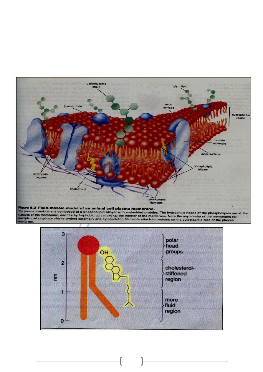
In 1972 , S. Singer and G. Nicolson proposed that membrane proteins are dispersed
and individually inserted into the phospholipids bilayer ,so the membrane is a
mosaic of protein molecules bobbing in a fluid bilayer of phospholipids: hence the
term fluid mosaic model
Fluid ----- not static, not solid sheets of molecules, molecules exchanging places with
neighbors.
Mosaics of structure and function—The P M and the M of the various organelles
have their unique collections of proteins ex. 50 kinds of proteins found in P M of R.
Blood.
Part of membrane is fluid phospholipids - bilayers in which protein molecules are
either partially or wholly embedded. The mosaic distribution (an irregular pattern) of
proteins is supported by E.M. of freeze-fractured membrane.
In a membrane, the hydrophilic heads of the phospholipids molecules face the
intracellular and extra cellular fluids. The hydrophobic tails face each other in the
membrane interior.
Phospholipids is an amphipathic molecule( has both a hydrophilic region &
hydrophobic ):
The other types of lipids in P.m.*Glycolipids the hydrophilic head is a variety of
sugars joined to form a straight or branch chain
19
And *Cholesterol reduces the permeability of the membrane to most biological
molecules; helps to keep the membrane fluid by hindering close packing of pho.
Lipids.
The fluidity of a phospholipids bilayer has the consistency of olive oil. Phospholipid
moves along the plan the membrane quite rapidly, proteins are much larger than
lipids.

21
Proteins:
Transmembrane proteins have hydrophilic region embedded within the have
hydrophilic region embedded within the membrane and hydrophilic regions that
project from both surfaces of the bilayer:
Many P.M. proteins are glycoproteins (have an attached carbohydrate chains).
Other proteins, on the cytoplasmic side or the other surface side of the membrane
*Peripheral membrane proteins.
The plasma membrane is asymmetrical; the two halves are not identical.
The carbohydrate chains of the glycolipids and glycoproteins form a carbohydrate
coat that envelops the outer surface of the plasma membrane.
The carbohydrate chains of the glycolipids and glycoproteins serve as the
"fingerprints" of the cells .The glycolipids and glycoproteins vary from species to
species , from individual to other of the same species and from cell to cell in the same
individual .
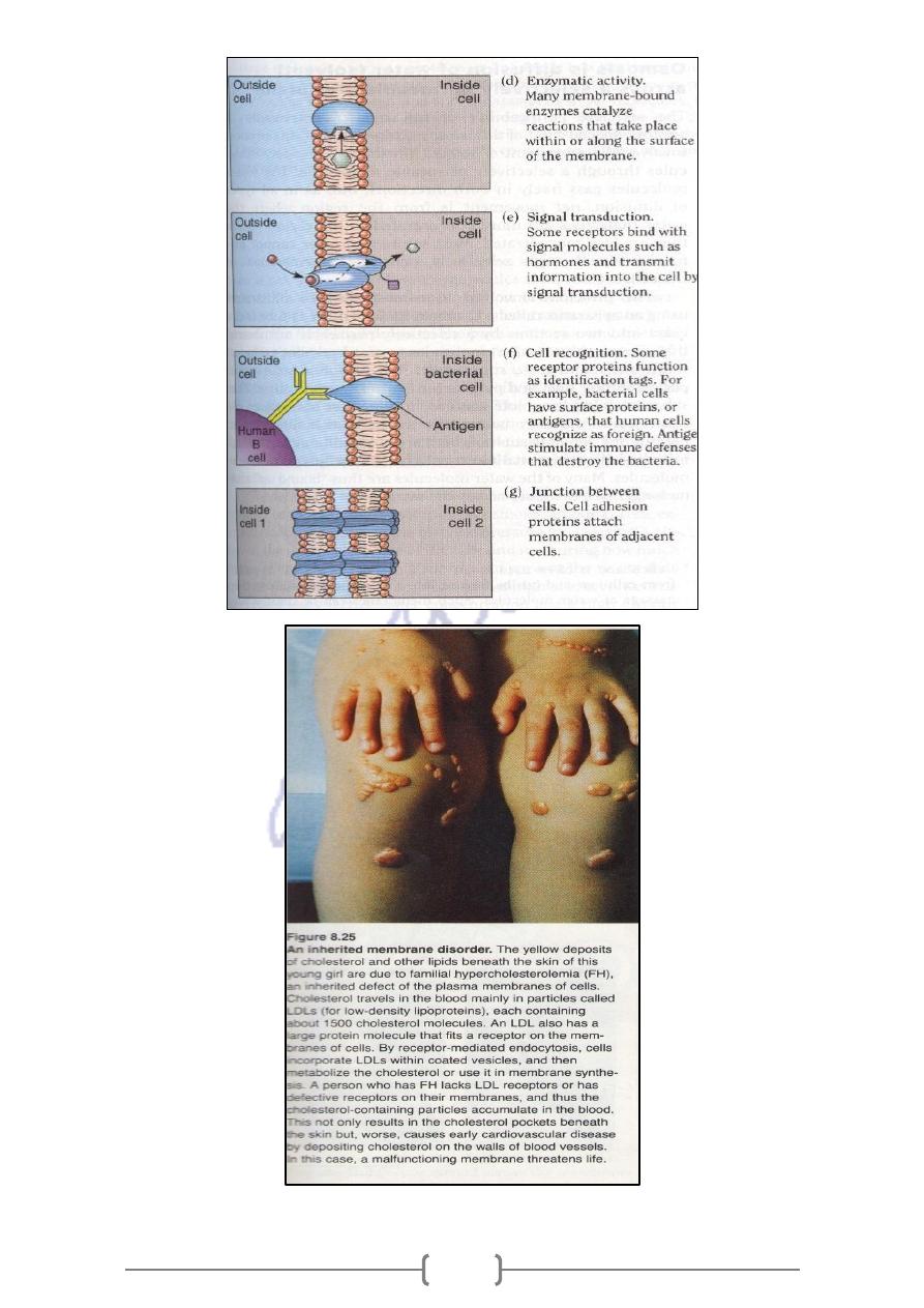
They make cell-cell recognition possible (the ability of a cell to determine if other
cells it encounters are alike or different from itself). Transplanted tissues are often
rejected by the body. This because the immune system is able to recognize that
tissue as foreign tissues. cells don't have the same glycolipids and glycoprotein , so
they are involved in marking the cells as belonging to a particular individual and
tissue.
Thus, the entire outside surface of the cell has a loose carbohydrate coat called the
Glycocalyx .
The important functions of carbohydrate moieties :
1- Many of them have a negative electrical charge that repels other negative
objects .
2- The glycoclyx of some cells attaches to the glycoclyx of the other cells, thus
attaching cells to one other
3- Many of carbohydrates act as receptor substances for binding hormones ex.
Insulin;
4- Some carbohydrate moieties enter into immune reactions .
Table 3.2 Functions of the Glycocalyx
Protection: Cushions the plasma membrane and protects it from physical and
chemical injury.
21
Immunity to Infection: Enables the immune system to recognize and selectively
attack foreign organisms
Defense Against Cancer :Changes in the glycocalyx of cancerous cells enable the
immune system to recognize and destroy them
Transplant Compatibility: Forms the basis for compatibility of blood transfusions,
tissue grafts, and organ transplants
Cell Adhesion :Binds cells together so that tissues do not fall apart
Fertilization: Enables sperm to recognize and bind to eggs
Embryonic Development: Guides embryonic cells to their destinations in the body.
How molecules cross the plasma membrane:
Plasma membrane is Semi permeable (allow some molecule to pass through it) some
molecules (lipid- soluble compound, water and gases) diffuses across the membrane
from the area of higher concentration to the area of lower concentration. No ATP
requires.
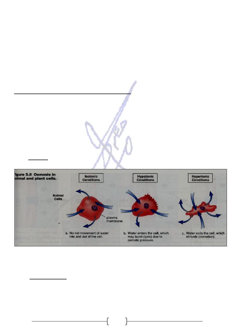
The diffusion of water across differentially permeable membrane is called Osmosis.
Osmosis occurs in living organism. ex. Water is absorbed from the human large
intestine , is retained by the kidneys , and is taken up by blood.
Tonicity: the strength of a solution in relationship to Osmosis.
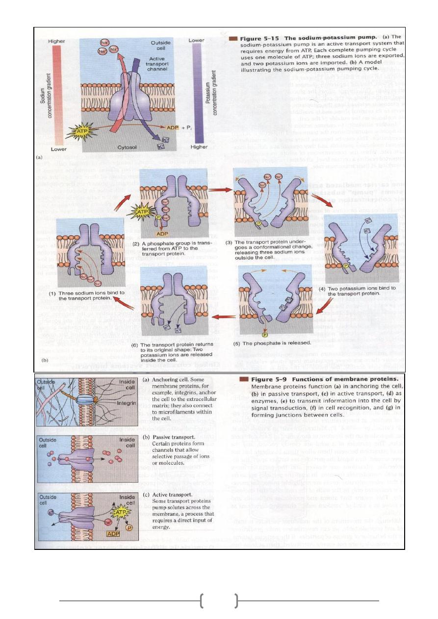
Other molecules are transported across the membrane by carrier proteins Facilitated
transport, a carrier proteins assists the movement of a molecule down its
concentration gradient .No energy is required.
Active transport , a carrier proteins acts as a pump that causes a substance to move
against its concentration gradient .The Na
+
-- K
+
pump carries 3Na
+
to the outside of
the cell and K to the inside of the cell. Energy in the form of ATP molecules is
required for active transport to occur
22
23
24
Functional Systems of the Cell:
What about the transport of molecules such as Polysaccharides or Polynucleotide?
They can enter and exit a membrane by Exocytosis and Endocytosis.
Endocytosis:
A- phagocytosis
B- Pinocytosis
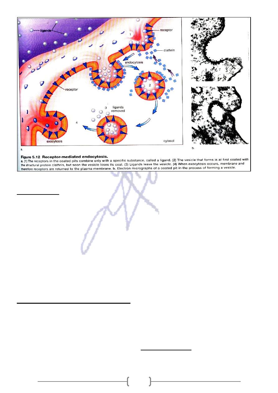
C- Receptor – mediated endocytosis
Pinocytosis is the only means by which most large macromolecules, such as most
protein molecules, can enter cells. In fact, the rate at which pinocytotic vesicles form
is usually enhanced when such macromolecules attach to the cell membrane
Figure
2-11
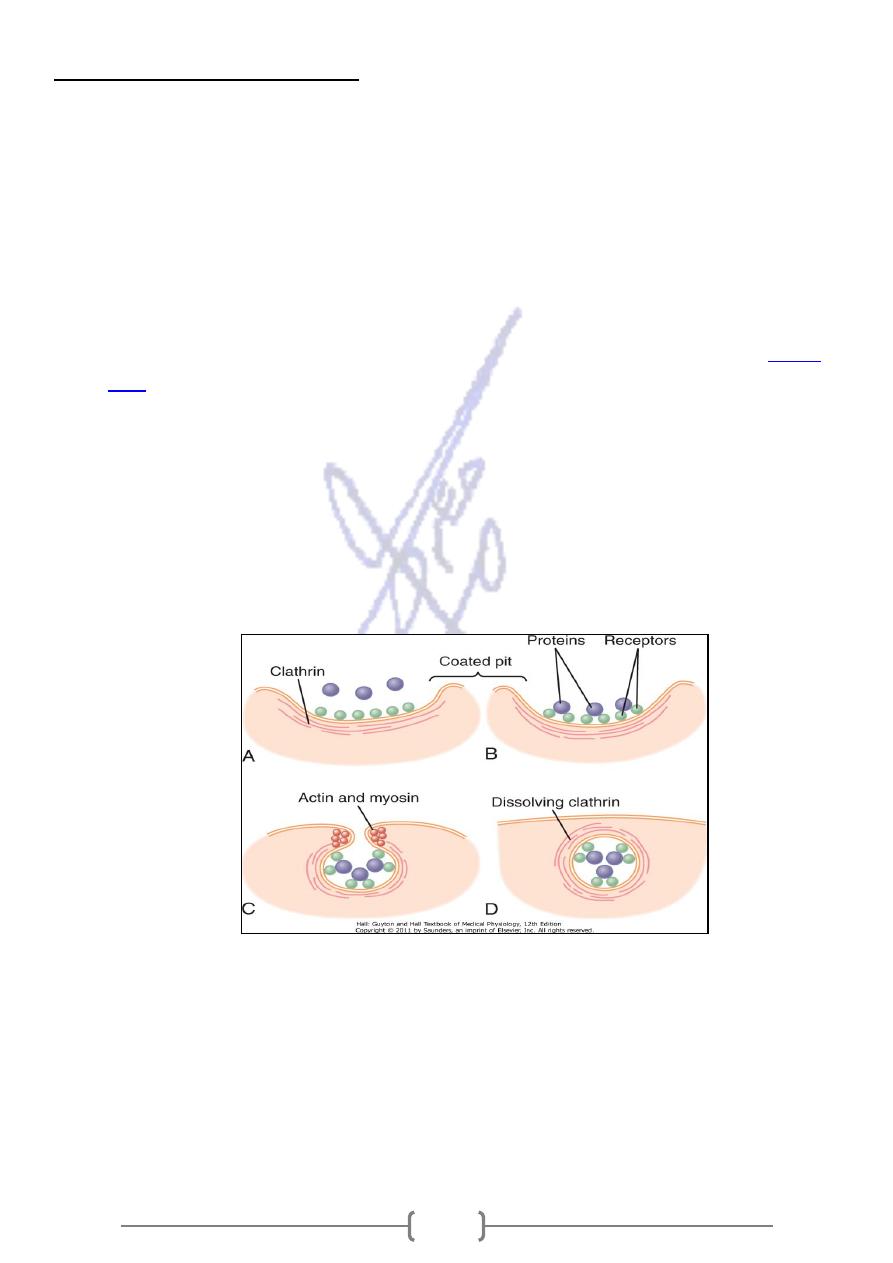
demonstrates the successive steps of pinocytosis, showing three molecules of
protein attaching to the membrane. These molecules usually attach to specialized
protein receptors on the surface of the membrane that are specific for the type of
protein that is to be absorbed. The receptors generally are concentrated in small pits
on the outer surface of the cell membrane, called coated pits. On the inside of the
cell membrane beneath these pits is to be absorbed. On the inside of the cell
membrane beneath these pits is a latticework of fibrillar protein calledclathrin, as
well as other proteins, perhaps including contractile filaments of actin and myosin
The protein molecules have bound with the receptors, the surface properties of the
local membrane change in such a way that the entire pit invaginates inward and the
fibrillar proteins surrounding the invaginating pit cause its borders to close over the
attached proteins, as well as over a small amount of extracellular fluid. Immediately
thereafter, the invaginated portion of the membrane breaks away from the surface
of the cell, forming a pinocytotic vesicle inside the cytoplasm of the cell.
25
Phagocytosis:
Phagocytosis occurs in much the same way as pinocytosis, except that it involves
large particles rather than molecules. Only certain cells have the capability of
phagocytosis, most notably the tissue macrophages and some of the white blood
cells.
Phagocytosis is initiated when a particle such as a bacterium, a dead cell, or tissue
debris binds with receptors on the surface of the phagocyte. In the case of bacteria,
each bacterium is usually already attached to a specific antibody, and it is the
antibody that attaches to the phagocyte receptors,. This intermediation of antibodies
is called opsonization.
Phagocytosis occurs in the following steps:
The cell membrane receptors attach to the surface ligands of the particle.
The edges of the membrane around the points of attachment evaginate outward
within a fraction of a second to surround the entire particle; then, progressively more
and more membrane receptors attach to the particle ligands. All this occurs suddenly
in a zipper-like manner to form a closed phagocytic vesicle.
Actin and other contractile fibrils in the cytoplasm surround the phagocytic vesicle
and contract around its outer edge, pushing the vesicle to the interior.
26
The contractile proteins then pinch the stem of the vesicle so completely that the
vesicle separates from the cell membrane, leaving the vesicle in the cell interior in
the same way that pinocytotic vesicles are formed.
Locomotion of Cells:
THE most important type of movement that occurs in the body is that of the muscle
cells in skeletal, cardiac, and smooth muscle, which constitute almost 50 percent of
the entire body mass. Two other types of movement-ameboidlocomotion and ciliary
movement-occur in other cells.
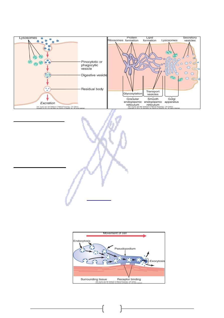
Ameboid Movement
Typically, ameboid locomotion begins with protrusion of a pseudopodium from one
end of the cell. The pseudopodium project far out, away from the cell body, and
partially secures itself in a new tissue area. Then the remainder of the cell is pulled
toward the pseudopodium.
Figure 2-16
demonstrates this process, showing an
elongated cell, the right-hand end of which is a protruding pseudopodium. The
membrane of this end of the cell is continually moving forward, and the membrane
at the left-hand end of the cell is continually following along as the cell moves.
27
Types of Cells That Exhibit Ameboid Locomotion:
The most common cells are the white blood cells when they move out of the blood
into the tissues to form tissue macrophages. Other types of cells, fibroblasts move
into a damaged area to help repair the damage and even the germinal cells of the
skin,, move toward a cut area to repair the opening. Finally, cell locomotion is
especially important in development of the embryo and fetus after fertilization of an
ovum. For instance, embryonic cells often must migrate long distances from their
sites of origin to new areas during development of special structures.
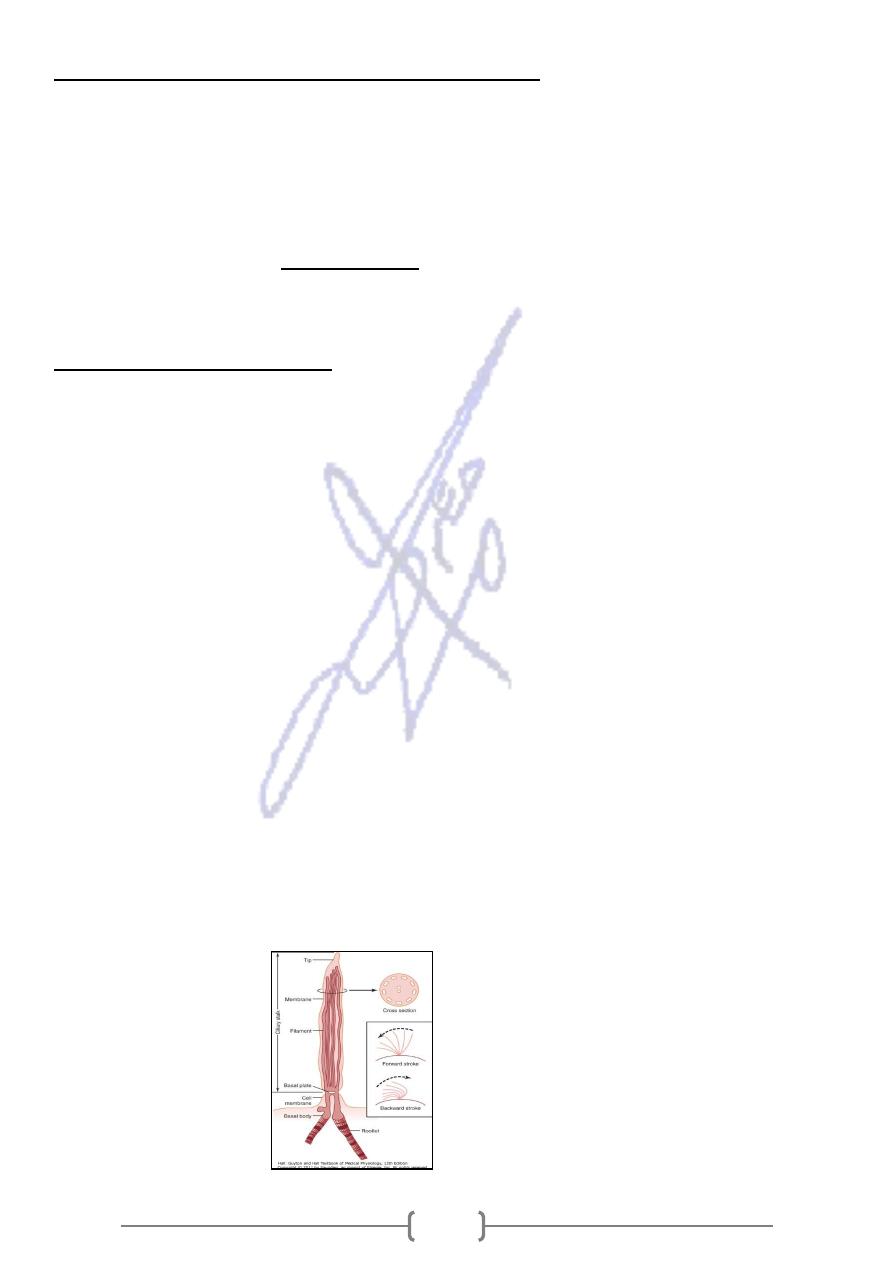
Cilia and Ciliary Movements
A second type of cellular motion, ciliary movement, is a whiplike movement of cilia
on the surfaces of cells. This occurs in only two places in the human body: on the
surfaces of the respiratory airways and on the inside surfaces of the uterine tubes
(fallopian tubes) of the reproductive tract. In the nasal cavity and lower respiratory
airways, the whiplike motion of cilia causes a layer of mucus to move at a rate of
about 1 cm/min toward the pharynx, in this way continually clearing these
passageways of mucus and particles that have become trapped in the mucus. In the
uterine tubes, the cilia cause slow movement of fluid from the ostium of the uterine
tube toward the uterus cavity; this movement of fluid transports the ovum from the
ovary to the uterus.
The cilium moves forward with a sudden, rapid whiplike stroke 10 to 20 times per
second, bending sharply where it projects from the surface of the cell. Then it moves
backward slowly to its initial position. The rapid forward-thrusting, whiplike
movement pushes the fluid lying adjacent to the cell in the direction that the cilium
moves; the slow, dragging movement in the backward direction has almost no effect
on fluid movement. As a result, the fluid is continually propelled in the direction of
the fast-forward stroke. Because most ciliated cells have large numbers of cilia on
their surfaces and because all the cilia are oriented in the same direction, this is an
effective means for moving fluids from one part of the surface to another.
28
ANIMAL CELLS HAVE AN EXTRACELLULLAR MATRIX:
Is a meshwork of insoluble proteins with carbohydrate chains (glycoprotiens).
The Ex. Matrix fills the spaces between animal cells & supports them.
The Ex. Matrix influences the development, migration, shape, & function of the cells
.Collagen & Elastin fibers are the structural component of extra cellular matrix.
Fibroectins & laminins are two adhesive proteins that play a dynamic role in
influencing the behavior of the cells. They form "highways" that direct the migration
of cell during development. Laminins were found to be necessary for the production
of the milk by the mammary gland cell.
Proteoglycan are glycoproteins that are composed of carbohydrate chains
containing amino sugars .Pro. Provide a packing gel that joins the various proteins in
the matrix.
Animal cells have Junction:
Three types of junctions are seen between animal cells.
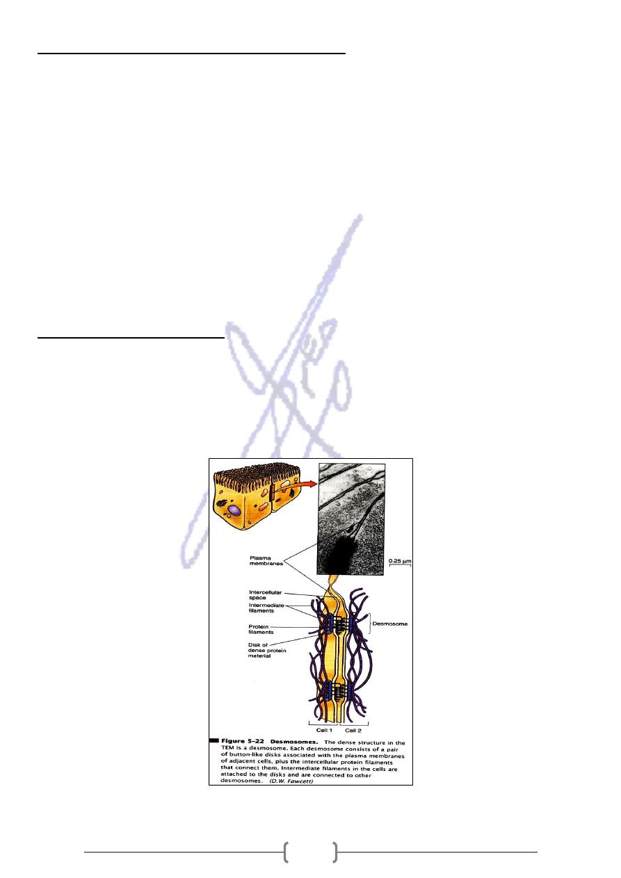
1) Adhesion junctions ( desmosom )
Desmosomes spot weld adjacent animal cells together .It found in heart , stomach &
bladder .
29
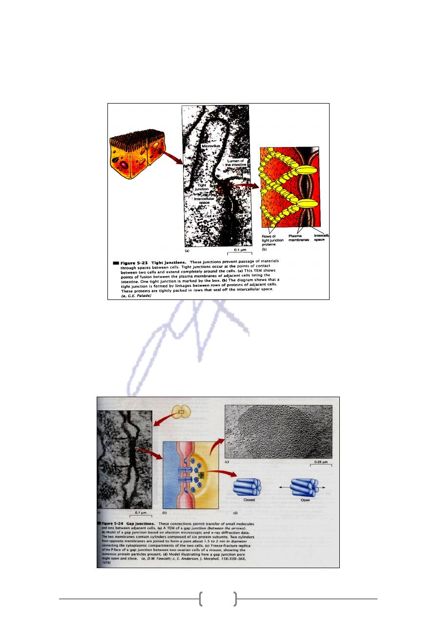
2) Tight Junction
Seal membranes of adjacent animal cells together, preventing substances from
moving through the spaces between the cells; in the intestine the digestive juices
stay out of the body, and the kidneys the urine stay within the kidney tubules.
3) Gap junctions
Are proteins complexes form channels in membranes, allowing communication
between cytoplasm of adjacent animal cells by channel is lined by six plasma
membrane proteins; in heart muscle & smooth muscle, because they permit a flow of
ions that is required for the cells to contract.