44
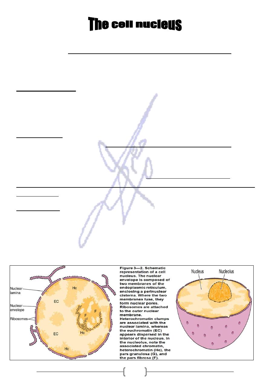
* They are appears as a round or elongated structure, usually in the center of the cell. The
.
matrix
and nuclear
nucleolus,
chromatin,
envelope,
the nuclear
nucleus comprises
* The irregular features plus the capacity to invade neighboring tissues used by
pathologists to estimate the degree of malignancy of a tumor.
Nuclear envelope
* Is a thin line of heterochromatin layer surrounding the nucleus and binds to the
internal surface of the nuclear envelope .It composes of two parallel unit
membranes separated by a narrow ( 40-70 –nm ) space called perinuclear cisterna .
is a protein structure which associated with the internal
Fibrous lamina,
called lamins
polypeptides,
is composed of three
It
envelope.
membrane of nuclear
that form part of the nuclear matrix .The chromatin has a definite organization within
the nucleus.
functions,
The nuclear envelope
.
outer membrane
Polyribosome attached to the
*
chains and segregating them in the perinuclear cistern between its
synthesizing polypeptide
mbranes
me
two
n the inner and outer membranes
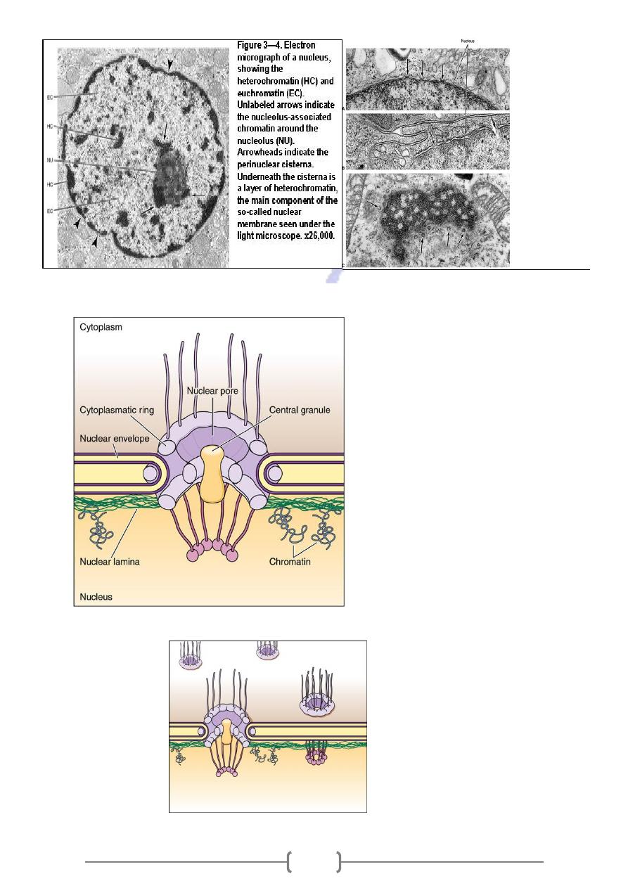
are circular gaps found betwee
Nuclear pores;
fuse. They provide pathways between the nucleus and the cytoplasm and are not
open but bridged an electron – dense membrane that forms a single-layered
diaphragm of protein .All pores are permeable to some macromolecules ( e.g. ,
mRNA, cytoplasmic protein ) .
45
Figure 3
–6. Electron
micrographs of nuclei
showing their envelopes
composed of two
membranes and the
nuclear pores (arrows).
A, B: Transverse
sections; C: a tangential
section. Chromatin,
frequently condensed
below the nuclear
envelope, is not usually
seen in the pore regions.
x80,000.
•
Figure 3
—5. Illustration
to show the structure,
the localization, and the
relationship of the
nuclear lamina with
chromosomes. The
drawing also shows that
the nuclear pore
complex is made of 2
protein rings in an
octagonal organization.
From the cytoplasmic
ring, long filaments
penetrate the cytosol,
and from the intranuclear
ring arise filaments that
constitute a basketlike
structure. The presence
of the central cylindrical
granule in the nuclear
pore is not universally
accepted.
Figure 3
—8. Simplified
representation of 2
nuclear pore
complexes. In this
model, the final nuclear
portion is seen to be a
more continuous
structure, in the shape
of a ring.
46
chromatin
as
appears as coarse granules (
visible in the light microscope,
is
Hetrochromatin
-
1
basophilic clumps of nucleoprotein)
is visible as an organized structure only in E.M.
Euchromatin
-
2
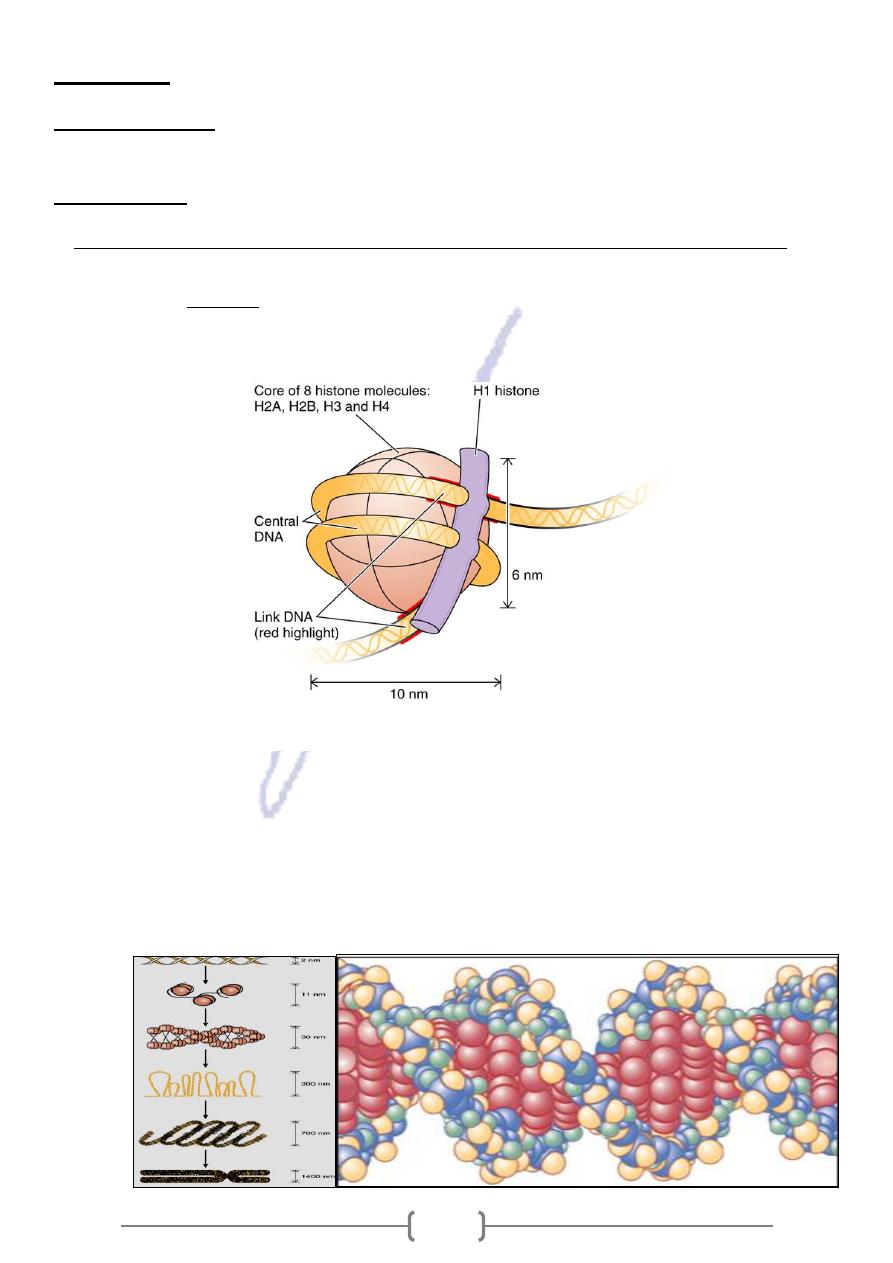
Chromatin is composed of coiled strands of DNA bound to basic proteins (histones)
*
* The basic structural unit of chromatin is the NUCLEOSOME ,which consists of a core of a
: two copies each of histones H2A, H2B , H3 & H4 around which are
histones
four types of
wrapped 166 DNA base pairs
Figure 3
—9. Schematic
representation of a
nucleosome. This structure
consists of a core of 4 types
of histones (2 copies of
each)
–H2A, H2B, H3, and
H4
–and one molecule of H1
or H5 located outside the
DNA filament.
* A further 48-base-pair segment forms a link between adjacent nucleosomes, and
another type of histone (H1 or H5) is bound to this DNA (BEAD –AN –STRING). The
next higher order of organization is The 30-nm fiber (A SOLENOID ),in which
nucleosomes become coiled around an axis , with six nucleosomes per turn, to form
the 30-nm chromatin fiber
47
* During mitosis and meioses there are higher orders of coiling in condensation of
chromatin into chromosomes .Within the chromatin, the precursors of the (mRNA,
rRNA, and tRNA ) are synthesized .
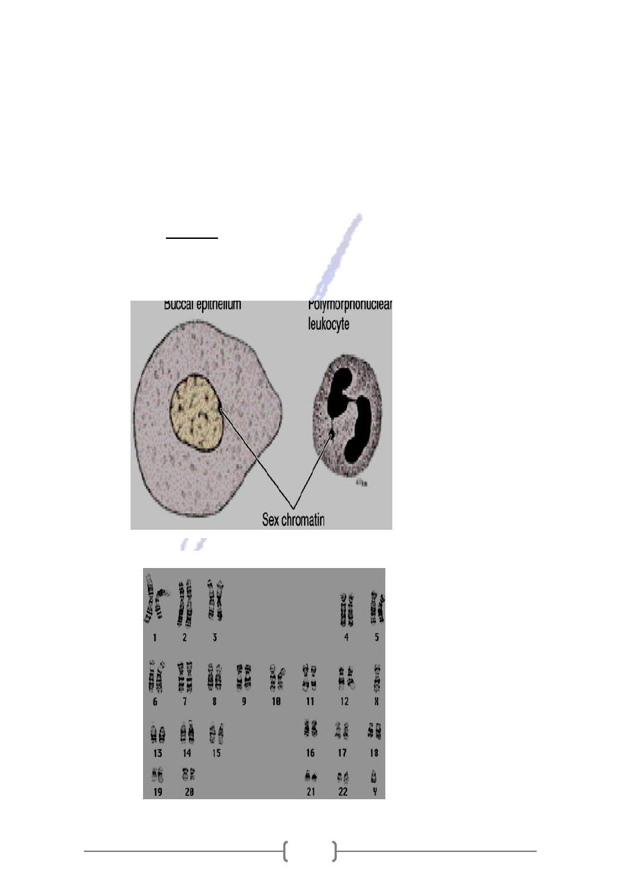
* In general, cells with light nuclei are more active than those with condensed, dark
nuclei. This chromatin clump is the SEX CHROMATIN and is one of the pair of X
chromosomes that visible in female cells during interphase. It remains tightly
coiled and visible and genetically inactive, while the other X chromosome is
uncoiled and not visible
chromosomes appears as a small granule attached to the
cells sex
In human epithelial
*
nuclear envelope. Blood smears are also often used, in which case the sex chromatin
appears as a drumstick-like appendage to the nuclei of the neutrophilic leukocytes
Figure 3
—11.
Morphologic
features of sex
chromatin in human
female oral (buccal)
epithelium and in a
polymorph nuclear
leukocyte. In the
epithelium, sex
chromatin appears
as a small, dense
granule adhering to
the nuclear
envelope. In the
leukocyte, it has a
drumstick shape.
Figure 3
—12.
Human karyotype
preparation made
by means of a
banding technique.
Each chromosome
has a particular
pattern of banding
that facilitates its
identification and
also the relationship
of the banding
pattern to genetic
anomalies. The
chromosomes are
grouped in
numbered pairs
according to their
morphologic
characteristics.
48
NUCLEOLUS
* Is a spherical structure that is rich in rRNA and protein .The nucleolus consists of
three components :
1- nucleolar organizer DNA –sequences of bases that code for rRNA .
2- Pars fibrosa –ribonucleoprotein fibers, which consist of primary transcripts of rRNA
genes.
3- Pars granulosa –consists of 15-20 nm granules (maturing ribosomes). Proteins,
synthesized in the cytoplasm, become associated with rRNAs in the nucleolus.
Heterochromatin is attached to the nucleolus (nucleolus associated chromatin).In nucleolus
they receive proteins and organized into small& large ribosomal subunits, which migrate to
cytoplasm through the nuclear pores
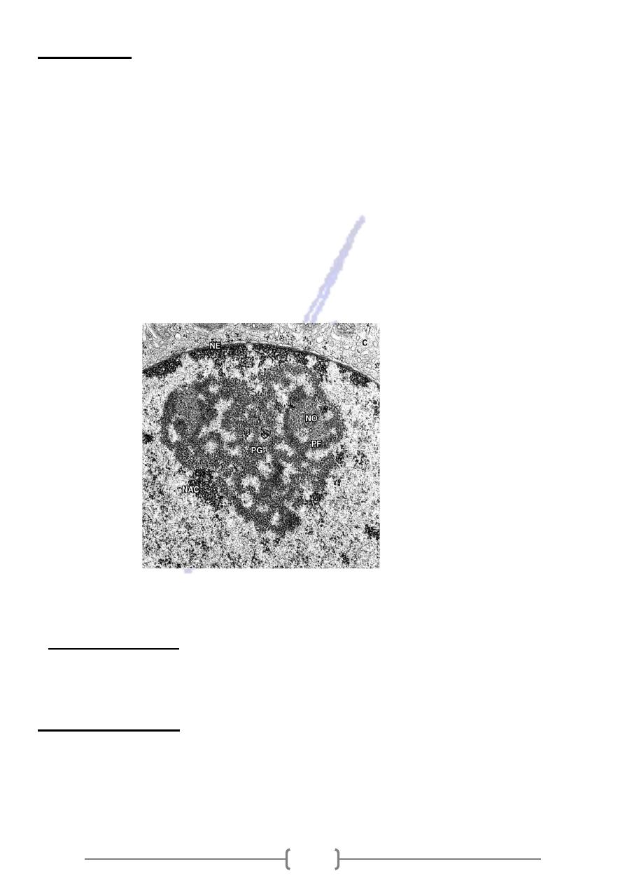
Figure 3
—14. Electron
micrograph of a
nucleolus. The
nucleolar organizer
DNA (NO), pars fibrosa
(PF), pars granulosa
(PG), nucleolus-
associated chromatin
(NAC), nuclear
envelope (NE), and
cytoplasm (C) are
shown.
* The study of sex chromatin has wide applicability to medicine as in
hermaphroditism, pseudohermaphroditism,
and other
abnormalities, azoospermia
in which testicular
Klinefelters syndrome
*
symptoms are associated with the presence of XXYchromosomes in the cell.
MATRIX
NUCLEAR
* Is the component that fills the space between the chromatin and the nucleoli in the
nucleus .It composes of proteins, metabolites, and ions .There is a fibrillar
structure forming the Nucleoskeleton . It probably contributes to the formation of
protein base to which DNA loops are bound.