163
They responsible for 1- providing and maintaining form in the body .
2- Provide a matrix that connects and binds the cells and organs to give support to the body . Is
formed by cells , fibers and ground substance.
The major constituent of connective tisue is Extracellular matrix
A- different combination of protein fibers( collagen ,reticular ,and elastic ) .
B- ground substance . Is hydrophilic ,viscous complex of
1- ( glycosaminoglycans and proteoglycans)
2- glycoproteins ( lamanin and,fibronectin) , which are binding to receptor proteins (integrins)
on the surface on cells and to other matrix Components.
The origin of connective tissue
Mesoderm (embryonic tissue) Mesenchyme tissue Mesodermal cell migrate
from their origin site, surrounding and penetrating developing organs
Cells of connective tissue
Table 5–1. Functions of connective tissue cells.
Cell Type
Representative Product or Activity
Representative Fun.
Fibroblast,
chondroblast,
osteoblast,
odontoblast
Production of fibers and ground substance
Structural
Plasma cell
Production of antibodies
Immunologic (defense)
Lymphocyte
(several types)
Production of immunocompetent cells
Immunologic (defense)
Eosinophilic
leukocyte
Participation in allergic and vasoactive reactions,
modulation of mast cell activities and the
inflammatory process
Immunologic (defense)
Neutrophilic
leukocyte
Phagocytosis of foreign substances, bacteria
Defense
Macrophage
Secretion of cytokines and other molecules,
phagocytosis of foreign substances and bacteria,
antigen processing and presentation to other cells
Defense
Mast cell and
basophilic
leukocyte
Liberation of pharmacologically active molecules
(eg, histamine)
Defense (participate in
allergic reactions)
Adipocyte
Storage of neutral fats
Energy reservoir, heat
production
164
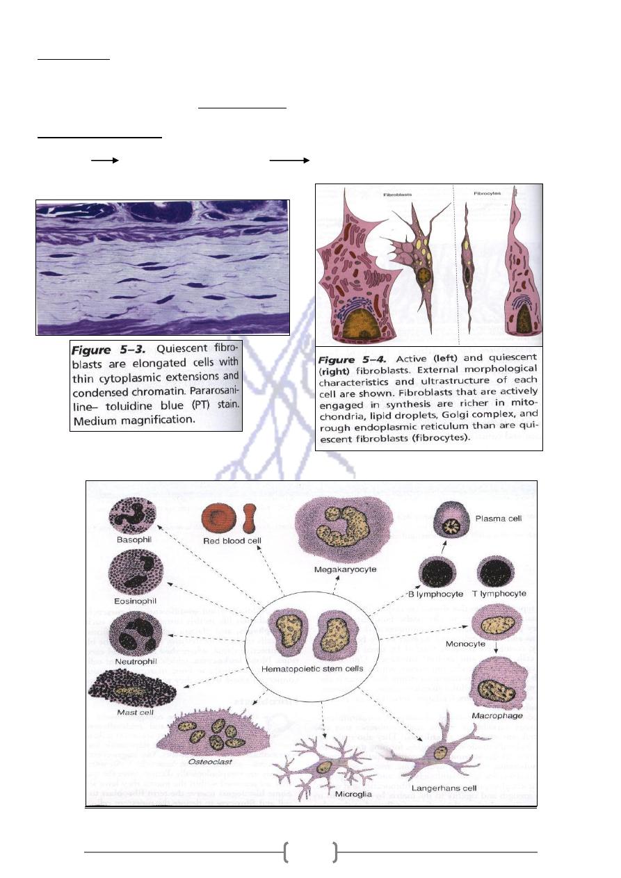
Fibroblasts
1- Synthesize collagen , elastin ,glycosaminoglycans ,proteoglycans & multiadhesive glycoproteins.
that influence cell growth and differentiation
Growth factors
Involved in production of
-
2
Two stages of activity
Fibrocyte
1- Active Fibrobast 2- Quiescent
165
Mediccal application
When tissues are destroyed by inflammation or traumatic injury the main cell type involved in
repair is Fibroblast.
Fibrocyte reverts to Fibroblasts state and its synthetic activities are reactivated during wound
both fibroblasts and smooth muscle observed
,a cell with features of
myofibroblast
healing .The
during wound healing .Its activity is responsible for wound closure ,this process call Wound
contractio
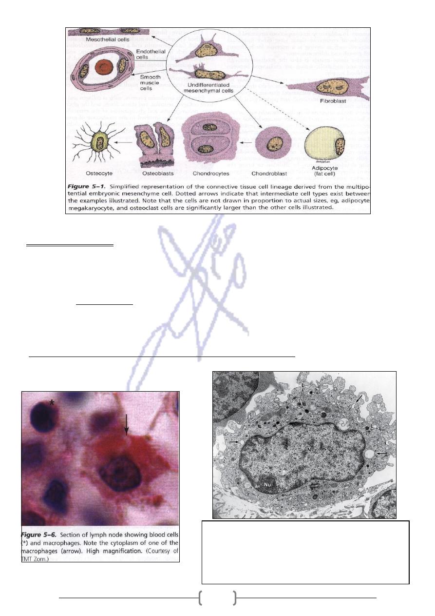
Macrophages & the mononuclear phagocyte system
---
2
Characteristic features of macrophages seen in this TEM of
one such cell are the prominent nucleus (N) and the
nucleolus (Nu) and the numerous secondary lysosomes (L).
The arrows indicate phagocytic vacuoles near the
protrusions and indentations of the cel surface. X10,000.

166
They are characterized by an irregular surface , a morphological expression of their active
pinocytotic and phagocytic activities .They have a well–developed Golgi complex, many
Lysosomes ,and R.E.R.
Bone marrow Monocyt circulate in blood cross the wall of venules and
capillaries to penetrate the connective tissue, where they mature and acquire
morphological features of Macrophage
application
Medical
Epithelioid cell
When they are stimulated increase in size & clusters form
-
1
multinuclear giant cells
Several Macrophages may fuse to form
-
2
3- Macrophages participate in cell- mediated resistance to infection by bacteria, viruses, protozoa,
fungi ,and metazoans( parasitic worm)
4- Macrophages are also secretory cells that produce enzymes eg,collagenase & cytokiwhich
they exhibit increased tumor cell –killingnes
5- They participate in the processes of partial digestion and presentation of antigen to other cells
The langerhans cell in the dermis .
ex.
5
-
They have an important role in removing cell debris and damaged extracellular
components formed during the physiological involution processes. Ex .during
pregnancy the uterus increases in size and after parturition ,some of its tissue are
destroyed by the action of macrophages
Table 5–2.
Distribution and main functions of the cells of the mononuclear phagocyte system.
Cell Type
Location
Main Function
Monocyte
Blood
Precursor of macrophages
Macrophage
Connective tissue, lymphoid
organs, lungs, bone marrow
Production of cytokines, chemotactic
factors, and several other molecules that
participate in inflammation (defense),
antigen processing and presentation
Kupffer cell
Liver
Same as macrophages
Microglia cell
Nerve tissue of the central
nervous system
Same as macrophages
Langerhans cell
Skin
Antigen processing and presentation
Dendritic cell
Lymph nodes
Antigen processing and presentation
Osteoclast
Bone (fusion of several
macrophages)
Digestion of bone
Multinuclear
giant cell
Connective tissue (fusion of
several macrophages)
Segregation and digestion of foreign bodies
167
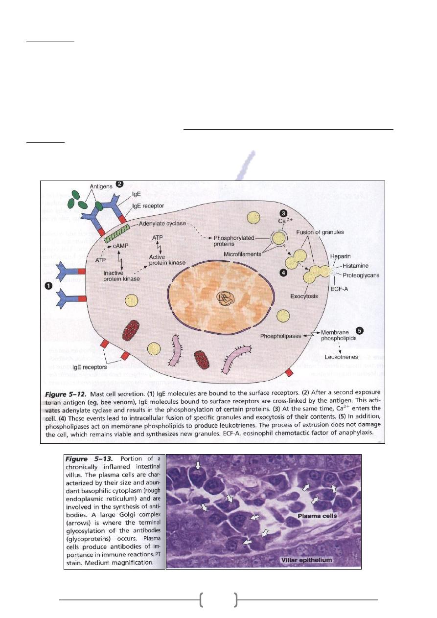
Mast cells
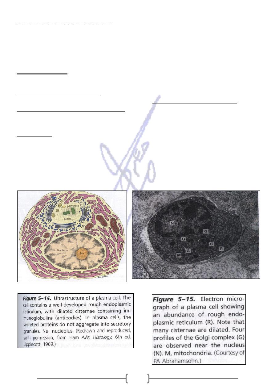
Are oval to round connective tissues cells ,cytoplasm is filled with basophilic granules that
give a heterogeneous in appearance with a prominent scroll-like substructure ,that contains
Histamine & Heparin . They are abundant in the dermis & in the digestive & respiratory tracts.
They originate from progenitor cells in bone marrow, circulate in the blood , cross the wall of
venules& capillaries & penetrate the tissue , they proliferate & differentiate.
the storage of chemical mediators of the inflammatory
cells is
The principal function of mast
response .
The surface of mast cells contains specific receptors for immunoglobulin E (igE)
,which produced by plasma cells.
168
There are two populations of mast cells
1- Connective tissue mast cell
Is found in the skin & peritoneal cavity ; their granules contain the anticoagulant heparin.
2- Mucosal mast cell
Is present in intestinal mucosa and in the lungs ; their granules contain chondroitin sulfate .
Medical application
Release of the chemical mediators stored in mast cells promotes the allergic reaction known as
Mediate hypersensitivity reactions.
immunoglobulin E (IgE), a type of
The surface of mast cells contains specific receptors for
.Most IgE molecules are bound to the surface of
immunoglobulin produced by plasma cells
mast cells and blood basophiles ;very few remain in the plasma.
Plasma cells
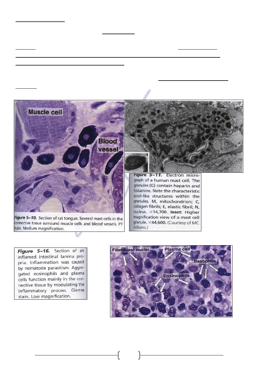
Are large cells ,have a basophilic cytoplasm due to their richness in R.E.R.The nucleus is spherical
and eccentrically placed ,containing compact ,coarse heterochromatin alternating with lighter areas
of approximately equal size .this configuration resembles the face of a clock .
169
Medical application
is a glycoprotein that interacts specifically with
of antibodies
responsible for the synthesis
They are
an antigenic determinant.
uch as proteins,
A molecule that is recognized by cells of the immune system s
antigens
polysaccharides, and nucleoproteins) or molecules belonging to whole cells (bacteria,
infected cells).
-
tumor cells, or virus
protozoa,
The cells of the immune system do not recognize and react to the whole antigen molecule but
as antigenic determinants or
en known
instead react to small molecular domains of the antig
epitopes

171
Adipose Cells
Adipose cells (adipocytes; L. adeps, fat, + Gr. kytos) are connective tissue cells that have become
specialized for storage of neutral fats or for the production of heat. Often called fat cells, they are
discussed in detail in Chapter 6: Adipose Tissue.
Leukocytes
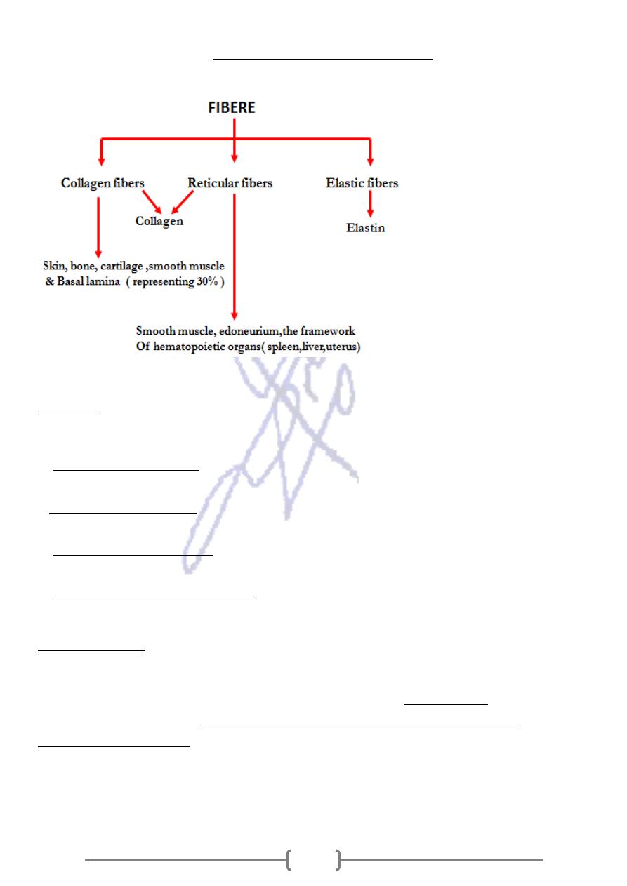
The normal connective tissue contains leukocytes that migrate from the blood vessels by diapedesis.
Leukocytes (Gr. leukos, white, + kytos), or white blood corpuscles, are the wandering cells of the
connective tissue. They migrate through the walls of capillaries and postcapillary venules from the
blood to connective tissues by a process called diapedesis. This process increases greatly during
inflammation (Figure 5–16). Inflammation is a vascular and cellular defensive reaction against
foreign substances, in most cases pathogenic bacteria or irritating chemical substances. The classic
signs of inflammation were first described by Celsus (first century A.D.) as redness and swelling
with heat and pain (rubor et tumor cum calore et dolore). Much later, disturbed function (functio
laesa) was added as the fifth cardinal sign.
Inflammation begins with the local release of chemical mediators of inflammation, substances of
various origin (mainly from cells and blood plasma proteins) that induce some of the events
characteristic of inflammation, eg, increase of blood flow and vascular permeability, chemotaxis,
and phagocytosis.
Chemotaxis
Is the phenomena by which specific cell types are attracted by some molecules ,is responsible for
the migration of large quantities of specific cell types to regions of inflammation .as leukocytes
,cross the venules and capillaries by Diapedesis ,invading the inflamed area.
171
Fibers of connective tissue
Collagen
Based on their structure and function ,they can be classified into the following groups:
: They form structures such as bones ,dentin ,tendons ,organ
Collagens that form fibers
-
1
capsules and dermis .
: Short structures that bind collagen fibrils to one another and
assciated collagen
-
Fibrile
-
2
other components of extracellular matrix
: Assemble in a meshwork that constitutes the structural
networks
Collagen that form
-
3
component of the basal lamina.
: Is present in anchoring fibrils that bind collagen
Collagen that form anchoring fibrils
-
4
fibers to the basal lamina
Collagen synthesis
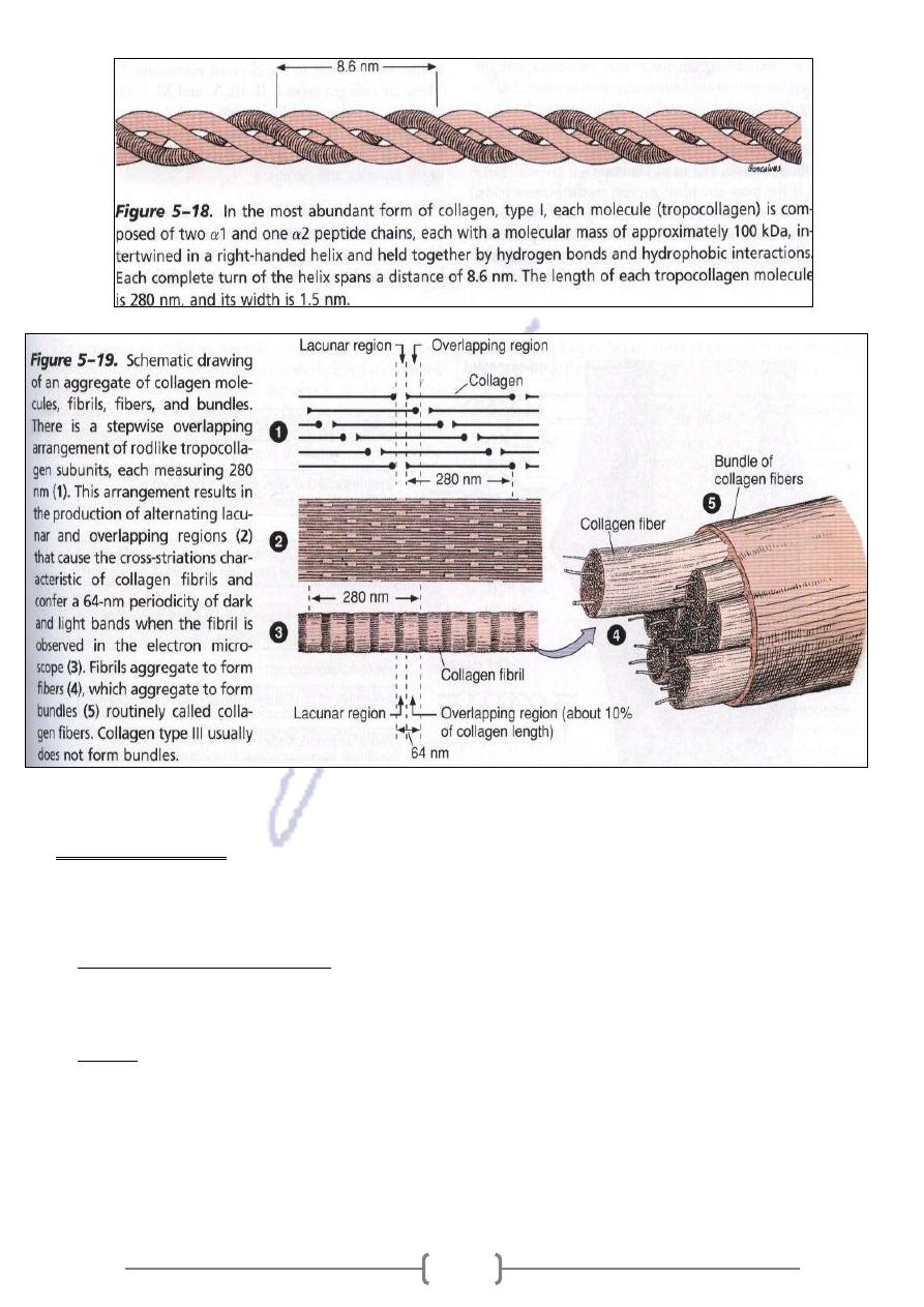
Fibroblast ,Chondroblast , Osteoblast , and Odontoblast producing collagen protein. The
(280 nm in
Tropocollagen
protein unit that form collagen fibrils is elongated molecule called
polypeptide chain
t
Tropocollagen consist of three sub uni
length and 1.5 nm in width) .
intertwined in a triple helix
Differences in chemical structure of these polypeptide chains are responsible for the
various type of collagen
The transverse striations of collagen fibrils are determined by overlapping arrangement of the
tropocollagen molecules
172
Medical application
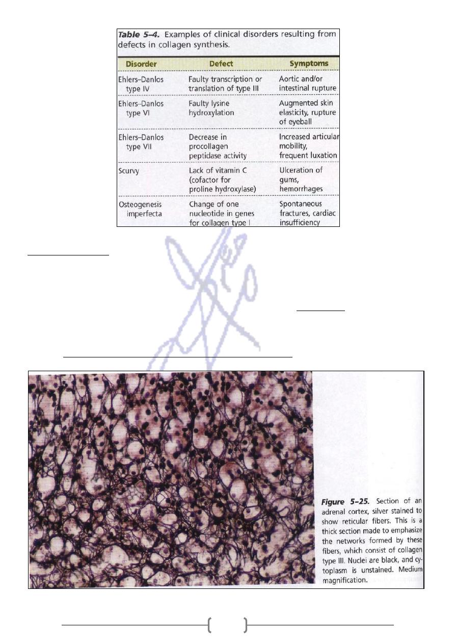
A large number of pathological conditions are directly attributable to insufficient or abnormal
collagen synthesis
(fibrosis) .It
n
accumulation of collage
Result from an excessive
progressive systemic sclerosis:
-
1
occurs in skin ,digestive tract muscle and kidney ,causing hardening and functional impairment of
implicated organ
mounts of collagen that form in scars of the skin
Is a local swelling caused by abnormal a
Keloid:
-
2
,especially in individuals of black African descent
Others disorders are illustrated in table 5-4.,
173
Reticular fibers
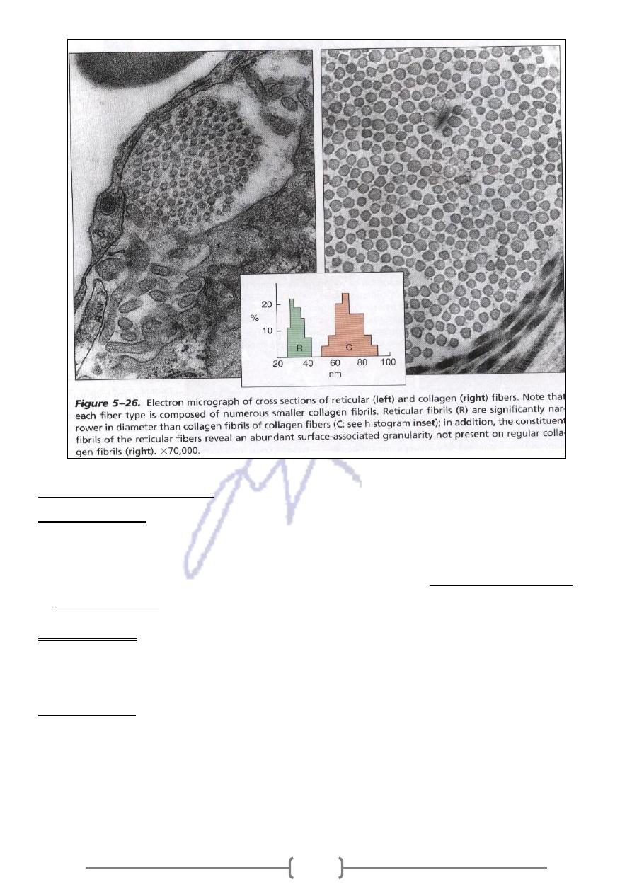
They consist of collagen type III, in association with other types of collagen, glycoprotein and
proteoglycans.
,because of their
argyrophilic
12% hexoses. These fibers are called
-
Reticular fibers contain 6
affinity for silver salts.
They are formed by loosely packed , thin fibrils bound together by interfibrillar bridges.They are
oietic organs
smooth muscle ,endoneurium ,and Hematop
found in
174
system
he elastic fiber
T
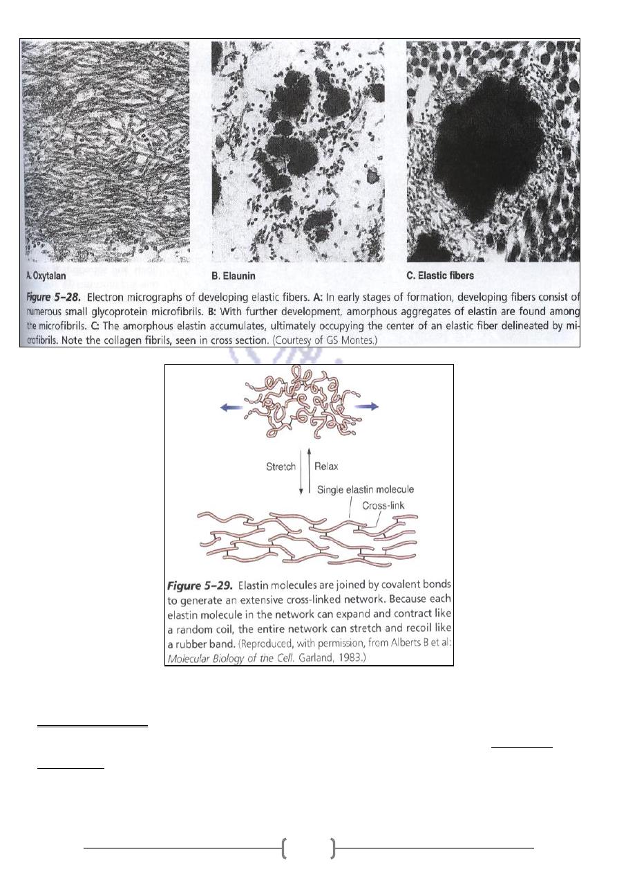
Oxytalan fibers
-
I
1- They are not elastic ,do not have elastin protein
2- Found in the zonule fibers of the eye & dermis.
&
fibromodulin
ding
inclu
They consist of a bundl of micro fibrils composed of glycoproteins,
-
3
molecule fibrilin
Elaunin fibers
-
II
An irregular deposition of the protein Elastin, appear between the Oxytalan fibers ,forming Elaunin
.They are found around sweat glands & in dermis .
Elastic fibers
-
III
Elastin gradually accumulates until it occupies the center of the fiber bundles, which are further
surrounded by a thin sheath of micro fibrils .They are rich in protein elastin ,they stretch easily in
response to tension
175
Medical application
MARFAN
Mutation in the fibrillin gene which it is necessary for deposition of elastin result in
, a disease characterized by a lack of resistance in the tissues rich in elastic fibers .
SYNDROM
Because the large arteries are in components of elastic system and because the blood pressure is
high in the aorta patients with disease often experience aortic rupture.
176
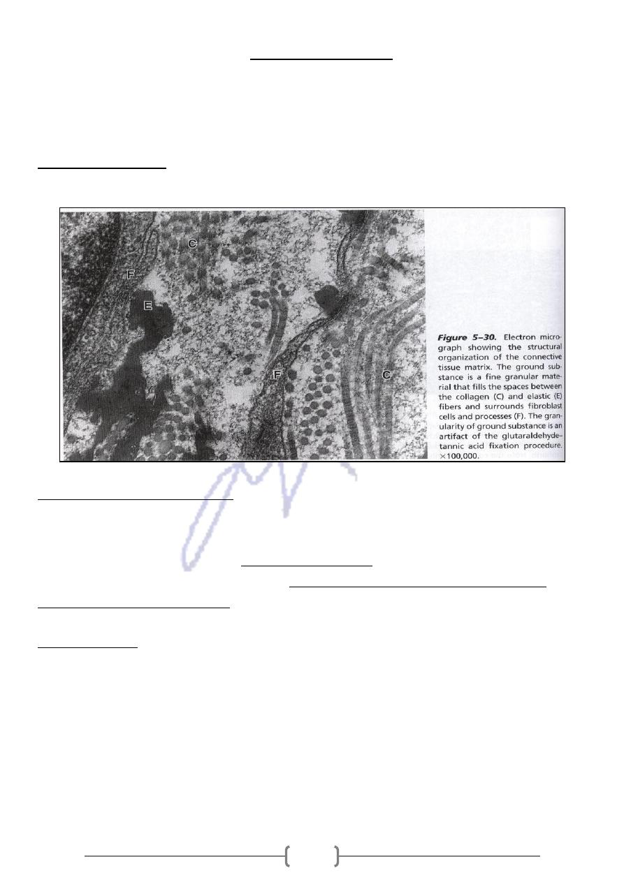
Ground substance
Is a highly hydrated ,colorless and transparent complex mixture of macromolecules. It fills the
space between cells & fibers of connective tissue
Act as both a lubricate & a barrier to penetration of invaders ,because of its viscosity.
Is formed mainly of three classes of components;
:
The ground substance
1- glycosaminoglycan ( GAG) 2- proteoglycan 3- multiadhensive glycoproteins
( GAG)
Glycosaminoglycan
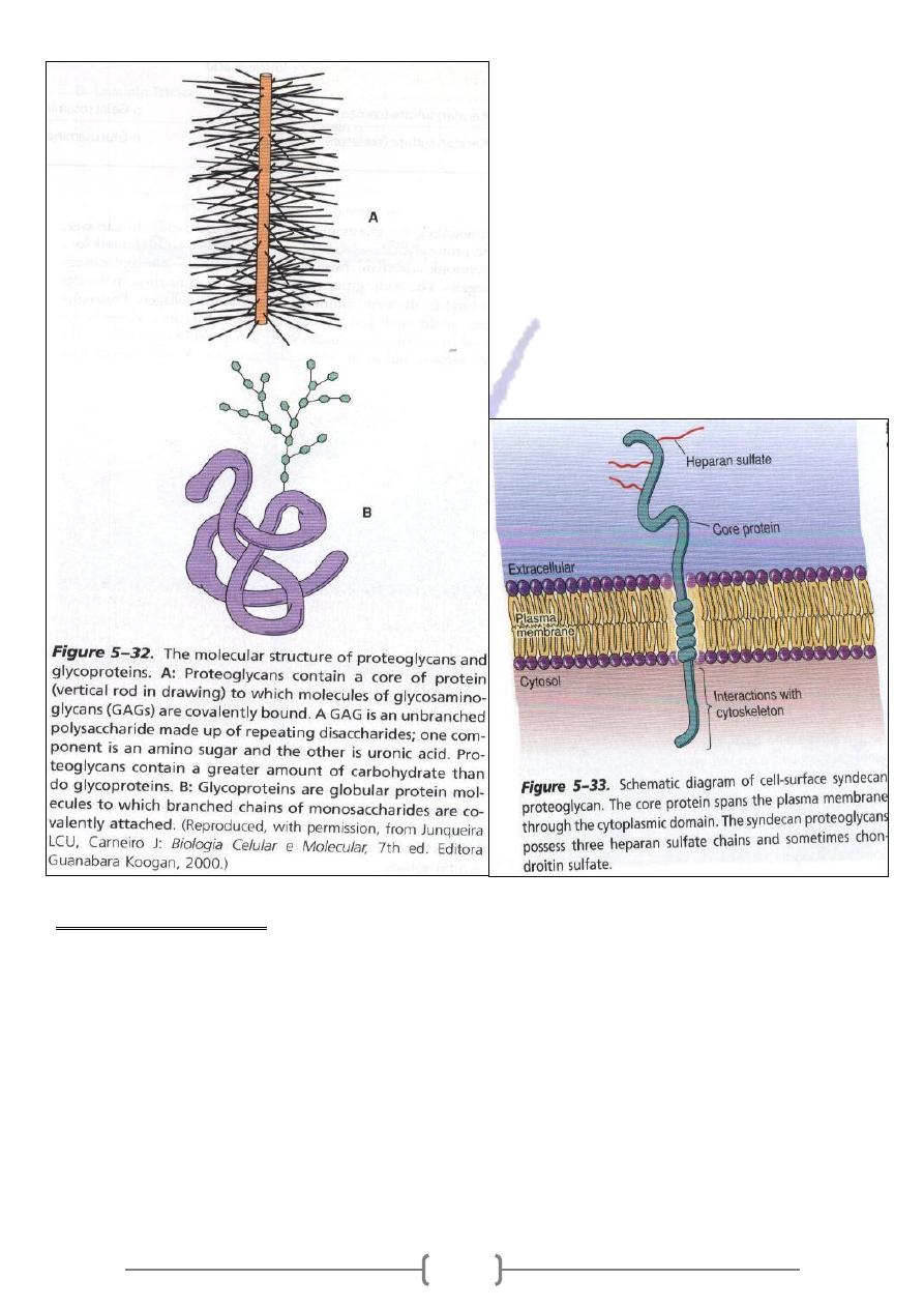
Are linear polysaccharides formed by repeating disaccharide unit usually composed of a
uronic acid & a hexosamine .This linear chains (exception of hyaluronic acid ) are bound
proteoglycan molecule .
covalently to protein core forming a
the abundance of hydroxyl ,carboxyl & sulfat
These molecule are hydrophilic because of
groups in the carbohydrate moiety.
Proteoglycans
Are composed of a core protein associated with the four main GAG s ; Dermatan sulfate ;
Chondroitin sulfate ;Keratan sulfate ;& Heparan sulfate. They are distinguished by their
molecular diversity and can be located in cytoplasmic granules such Heparin of mast cells, in
the cell surface, and the extracellular matrix
177
Proteoglycans functions
1- Structural components of the extracellular matrix .
2- Anchoring cells to the matrix .
3- Both extracellular & and surface proteoglycan also bind many protein growth factor .
4- Because of their viscosity , they are acted as barrier to the penetration of bacteria and others
microorganism . Bacteria that produce hyaluronidase ,an enzyme that hydrolyzed hyaluronic acid
and other glycosaminoglycan, have great invasive power because they reduce the viscosity of
connective tissue ground substance

178
Multiadhesive glycoproteins
.
Are compounds that contain a protein moiety to which carbohydrates are attached
1- The protein moiety usually predominates and do not contain the linear polysaccharides
2- The carbohydrate moiety is frequently a branched structure .
Glycoprotein has an important in the adhesion of cells to their substrate and interaction
between neighboring adult and embryonic cells.
.
Several glycoprotein have been isolated from connective tissues
It has
.
lasts and some epithelial cells
Is a glycoprotein synthesized by fibrob
Fibronectin :
-
1
binding sites for cells ,collagen ,and GAG.
the
to
Is a large glycoprotein that participates in the adhesion of epithelial cells
Laminins :
-
2
basal lamina a structure rich in laminins
Medical application
Both Fibronectin & Laminin participate in embryonic development and the increased
ability of cancer cells to invade other tissues has been postulate
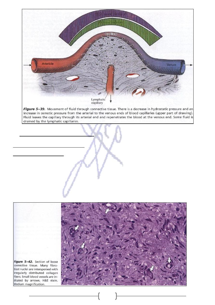
In addition to ground substance ,there is a very small quantity of Tissue fluid –
Which it is similar to blood plasma in it content ion and diffusible substances.
In some pathological conditions ,the quantity of tissue fluid may increase ,causing
Edema ,which characterized by enlarged spaces ,caused by the increased in liquid
,between the components of connective tissue .
Edema may result from
1- Venous or lymphatic obstruction .
2- Decrease in venous blood flow ( eg. congestive heart failure )
3- Obstruction of lymphatic vessels due to parasitic plugs or tumor cells and chronic starvation.
4- Protein deficiency in a lack of plasma protein and a decrease in colloid pressure.
5- Increased permeability of the blood capillary or post capillary venule endothelium
resulting from chemical or mechanical injury
179
Types of connective tissue
connecctive tissue proper
-
1
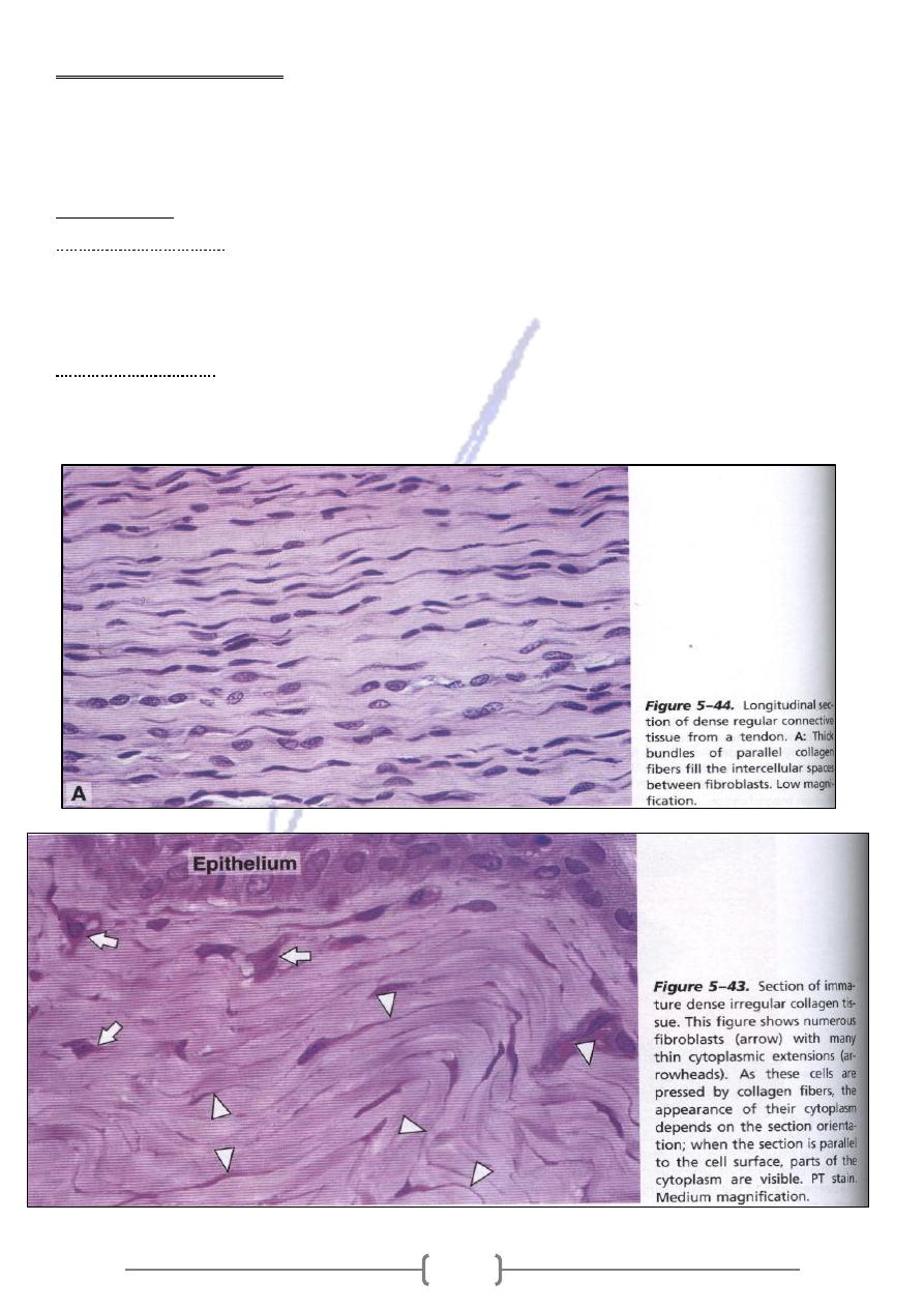
connective tissue
loose
-
A
1- Supports many structures that are normally under pressure and low friction .
2- It fills spaces between groups of mucle cells .
3- Supports epithelial tissue .
4- Forms a layer that sheathes the lymphatic and blood vessels .
5- It is found in Papillary layer of the dermis , in the hypodermis , in glands & mucous membranes
supporting to epithelial cells.
181
denes connective tissue
-
B
It adapted to offer resistance and protection ,it is the same components of loose c .t., .There are
fewer cell and a clear predominance of collagen fibers .Is less flexible and far more resistant to
stress than is loose c. t.
It is known as:
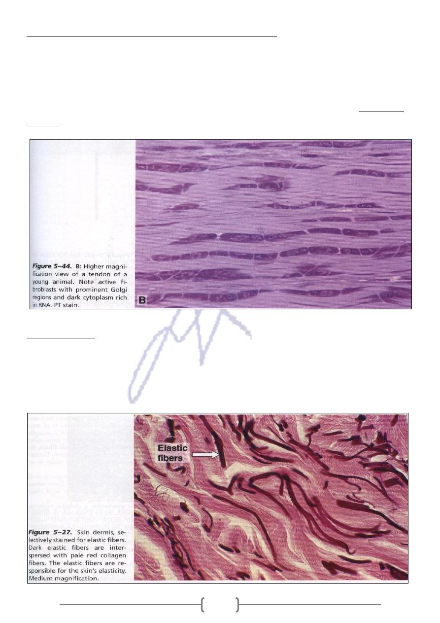
irregular :
Dense
-
1
The collagen fibers are arranged in bundles without a definite orientation .
The collagen fibers form a three dimensional network .
Provide resistance to stress from all directions .It is found in DERMIS.
lar:
Dense regu
-
2
The collagen fibers aligned with the linear orientation of fibroblasts .
Provide resistance to stresses exerted in same direction
181
Tendons are the most common example of denese regular C. T.
1- A re elongated cylindrical strictures attach striated muscle to bone .
2- They are white and inextensible .
3- They have parallel ,closely packed bundles of collagen.
4- Their fibroblasts contain elongated nuclei parallel to fibers.
(secondary
es
The collagen bundles of the tendons ( primary bundles ) aggregate into larger bundl
,that are enveloped by loose connective tissue.
bundles)
Elastic tissue
Is composed of bundles of thick ,parallel elastic fibers ,between these fibers is occupied thin
collagen fibers and flattened fibroblasts .
It is yellow color , great elasticity .
It is present in the yellow liquments of the vertebral column.
182
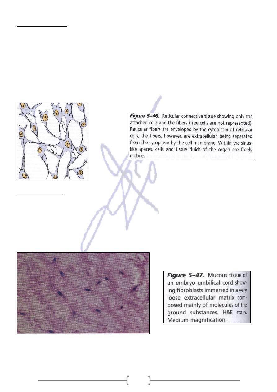
Reticular tissue
1- It forms three dimensional networks that support cells .
2- Specialized loose connective tissue consisting of reticular fibers associated with specialized
fibroblasts called Reticular cells It forms three dimensional networks that support cells.
3- Provides the architectural framework for hematopoietic organs & lymphoid organs ( bone
marrow ,lymph nodules ,and nodes and spleen)
Reticular cells are dispersed along this framework and cover the reticular fibers and ground
substance within cytoplasmic processes.
Mucous tissue
1- It is found in umbilical cord Where it is referred to as Wharton's jelly & found in the pulp of
young teeth.
2- Has an abundance of ground substance composed of hyaluronic acid .
3- It is a jelly tissue containing very few fibers .
4-The cells are mainly fibroblasts
