191
Bone tissue supports fleshy structure & protects vital organs such as cranial & thoracic
cavities ,and harbors the bone marrow
is highly vascularized & metabolically very active .
It serves as a reservoir of calcium phosphates , and other ions
Give the mechanical & metabolic functions to skeleton
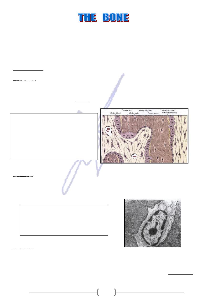
Bone tissue composes of bone matrix & three types of cells Osteocytes, Osteoblasts & Osteoclasts.
BONE CELL
collagen ,
(
the synthesis of the organic components of bone matrix ,
. Are responsible for
Osteoblasts
proteoglycan and glycoproteins ).# They are located at the surface of bone tissue their shape ranged
between cuboidal to columnar to flatten shape according to their activity. Osteoblasts are gradually
surrounded by new matrix and become Osteocytes and lacuna is formed .Matrix compoents are
).
(not yet calcified
Osteoid
called
secreted at the cell surface ,
Derive from Osteoblasts , lie in lacunae , situated between lamellae of matrix .Only one
:
Osteocytes
osteocyte is found in each lacuna .
They have a thin cylindrical matrix canaliculi by which the molecules are passed from cell to cell. Some
molecular exchange between cells takes place through extra cellular substance
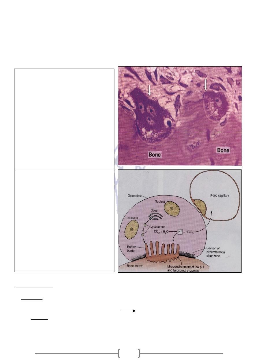
Osteoclast
Are very large ,branched motile cells contain from 5-50 or more nuclei
They lie within enzymatically etched depressions in the matrix known as
howship’s lacunae, are
derived from the fusion of bone marrow_ derived mononucleotide cells.
uffled border
R
, forming a
rregular projections
nto i
forming bone matrix is folded i
-
In active, the surface
Fig.8-3 :
Events that occur during
intramembranous ossification. Osteoblasts are
synthesizing collagen, which forms a strand of
matrix that traps cells. As this occurs, the
osteoblasts gradually differentiate to become
osteocytes. The lower part of the drawing
shows an osteoblast being trapped in newly
formed bone matrix.
Fig.8-1 :
Section of bone tissue showing an
osteocyte with its cytoplasmic processes
surrounded by matrix. The ultrastructure of the
cell nucleus and cytoplasm is compatible with a
low level of protein synthesis.
192
Clear zone is cytoplasmic zone that surround the ruffled border, which is devoid of organelles, yet
rich in actin filament .This zone is a site of adhesion of the osteoclast to bone matrix, in which bone
resorption occurs
They secretes collagenas and other enzymes
pump proton into the microenvironment .Their activity is controlled by cytokines and hormone
Ruffled borders are related to the activity, of osteoclasts
Bone matrix
matter represents 50% of dry weight of bone matrix : calcium& phosphorus are especially
Inorganic
abundant ,bicarbonate , citrate ,magnesium, potassium & sodium are also found Calcium & Phosphorus
Hydroxyapatite crystals with Ca10(PO4)6 (OH)2
matter in bone matrix is a type I collagen& ground substance (proteoglycan
organic
The
aggregates & glycoprotein ).
Mineral & collagen fibers is responsible for the hardness & resistance of bone tissue.
After decalcified the bone ,its shape is preserved, but it becomes as flexible as a tendon .
Fig.8.4 : Section showing three
osteoclasts (arrows) digesting bone
tissue. The osteoclast is a large cell with
several nuclei and a ruffled border close
to the bone matrix. Note the clear
compartment where the process of bone
erosion occurs. This compartment is
acidified by a proton pump localized in
the osteoclast membrane. It is the place
of decalcification and matrix digestion
and can be compared to a giant
extracellular lysosome. Chondroclasts
found in eroded regions of epiphyseal
calcified cartilage are similar in shape to
osteoclasts.
Fig.8.5 : Bone resorption. Lysosomal
enzymes packaged in the Golgi complex
and hydrogen ions produced are released
into the confined microenvironment created
by the attachment between bone matrix and
the osteoclast's peripheral clear zone. The
acidification of this confined space
facilitates the dissolution of calcium
phosphate from bone and is the optimal pH
for the activity of lysosomal hydrolases.
Bone matrix is thus removed and the
products of bone resorption are taken up by
the osteoclast's cytoplasm, probably
digested further, and transferred to blood
capillaries.
193
Removable the collagenous
– also leaves the bone with its original shape however, it becomes
fragile, breaking & crumbling easily when handle.
Medical application
in which mineralization is impaired .
Osteomalacia ,
osteoclasts
zed by dense, heavy bones, the
The genetic disease ,which is characteri
Osteopetrosis
lack ruffled borders ,and resorption is defective
steum
Periosteum & endo
The periosteum consists of an outer layer of collagen fibers & fibroblasts .Sharpey's fibers
,penetrate the bone matrix ,bining the periosteum to bone . In the
Osteoprogenitor cells
inner ,cellular layer of the periosteum is composed of fibroblast like cells called
,they have a potential to divide by mitosis & differentiate into Osteoblasts. Osteoprogenitor cells play a
prominent role in bone growth & repair
The endosteum lines all internal cavities within the bone & is composed of a single layer of
flattened Osteoprogenitor cells & very small amount of connective tissue .
The principle functions of periosteum & endosteum are nutrition of osseous tissue &
provision of a continuous supply of new osteoblast for repair or growth of bone .
Types of bone
which shows dense area without cavities ,and
Compact bone
There are two principle types of bones
which shows area with numerous interconnecting cavities .
Cancellous bone
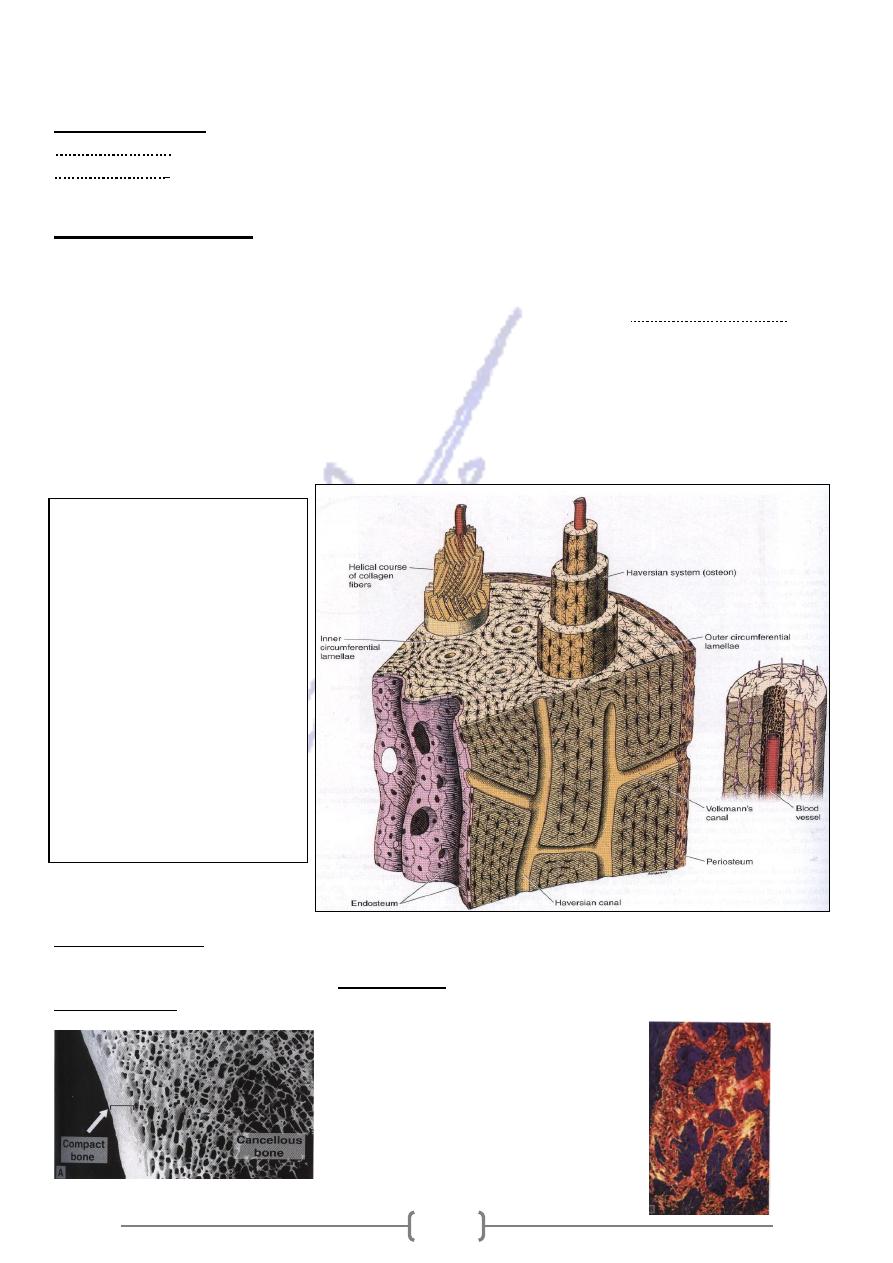
Fig.8.6 : Schematic drawing of
the wall of a long-bone
diaphysis showing three types
of lamellar bone: haversian
system and outer and inner
circumferential lamellae. (For
interstitial lamellae, see Figure
8–10.) The protruding
haversian system on the left
shows the orientation of
collagen fibers in each lamella.
At the right is a haversian
system showing lamellae, a
central blood capillary (there are
also small nerves, not shown),
and many osteocytes with their
processes.

194
According to shape ;
are composed of spongy bone covered by a thin
Epiphyses
The bulbous ends called
:
Long bones
layer of compact bone
The cylindrical part _ Diaphysis is composed of compact bone with a small component of spongy bone
on its inner surface around the bone marrow cavity .
Have a core of spongy bone completely surrounded by compact bone .
:
Short bones
separated
,
ct bones called plates ( table)
layers of compa
Form the calvarias have two
:
Flat bones
by a layer of spongy bone called the diloe.
According to development rate
Primary ,immature ,woven bone: Is the first bone tissue to appear in embryonic development and in
repair processes .It is temporary, is replaced in adults by secondary bone tissue except in few places in
the body ( near the structures of the flat bones of the skull, in tooth socket ).
They have irregular array of collagen fibers .
They are a lower mineral content & higher proportion of osteocytes than in secondary bone T.
Sesondary bone tissue : It is found in adults.
collagen fibers arranged in lamellae , which are paralled to each other or concentrically
organized around a vascular canal.
The concentric lamellae of bone surrounding a canal containing blood vessels ,nerve & loose
Haversian system ,or Osteon
connective tissue is called
Lacunae containing osteocytes are found between or within the lamellae .
Surrounding each haversian systems is cementing substance Which consists of mineralized with few
collagen
In long bone , the lamellae exhibit a typical organization consisting of Havesian systems , outer
circumferential lamellae , inner circumferential & interstitial lamella .
,is long .bifurcated cylinder parallel to the axis of the diaphysis .It communicate with
Haversian system
the marrow cavity ,the periosteum, and one another through transverse or oblique Volkmann's canals .
Each system is formed by successive deposits of lamella ,starting inward from the periphery ,so the
most recently formed lamella is the one closest to the central canal.
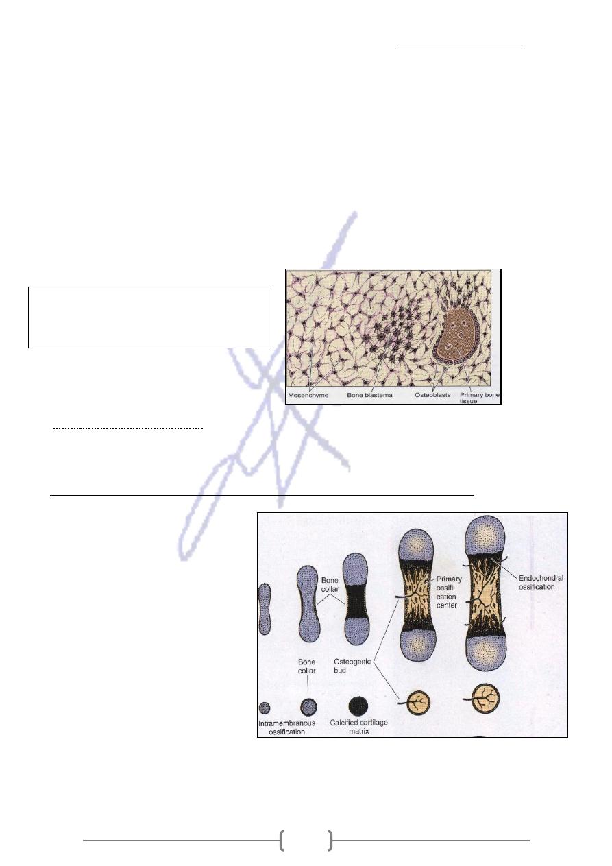
Histogenesis
Bone can be formed in two ways :
alization of matrix secreted by
: By which direct mener
Intramembranous Ossification
Ostoblasts. is the source of most of the flat bones ,it takes place within Mesenchymal tissue . The
frontal & partial bones of the skull-as well as parts of the occipital , temporal bones , the mandible &
maxilla are formed by this way .
Fig.8.7 : A: Thick section of bone illustrating the cortical
compact bone and the lattice of trabeculae of cancellous bone.
(Courtesy of DW Fawcett.) B: Section of cancellous (spongy)
bone with its characteristic random disposition of collagen fibers.
Picrosirius-polarized light (PSP) stain. Low magnification.
195
The process begin when
A primary ossification center.
The starting point for ossification is called
1) Group of mesenchymal cells differentiate into osteoblasts.
2) Osteoblasts produce bone matrix & calcification follows ,resulting in the encapsulation of
some osteoblast which then become osteocytes
3) These islands of developing bone form wall that delineate elongated cavities containing
capillaries, bone marrow cells & undifferentiated cells .
4) Several such groups arise almost simultaneously at the ossification center .
5) Fusion of the walls gives the bone a spongy structure .
6) The connective tissue that remain among the bone walls is penetrated by growing blood
vessels & additional undifferentiated mesenchmal cells .
7) The ossification centers of a bone grow radially & finally fuse together , replacing the original
connective tissue .
8) The fontanelles of newborn infants, for example , are soft areas in the skull that are not yet ossified .
Endochodrale Ossification :
It takes place within a piece of Hyaline cartilage .
These type of ossification is principally responsible for the formation of short and long bones .
vents.
Endochondral Ossification of long bone consists of the following sequence of e
The ( bone collar ) which is produced
by intramembranous ossifications within
the local perichondrium represent the first
bone tissue appear.
the local cartilage undergoes a
degenerative in process of programmed
cell death with cell enlargement
(hypertrophy) and matrix calcification .
This process begins at the central
portion of the cartilage model (diaphysis)
,which previously perforate by osteoclasts
. The blood vessels penetrate the
perforation region bringing
osteoprogenitor cells to this region.
Osteoblasts adhere to be calcified cartilage matrix and produce continuous layers of primary bone that
surround the cartilaginous matrix remnants, so the primary Ossification center is produced
Fig. 8.9 : The beginning of intramembranous
ossification. Mesenchymal cells round up
and form a blastema, from which osteoblasts
differentiate, producing primary bone tissue.
196
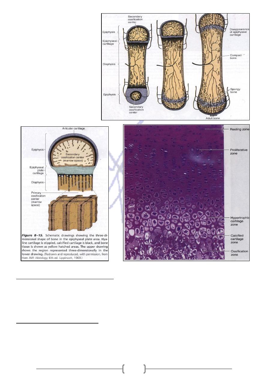
Then the secondary ossification
center, appear at the swellings in
the extremities of the cartilage
model( epiphyses) . During their
expansion and remodeling the
primary & secondary oss. Centers
produced cavities that gradually
filled with bone marrow .
Epiphyseal cartilage (epiphyseal
plate) ,which connects the two
epiphyses to the diaphysis .It is
responsible for the growth in length
of bone and Disappears in adults.
Bone growth and remodlingbone
Bone growth is associated with partial resorption of preformed tissue and the simultaneous laying down
of new bone .This process permits the shape of the new bone to be maintained ,while it grows .bone
remodeling in young children can be 200 times faster than the rate in the adult .Bone responds to the
growth of the brain and forms a skull of adequate size .
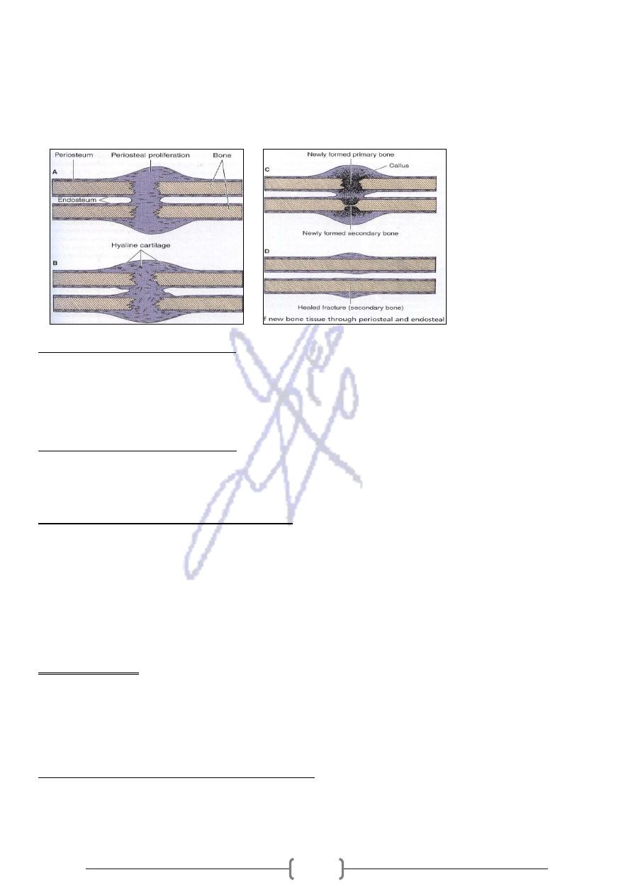
Medicale aplication (Fractur repair)
Bone matrix is destroyed .
Bone cells adjoining the fracture die .
The damaged blood vessels produce a localized hemorrhage and form a blood clot.
During repair ,the blood clot ,cells and damaged bone matrix are removed by macrophages
197
The periosteum and endosteum around the fracture respond with intense proliferation producing a
tissue that surrounds the fracture and penetrates between the extremities of fractured bone .
Primary bone is formed by endochondral and intramembranous ossification .
The extremities of the fractured bone ,forming a Bone callus .
The primary bone tissue of the callus is gradually resorbed and replaced by secondary tissue,
remodeling the bone and restarting its original structure
Internal Structure Of Bones
Despite its hardness,bone is capable of changes in its internal structure in response to the various
sresses it is subjected ,for ex. The positions of the teeth in the jaw bone can be modified by lateral
pressures produced by Orthodontic appliances.
Metabolic Role Of The Bone
The skeleton contains 99% of the total calcium of the body .The concentration ions in the blood &tissue
is stable because of continuous interchange between blood calcium & bone calcium .
Bone calcium mobilized by two mechanisms .
The first is the simple transfer of ions from hydroxypatite crystals to interstitial fluid then pass into the
blood .It takes place in spongy bone .
The second mechanism ,depend on the action of hormones on bone .
Parathyroid hormone promotes Osteoclastic resorption of the bone matrix with liberation of calcium .
This hormone act on osteoblast receptors ,which start the secretion of an Osteoclast-stimulating factor
Another hormone, Calcitonin , which is synthesized mainly by the parafollicular cell of the thyroid
gland , Inhibits matrix resorption.
Medical aplication
Decalcification of bone ,due to nutritional deficiency of calcium .Decalcification may also be caused by
excessive production of Parathyroid hormone (hyperparathyriodism), which results in creased
osteoclastic activity ,intense resorption of bone, elevation of blood Ca and PO4 levels,and abnormal
deposits of calcium in several organs, mainly the kidney & arterial walls.
encies on Bone Tissue
Effects of Nutritional Defici
Calcium deficiency in children causes Rickets , A disease in which the bone matrix does not
calcify normally & the epiphyseal plate distorted by the body weight & muscular activity .
198
Osteopetrosis , a disease caused by a defect in osteoclast Function that results in overgrowth
,thickening ,and hardening of bones .
Osteoporosis , is an imbalance in skeletal turnover so that bone resorption exceed bone
formation ,frequently found in, immobilized patients and postmenopausal women .
Hormones Acting On Bone Tissue
The anterior lobe of pituitary synthesizes hormone has an effect on growth ,especially on the epiphyseal
cartilage .Lack of growth hormone during the grow thing years causes Pituitary Dwarfism ; an excessive
growth of the long bones , resulting in Gigantism . The sex hormones , both male (androgens) & female
(astrogens),have a complex effect on bones . They influence the time of appearance and development
of ossification centers and accelerate the closure of epiphyses
Bone tumors
Bone tumor uncommon ( 0.5% of all cancer deaths) .The benign ( e.g. Osteoblastoma, osteoclastoma )
, while malignant (e.g. Osteosarcoma) .The lower end of the femur, the upper tibia ,and the upper
humerus are the most common locations. The most frequent bone metastases are from breast , lung
,prostate , kidney , & thyroid tumors
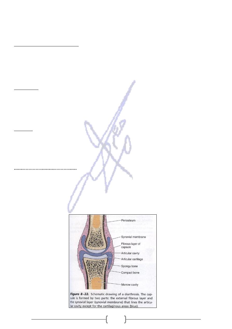
Joints
Joints are regions in which bones are capped and surrounded by connective tissues that hold the bones
together and determine the type and the degree of movement between them . There are classified as
Diarthroses ,in which there is free bone movement , or Synarthroses , in which very limited or no
movement occurs .
.
There are three of Synathroses
Synostosis ,in which bone are united by bone tissue and no movment takes place eg. Skull
bones in adults .
Synchodroses are articulation in which the bones are joined by hyaline cartilage (eg. The
epiphyseal plates ,in adult ,synchodrosis unites the first ribe to the sternum ) .
Diarthroses are joints that generally unite long bones and have great mobility ,such as the
elbow and knee joints.