Maxillary Sinus disease
Dr. Wafaa Khalil
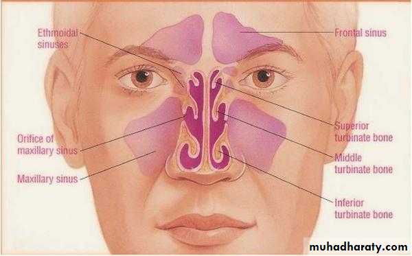
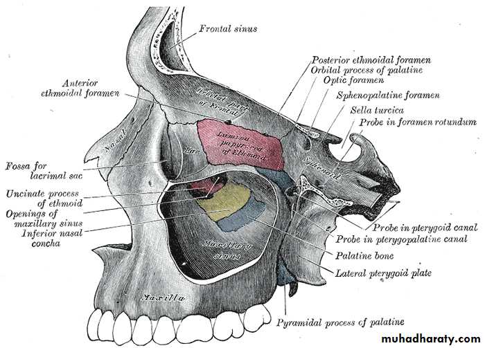
The maxillary sinus is a pneumatic space. It is the largest bilateral air sinus located in the body of the maxilla and opens in the middle nasal meatus of the nasal cavity with single or multiple openings are termed ostium maxillare, which found in a recess called hiatus semilunaris..
Maxillary Sinus
(Antrum of Higmore)The average dimensions of the sinus are approximately 3.5 (anteroposterior) x 3.2 (height) x 2.5 (width) cm
The average capacity of the maxillary sinus is about 15 ml
Development:• The maxillary sinuses are the only sizable sinuses present at birth
• They develop at the third month of intrauterine life, in the place existing between the oral cavity and the floor of the orbit.
• The maxillary sinus enlarges variably and greatly by pneumatization until it reaches the adult size by the eruption of the permanent teeth.
• Enlargement of the maxillary sinus is consequent to facial growth.
• Growth of the sinus slows down with decline of facial growth during puberty but continues throughout life.
Anatomy:
The maxillary sinus varies greatly in size, shape and position not only in different individuals but also in different sides of the same individual.It is pyramidal in shape having a base, an apex and four walls:
The base: lateral wall of the
nasal cavity.
The apex: directed laterally
towards the zygomatic
process of the maxilla.
The four walls:
• Anterior wall: facial surface of the maxilla.• Posterior wall: infratemporal surface of the maxilla.
• Roof: floor of the orbit.
• Floor: alveolar process of the maxilla.
Function of the maxillary sinus:
1. Lightening the weight of the skull.2. Resonance of voice.
3. Olfactory and respiratory modulations through regulation of the air pressure within the sinus during respiration.
4. Inspired air conditioning.
5. Craniofacial protection against mechanical trauma.
6. Production of the bactericidal enzyme (lysozyme) which may be significant in protection against bacterial infection of the nasal mucosa.
Blood Supply
Blood supply to the mucous membrane is from arteries which pierce the bone; and are derived from facial, maxillary, infraorbital and greater palatine arteries.
The veins accompany the arteries, and drain into anterior facial vein and then to pterygoid plexus of veins.
The lymphatic drainage of maxillary sinus is through the infraorbital foramen or through the ostium and then to submandibular and deep cervical lymph nodes.
Nerve Supply
From superior dental nerves (anterior, middle and posterior), and the greater palatine nerve.
Physiology
The sinuses are lined by respiratory epithelium; namely, the mucus secreting; pseudostratified, ciliated, columnar epithelium. It is also known as schneiderian membrane.
The mucociliary mechanism is useful means for removal of particulate matter, bacteria, etc. The cilia move the mucus and other debris towards the ostium
and subsequently discharged in the middle meatus.
Classification of max. Sinus disease
• Infections– Acute maxillary sinusitis
– Chronic maxillary sinusitis
• Cysts
– Antral lining cyst
– Odontogenic cyst
• Bone dysplasias
– Fibrous dysplasia
– Paget’s disease of bone
• Malignant tumors
– Squamous cell carcinoma
– Osteosarcoma
• Invasive tumors
– Salivary gland tumors/ Adenocarcinoma
– Basal cell carcinoma
• Benign tumors
– Ossifying fibroma
– Osteoma
– Odontogenic tumors
– Adenomas
MAXILLARY SINUSITIS
Infection or Inflammation ofthe maxillary sinus
Although the term sinusitis is commonly in use, the process may more accurately be described by the term rhinosinusitis because the nasal and sinus mucosal surfaces are contiguous and it would be impossible to have sinusitis without a coexisting rhinitis.
Sinusitis Classification
ACUTE SINUSITIS < 3 weeksSUBACUTE SINUSITIS 3 weeks-3 months
CHRONIC SINUSITIS > 3 months
Etiologic Organisms
Aerobic bacteria
Strep. pneumoniae (30)
Staph. aureus
Hemophilus influenzae (25 to 30)
Anerobes (10 % acute, 66 % chronic)
Peptostreptococcus, Bacteroides, Fusobacterium
Fungi (2 to 5)
Viruses (5 to 10)
SYMPTOMS
Nasal obstruction/blockageHeadache
Fever in acute condition only
Yellow or green-coloured mucus from the nose
Swelling of the face
Aching teeth in the upper jaw and facial pain / pressure
Loss of the senses of smell and taste (Hyposmia/anosmia)
Persistent cough due to postnasal drip
Halitosis
Generally feeling unwell
ETIOLOGY
Allergic responses
chemical irritation
Infections mechanical obstruction
Infected maxillary tooth
MAXILLARY SINUSITIS
FROM DENTAL ORIGIN1.Periapical abscess : secondary to direct extension of infectious or inflammatory processes through the apices of maxillary teeth into the sinus
2.Periodontal diseases: communication with the maxillary sinus via a periodontal pocket
3.Infected dental cyst: most common of all cysts of the oral region, cyst originate from epithelium rest of Malassez
4.Dental material in antrum: 1.Displacement of root extraction of third molar > second molar > canine
PA or occlusal film loss of lamina dura
2.Implant
3.Root canal overfilling
Infection following a sinus lift procedure appears to be more likely when there is preexisting osteomeatal inflammation
5.Oroantral communication: causes
Extraction Trauma
Tumour Cyst
Definition
Odontogenic sinusitis is the inflammation of mucosa of any of the paranasal sinuses. Inflammation of most or all of the paranasal
air sinuses simultaneously is known as pansinusitis.
Radiographs will reveal, either a totally opaque sinus or a fluid level.
Odontogenic Sinusitis
Management
• The extraction of the offending teeth carries a risk of perforation and a persistent fistula.
• Antibiotic prophylaxis and taken for at least five days postoperatively
• Decongestants: in the forms of nasal inhalations and drops.
Clinical Examination of Maxillary Sinus
a. Extraoral examination: Pain and tenderness, swelling over the prominence of cheek bonesb. Intraoral examination:
• Pain and tenderness, swelling over the maxilla between the canine fossa and the zygomatic buttress.
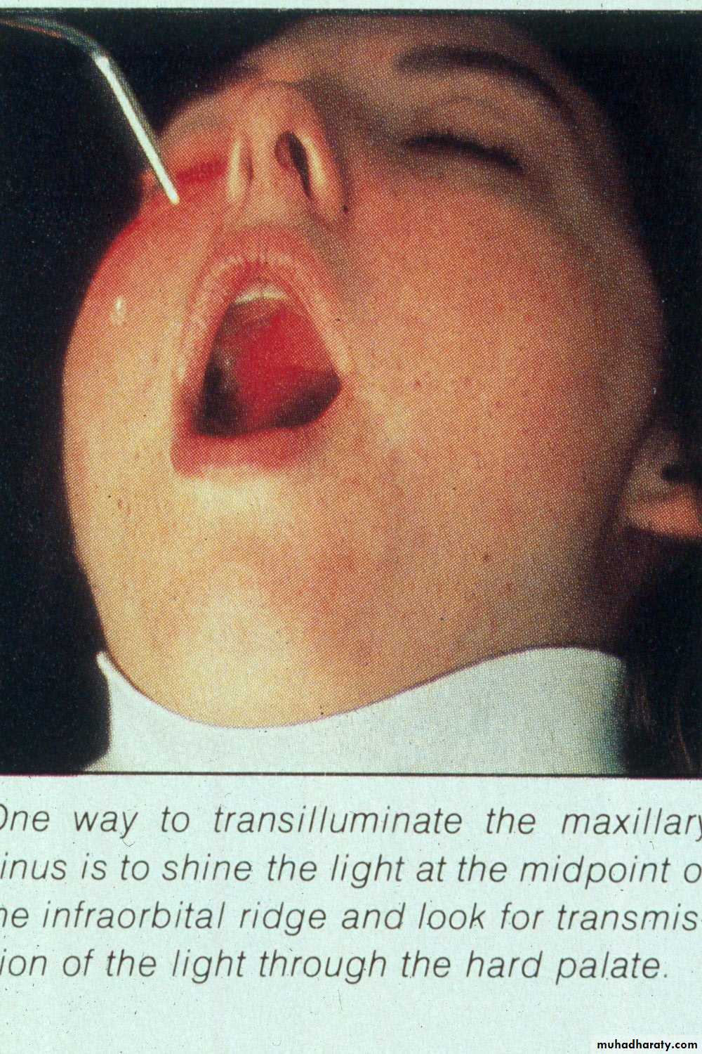
• Transillumination: The affected sinus shows decreased transmission of light; due to accumulation of fluid, debris, pus, and thickening of the sinus mucosa.
Radiology of Maxillary Sinus
• Extraoral views:
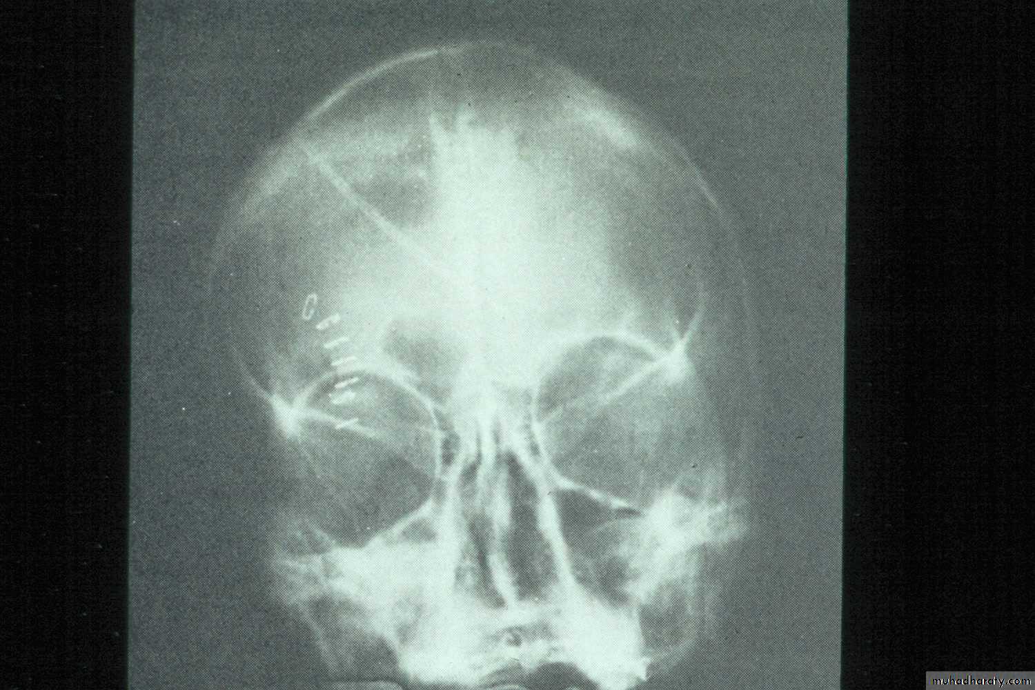
• Occipitomental View (15° OM): it’s called Water’s view. The presence of pus will produce a horizontal fluid level in this view; provided that there is air above it. As a measure of confirmation of the diagnosis, the view is repeated with the head tilted toward the side of pathology. The fluid level remains horizontal.
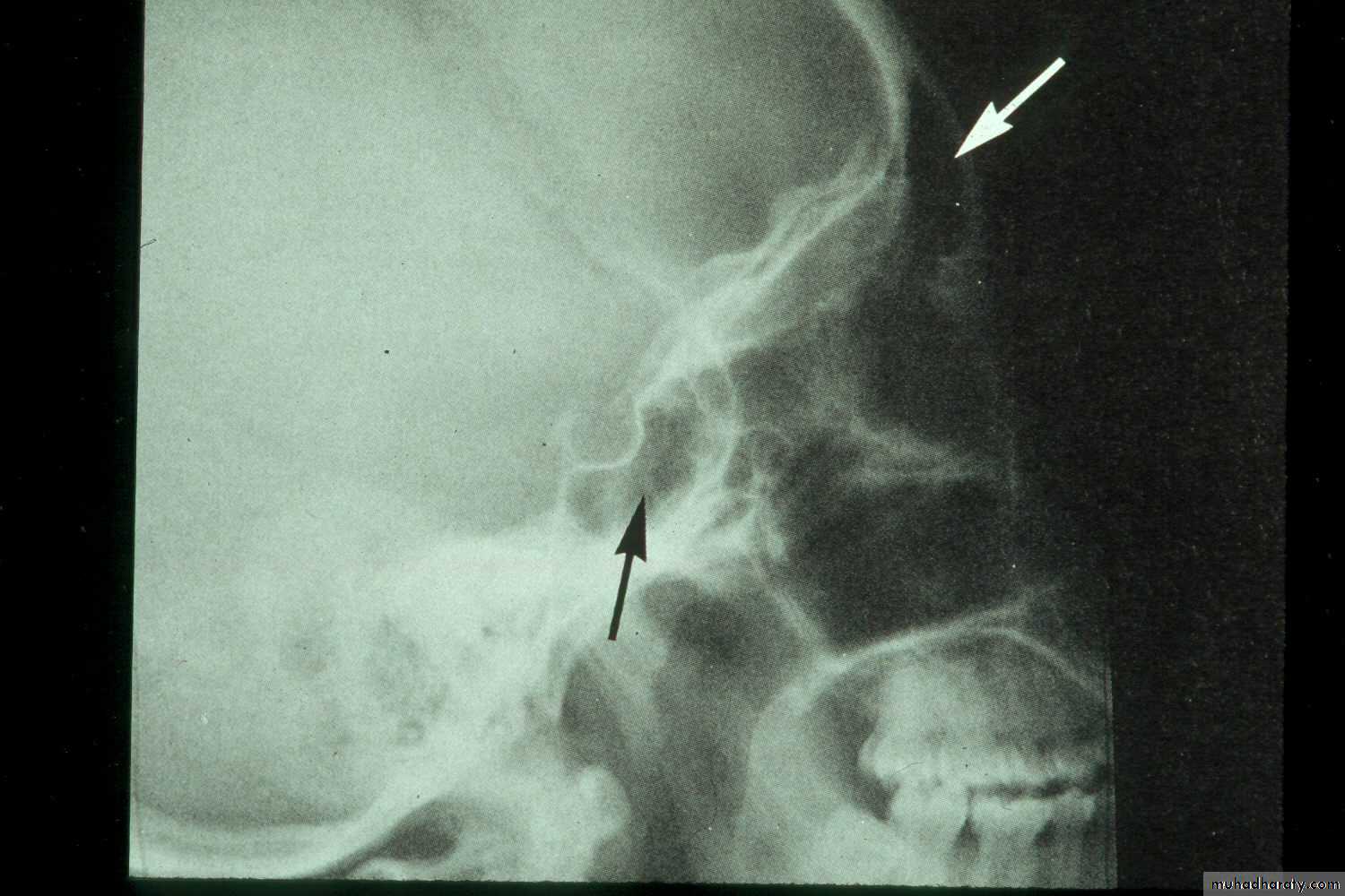
• Lateral Skull: (i) confirming the presence of fluid level and cyst (ii) in localizing a foreign body, e.g. root; particularly when the foreign body is located higher up in the sinus.
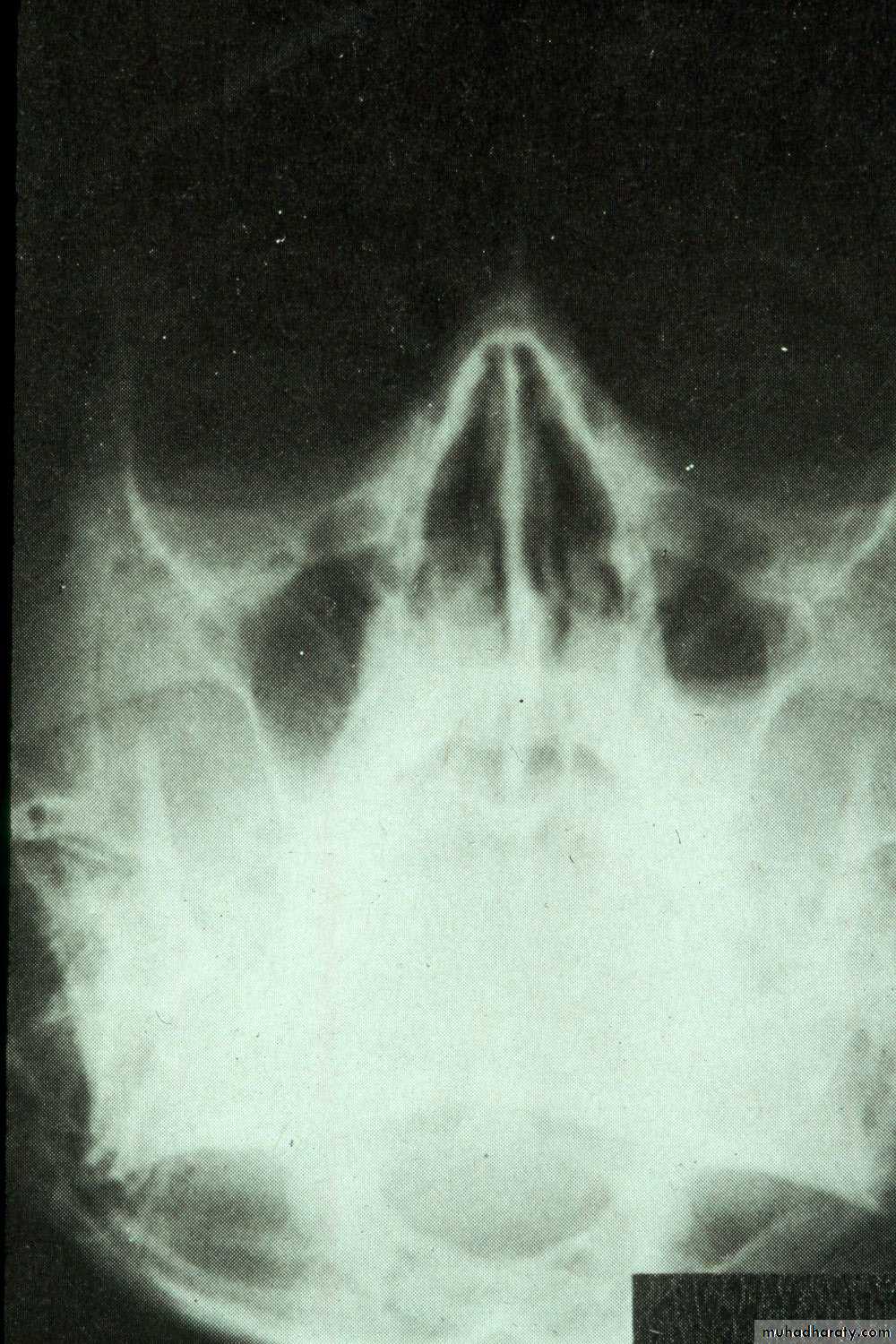
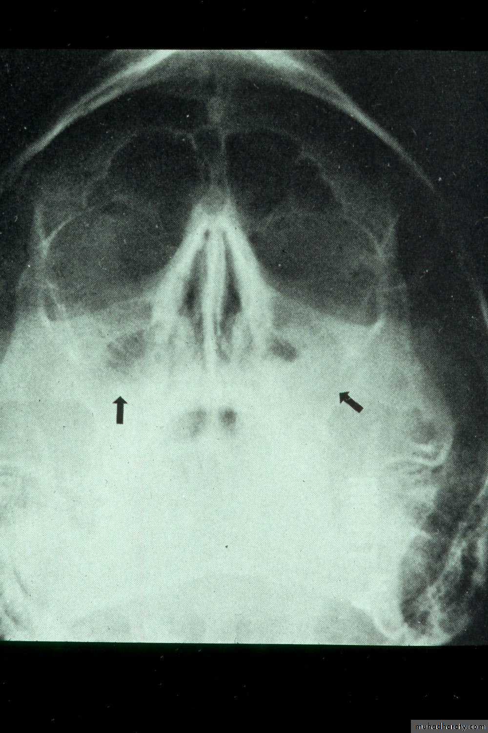
• Submentovertex View: This view is helpful in visualizing the posterior walls of maxillary sinus.
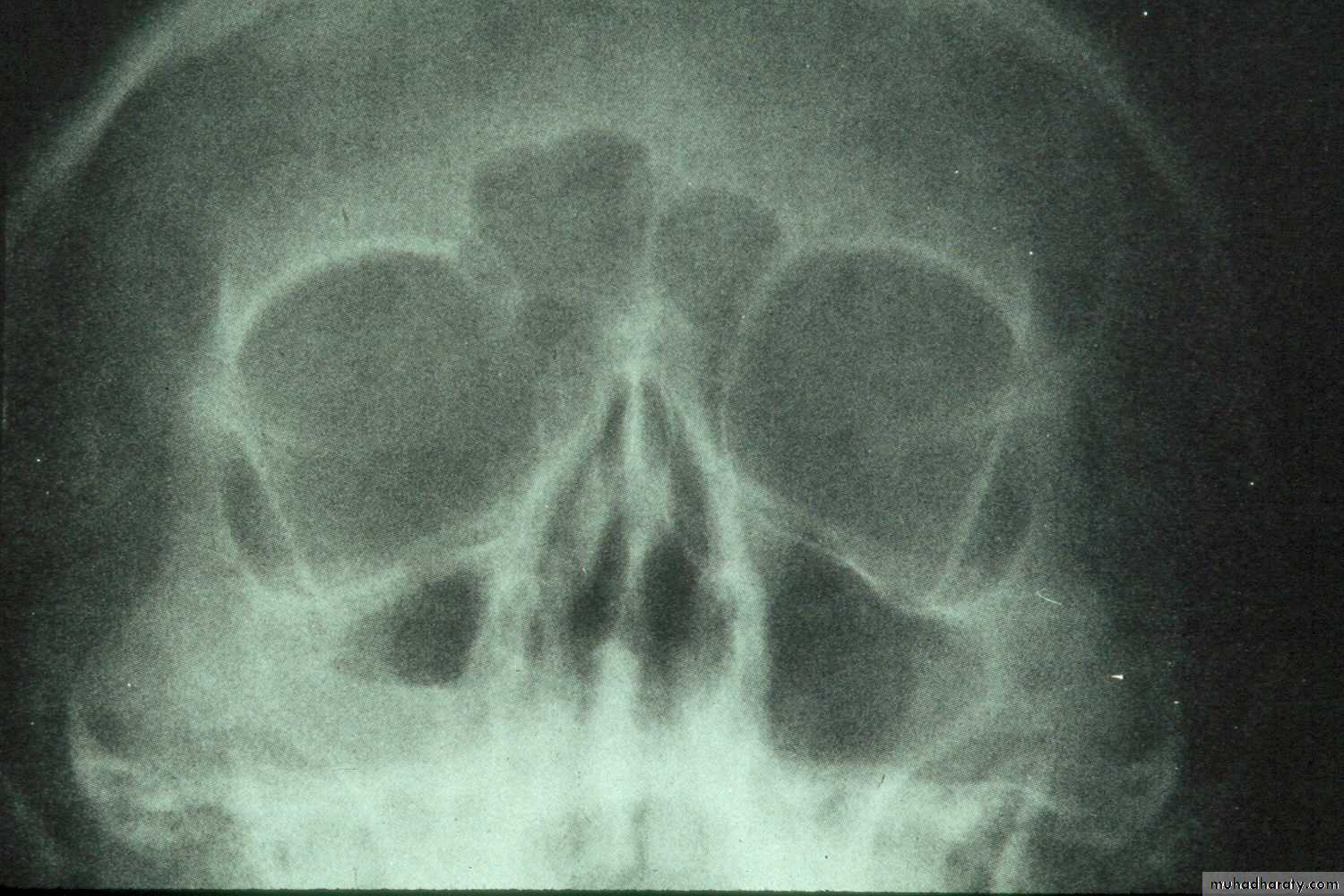
d. Occipitofrontal View: Is recommended to detect multisinusitis,
pansinusitis, if present.e. Tomography:This technique provides details of sinus structure.
(i) Solid masses within maxillary sinus such as osteoma; andantroliths,
(ii) early erosion of walls of maxillary sinus from malignant diseases.
f. Orthopantograph: is helpful in routine detection of lesions such as odontogenic and mucosal cysts of maxillary sinus.
2. Intraoral Views: (i) locating and retrieving foreign bodies in the sinus such as: teeth, roots, osseous fragments.
(ii) Careful planning of their surgical removal.
Acute Sinusitis
RadiographyRadiographic signs of sinus pathology :
Air fluid levels
Partial or complete opacification
Bony wall displacement
4 mm or more of mucosal wall thickening
Water’s view with air-fluid level in left maxillary sinus
Water’s view showing air-fluid level in right maxillary sinus and mucosal thickening in left maxillary sinus
Lateral view of normal frontal and sphenoid sinuses

Hypoplastic left frontal sinus and nosocomial right maxillary sinusitis
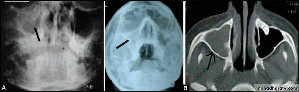
(A) PA view of Water’s position showing haziness of the (R) maxillary sinus, following extraction of upper right first molar 3 months back. Chronic maxillary sinusitis with oroantral fistula (B) PA view Water’s position and CT scan picture of another patient showing complete haziness of (R) maxillary sinus, indicating chronic maxillary sinusitis
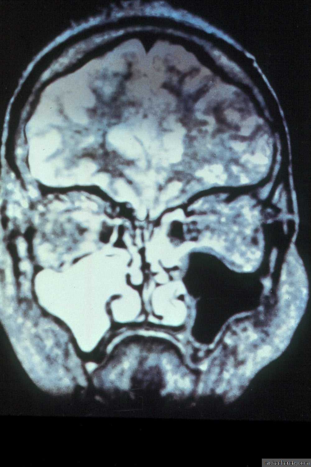
Coronal MRI scan showing maxillary sinusitis
Another diagnostic modality for sinusitis is nasal endoscopy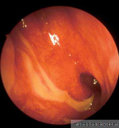
View into left nasal cavity demonstrates a polyp (P) extending from the middle meatus.
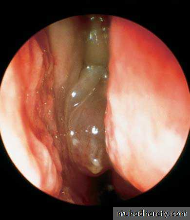
p
Purulent discharge from the middle meatus draining into the nasopharynx adjacent to the eustachian tube orifice.
Aspergillus fungal balls of the maxillary sinus. Note the fungal debris and mucosal edema.
General Treatment forAcute Sinusitis
• Oral antibiotic (Amoxicillin, Augmentin, Azithromycin)
• Topical and systemic decongestants as nasal drops or sprays e.g. Ephedrine sulphate
• Mucolytic agents: menthol, chlorbutol.
• Non-steroidal anti-inflammatory analgesic agents
• Other medication:
warm nasal saline irrigationsAntihistamine orally : if ellergy present
• Oral steroid: prednisolon
• Surgery (maxillary antrostomy, and Caldwell-Luc operation)
Oroantral Communication and Fistula
Definition• An oroantral perforation is an unnatural communication between the oral cavity and maxillary sinus.
b. An oroantral fistula is an epithelialized, pathological, unnatural communication between these two cavities.
Symptoms: Fresh Oroantral Communication:
• Escape of fluids: From mouth to the nose on the side of extraction.
• Unilateral epistaxis : It is due to blood in the sinus escaping through osteum into the nostril.
• Escape of air from mouth into the nose, on sucking, inhaling or drawing on a cigarette, or puffing the cheeks (Inability to blow cheeks.Passage of air into mouth on sucking).
• Enhanced column of air: Causes alteration in vocal resonance and subsequently change in the voice.
• Excruciating pain: In and around the region of the affected sinus, as the local anaesthesia begins to wear off.
Symptoms of Established Oroantral Fistula:
In late Stage• Pain
• Persistent, purulent or mucopurulent, foul, unilateral nasal discharge Postnasal drip
• Foul or foetid taste and smell.
• Possible sequelae of general systemic toxaemic condition : Fever, malaise, morning anorexia, frontal and parietal headaches
• Popping out of an antral polyp
Managment of Recently Created Communication
If the extraction demanded more force, however, the extraction was straight forward, and the examination of roots of the tooth revealed that a part of bony floor of antrum is seen adherent to the tooth, then the operator must examine the socket to establish any tear in the lining of sinus.If the fistula is large, it can be assessed from inspection;
In case, its patency is not obvious, the nose blowing test is useful. Compression of anterior nares, followed by gentle blowing of nose (with mouth open). Escape of air. bubbles, blood or pus, etc. may appear at the oral orifice. The site of recent extraction, should not be explored with an instrument, which lead to breakdown of the blood clot and the seal.
Treatment of Early Cases
Ideal treatment (i) is immediate surgery repair to achieve primary closure, and
(ii) simultaneous antibiotic prophylaxis to prevent sinus infection: Penicillin
and its derivatives until symptoms begin to subside.
(iii) Nasal decongestants: to encourage the drainage of pus and secretions.
(iv) Analgesics: Non-steroidal anti-inflammatory agents.
The immediate primary closure is done by a simple reduction of the buccal and palatal socket walls, to allow coaptation of buccal and palatal soft tissue flaps to close over the defect. A protective acrylic denture or splint can be used to provide a barrier to the inadvertent entry of food particles.
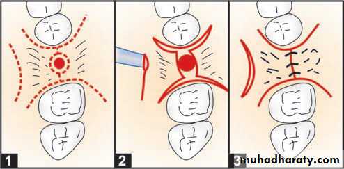
Closure of accidental oro-antral communication in the dentulous arch (1) Incisions are made around the teeth and antral opening. A relaxing incision is made on the palate (2) Mucoperiosteal flaps raised and the buccal and palatal alveolar walls are reduced with rongeur, (3) Interrupte suturing done
Treatment of cases seen more than 24 hours after accident
When a period of 24 hours has elapsed, the soft tissue margins of fistula often get infected. It is preferable to defer the surgical closure until gingival edges show sound healing, i.e. approximately three weeks.Surgical Procedures Used in Closure of Oroantral Fistula
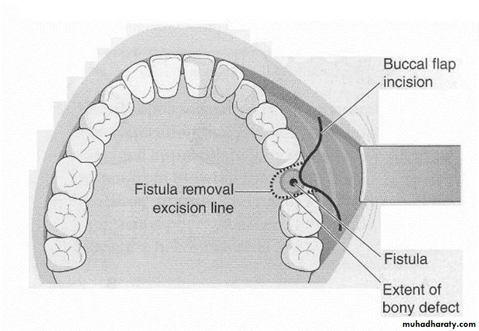
(i) Buccal advancement flap
(ii) Palatal rotational flap
(iii) Combination of both.
Advancement of buccal flap
General principle of flap design:• The free end of the flap should have an adequate blood supply.
• The buccal flap is so designed, that the base should be wider than the apex; to ensure adequate vascularization at the apex.
• Suture line is well-supported by sound bone.
• Mobilization of either buccal or palatal flap should be done in such a manner that there is no tension on the suture line.
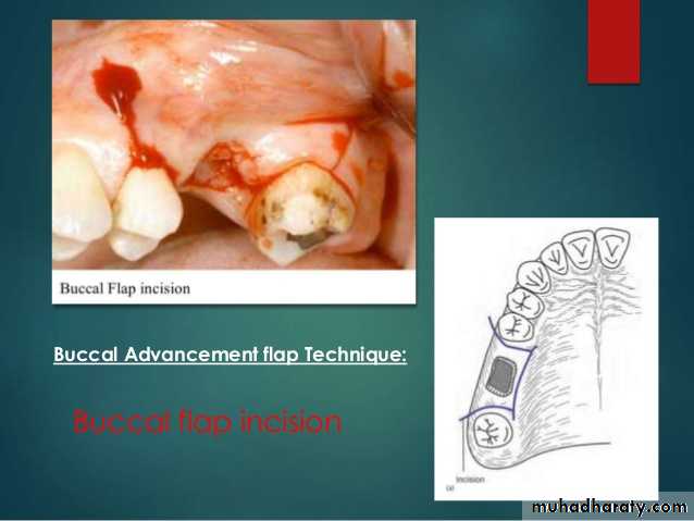
Advancement of buccal flap
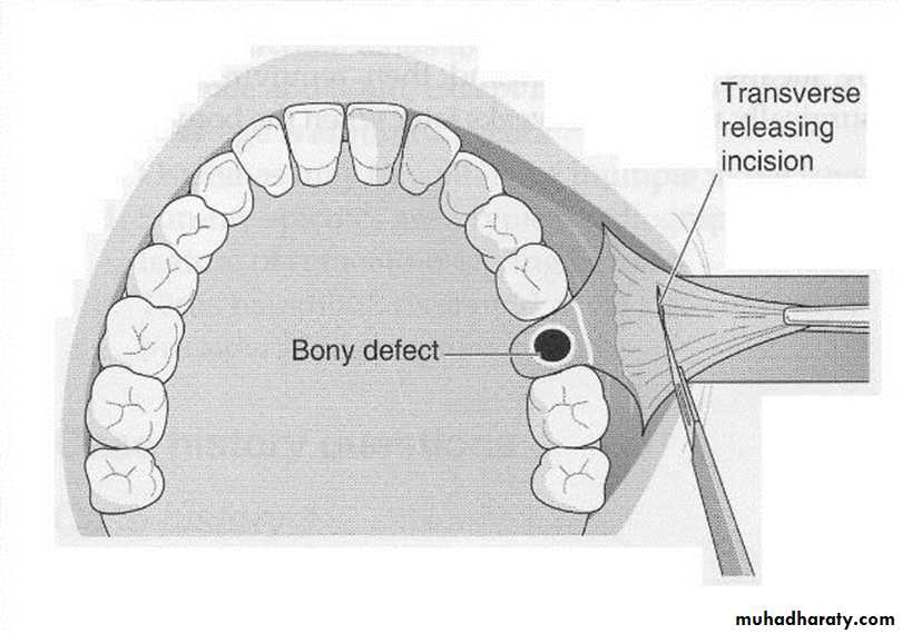
The buccal mucoperiosteal flap falls short of covering the fistula; the flap can be advanced. A horizontal incision is made in the periosteum, as high as possible. This will allow advancement of buccal flap.
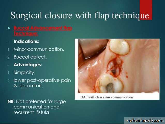
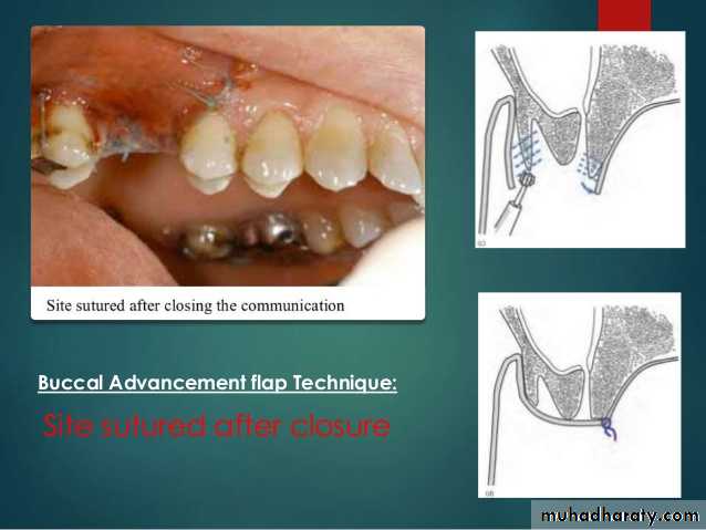
Buccal Advancement flap Technique: -
Indications:
• Minor communication.• Buccal defect.
Advantages:
1. Simplicity.
2. lower post-operative pain & discomfort.NB: Not preferred for large communication and recurrent fistula
Buccal Advancement flap Technique: -
Disadvantages:• Thin flap dehiscense.
• limited extent.• loss of vestibular depth.
• scaring may cause
• impaired mobility.
• In case, if antral pathology is present, Caldwell-Luc procedure should be carried out before the final closure of fistula.
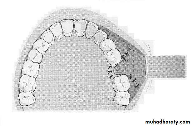
• After that closure of wound: The mucoperiosteal flap is sutured into position across fistula with interrupted sutures.
• Postoperative medications: (i) Antibiotics, (ii) Analgesics, and (iii)Nasal decongestants and inhalations.
• Restriction to soft diet.
• Instructions to patients: (i) to avoid sneezing, (ii) to explore the wound with tongue, or deliberately sucking air or fluid through it.
• Removal of sutures 7 to 10 days postoperatively.
• Patient is reviewed regularly to observe progress, clinically and radiologically.
Diagramatic illustration of advancement buccal flap
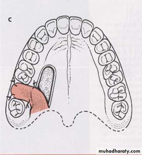
Palatal Pedicled Flap: Rotational Advancement Flap Operation
Advantage:• The palate gets its blood supply from greater palatine arteries that promotes satisfactory healing.
• Palatal flap is rotated across fistula so that the suture line rests on sound bone on buccal side of the orifice.
• Provides adequate mobility and tissue bulk to the flap.
• Do not affect the buccal vestibular height.
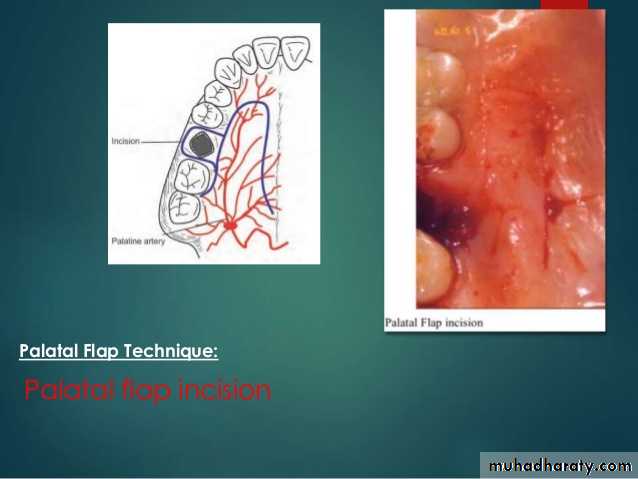
Palatal Pedicled Rotational Flap technique
Palatal flap incision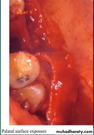
Palatal Pedicled Rotational Flap technique
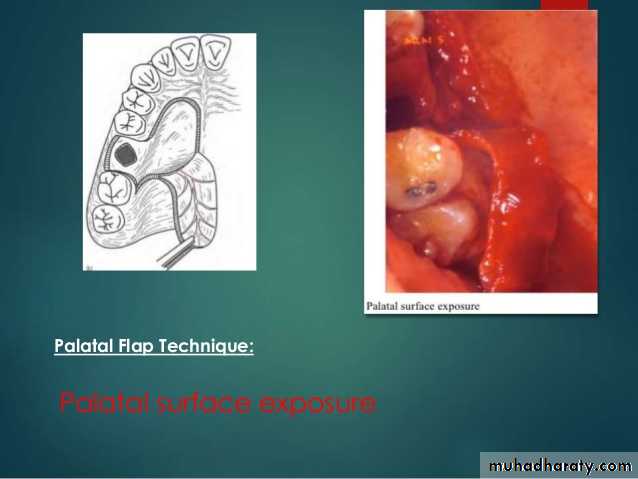
Palatal surface exposure
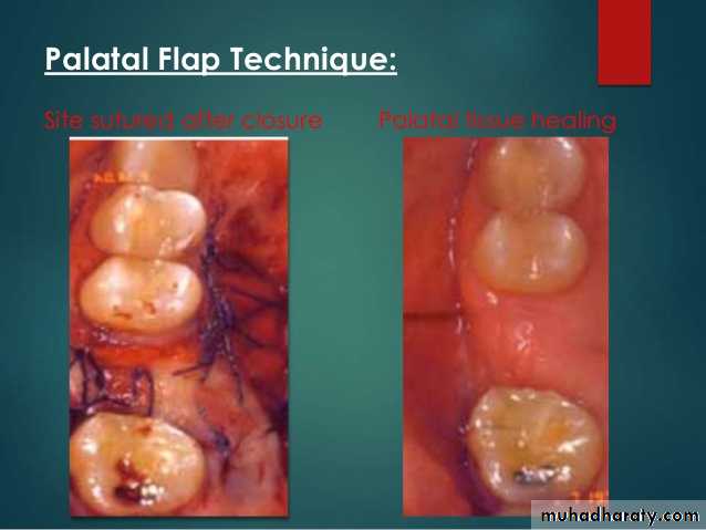
Site of suture after closure
Palatal tissue healingPalatal Pedicled Rotational Flap technique
Disadvantage
1- Denudation of palatal surface .2- Greater post-operative pain.
3- More difficult than buccal flap
4- Appearance of roughness at doner site (epithilization)
5- Possible flap necrosis.
6- Interfer with wearing partial denture for covering the hard palate.
Combination of Buccal and Palatal Flaps
To close larger defects by local flaps often leads to failure.
When there is a history of earlier repair with failure.
It helps to have two layered closure to improves the strength of the flaps, also to minimizes contraction and risk of infection