2nd type of Basic tissues
Connective Tissue (Co.T)Co. T Facts
• They are the most abundant and widely distributed tissue type in the body.• Connective tissues run the gamut for vascularity.
• Some tissues are avascular (Cartilage), some are poorly vascularized (dense Co. T) and some have rich blood supplies (bone).
Co. T. Facts
Co. T. can be rigid (bone), flexible (adipose), or fluid (blood).Unlike the tightly packed Epithelial tissues, living cells in Co.T are separated by a non-living extra-cellular matrix [ECM](Ground Substance and Fibers).
Due to the matrix, Co.T are able to bear weight, withstand tension, and endure abuses that no other tissues could tolerate.
Function of Co.T
Many specific functions. Its major functions includeBinding and support
Protection
Insulation
Transportation of substances
Components of Co.T
• Ground Substance (Matrix)
• Fibers
• Cells
Co.T Facts – Ground Substance
The ground substance is the unstructured material between cells that contains the fibers.The ground substance holds large amounts of fluid and serves as a medium through which nutrients and other substances can diffuse between blood vessels and the cells.
Ground Substance
Ground Substance
Co.T, FibersThere are 3 types of fibers prevalent in Co.T
A). Collagen fibers – are wide and wavy in appearance and generally stain pink. 79% of the protein in the body is collagen.
B). Elastic fibers – are thin flexible fibers made of protein elastin, that generally stain black.
C). Reticular fibers – are actually thin collagen fibers. They have a spider web appearance and appear black under stain.
Fiber Types
Reticular Fibers
Collagen FiberElastic Fiber
Co.T, The Cells
Each major type of Co. T has its own fundamental cell type in both immature and mature forms.• The following types of connective tissue are covered in this activity:
• Loose (areolar) connective tissue (delicate thin layers between• tissues; present in all mucous membranes)
• 2. Adipose tissue (fat)
• 3. Dense connective tissue (tendons/ligaments)
• 4. Hyaline cartilage (nose/ends of long bones/ribs)
• 5. Elastic cartilage (outer ear/epiglottis)
• 6. Fibrocartilage (between vertebrae/knee joints/pubic joint)
• 7. Bone (skeletal system)
• 8 Blood (bloodstream)
•
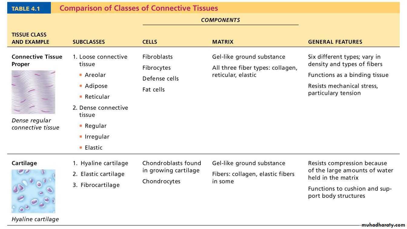
Table 4.1 Comparison of Classes of Connective Tissues (1 of 2)
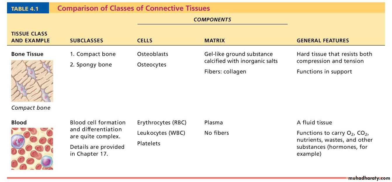
Table 4.1 Comparison of Classes of Connective Tissues (2 of 2)
Figure 4.8a Connective tissues.
(a) Connective tissue proper: loose connective tissue, areolar
Description: Gel-like matrix with allthree fiber types; cells: fibroblasts,
macrophages, mast cells, and some
white blood cells.
Function: Wraps and cushions
organs; its macrophages phagocytize
bacteria; plays important role in
inflammation; holds and conveys
tissue fluid.
Location: Widely distributed under
epithelia of body, e.g., forms lamina
propria of mucous membranes;
packages organs; surrounds
capillaries.
Photomicrograph: Areolar connective tissue, a
soft packaging tissue of the body (300x).
Epithelium
Lamina
propria
Fibroblast
nuclei
Elastic
fibers
Collagen
fibers
Figure 4.7 Areolar connective tissue: A prototype (model) connective tissue.
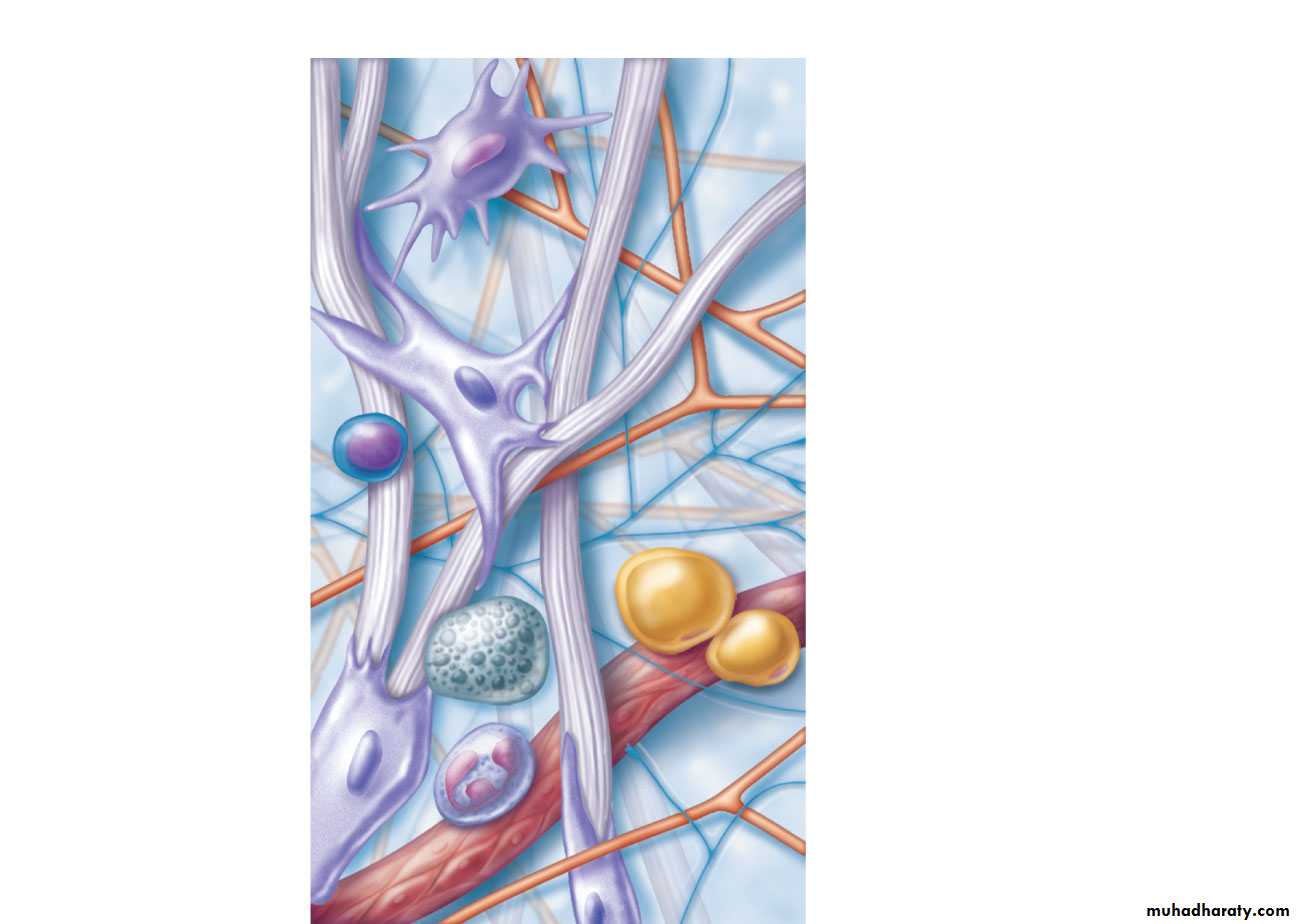
Macrophage
FibroblastLymphocyte
Fat cell
Mast cell
Neutrophil
Capillary
Cell types
Extracellular
matrix
Fibers
• Collagen fiber
• Elastic fiber
• Reticular fiber
Ground substance
Figure 4.8b Connective tissues.
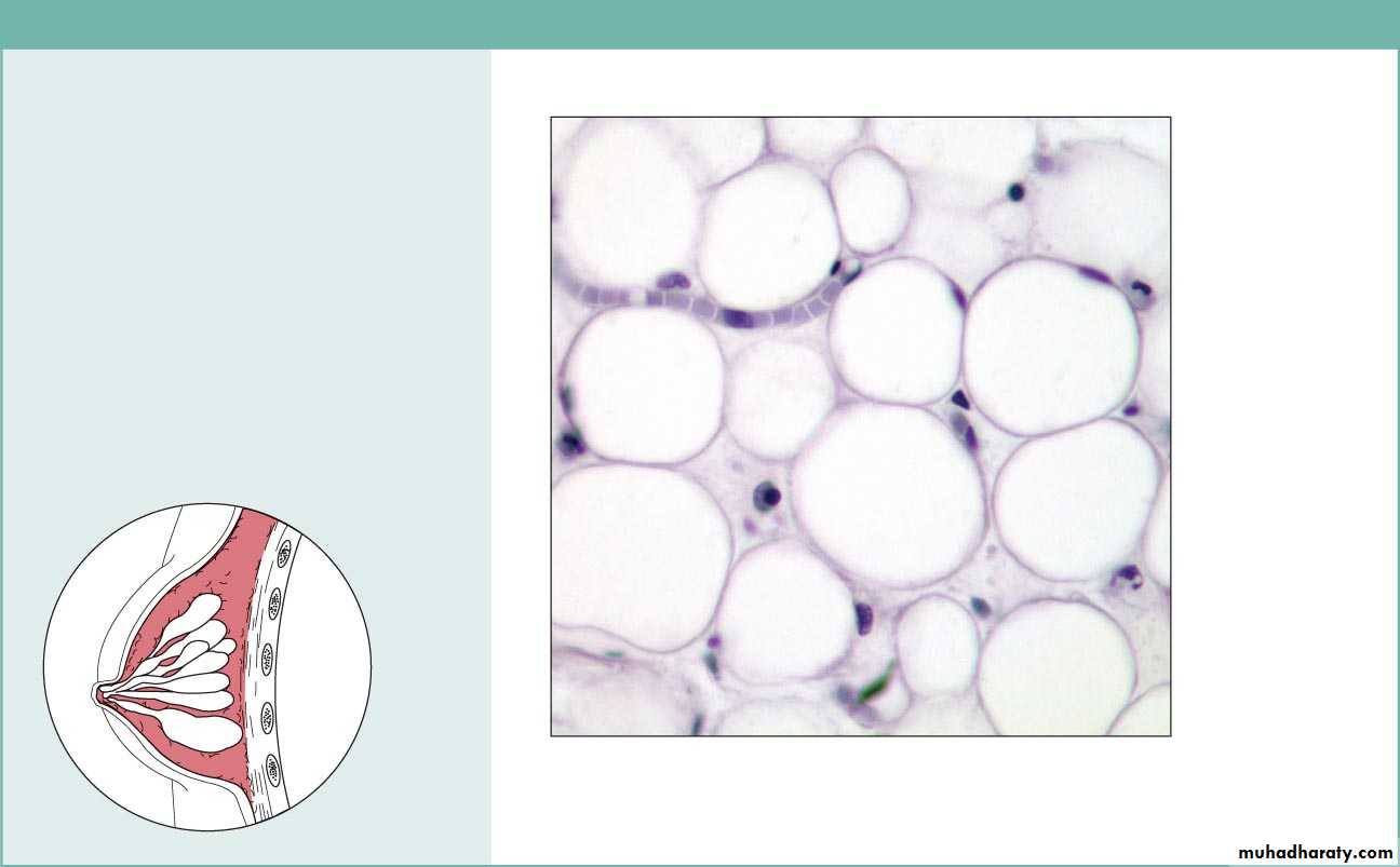
(b) Connective tissue proper: loose connective tissue, adipose
Description: Matrix as in areolar,but very sparse; closely packed
adipocytes, or fat cells, have
nucleus pushed to the side by large
fat droplet.
Function: Provides reserve food
fuel; insulates against heat loss;
supports and protects organs.
Location: Under skin in the
hypodermis; around kidneys and
eyeballs; within abdomen; in breasts.
Photomicrograph: Adipose tissue from the
subcutaneous layer under the skin (350x).
Nucleus of
fat cell
Vacuole
containing
fat droplet
Adipose
tissue
Mammary
glands
Figure 4.8c Connective tissues.
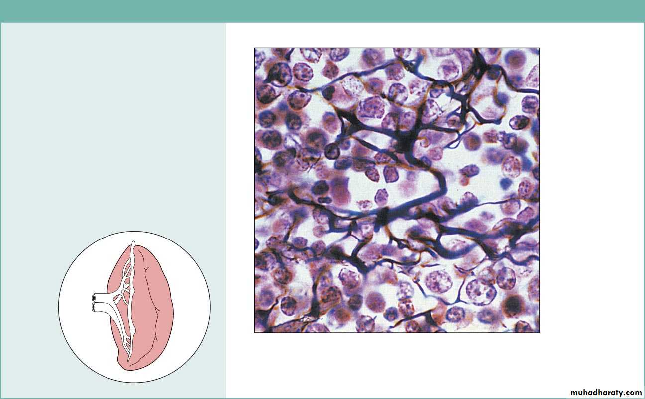
(c) Connective tissue proper: loose connective tissue, reticular
Description: Network of reticularfibers in a typical loose ground
substance; reticular cells lie on the
network.
Function: Fibers form a soft internal
skeleton (stroma) that supports other
cell types including white blood cells,
mast cells, and macrophages.
Location: Lymphoid organs (lymph
nodes, bone marrow, and spleen).
Photomicrograph: Dark-staining network of reticular
connective tissue fibers forming the internal skeleton
of the spleen (350x).
Spleen
White blood
cell
(lymphocyte)
Reticular
fibers
Figure 4.8d Connective tissues.

(d) Connective tissue proper: dense connective tissue, dense regular
Description: Primarily parallelcollagen fibers; a few elastic fibers;
major cell type is the fibroblast.
Function: Attaches muscles to
bones or to muscles; attaches bones
to bones; withstands great tensile
stress when pulling force is applied
in one direction.
Location: Tendons, most
ligaments, aponeuroses.
Photomicrograph: Dense regular connective
tissue from a tendon (500x).
Shoulder
joint
Ligament
Tendon
Collagen
fibers
Nuclei of
fibroblasts
Figure 4.8e Connective tissues.

(e) Connective tissue proper: dense connective tissue, dense irregular
Description: Primarilyirregularly arranged collagen
fibers; some elastic fibers;
major cell type is the fibroblast.
Function: Able to withstand
tension exerted in many
directions; provides structural
strength.
Location: Fibrous capsules of
organs and of joints; dermis of
the skin; submucosa of
digestive tract.
Photomicrograph: Dense irregular
connective tissue from the dermis of the
skin (400x).
Collagen
fibers
Nuclei of
fibroblasts
Fibrous
joint
capsule
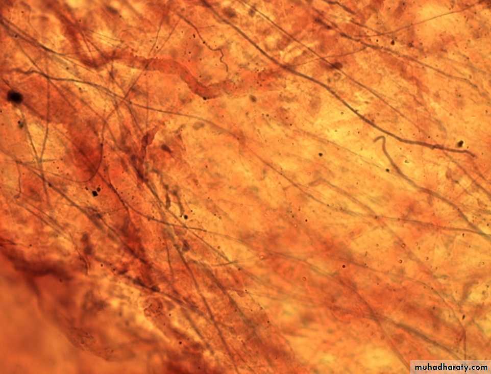
Figure 4.8f Connective tissues.
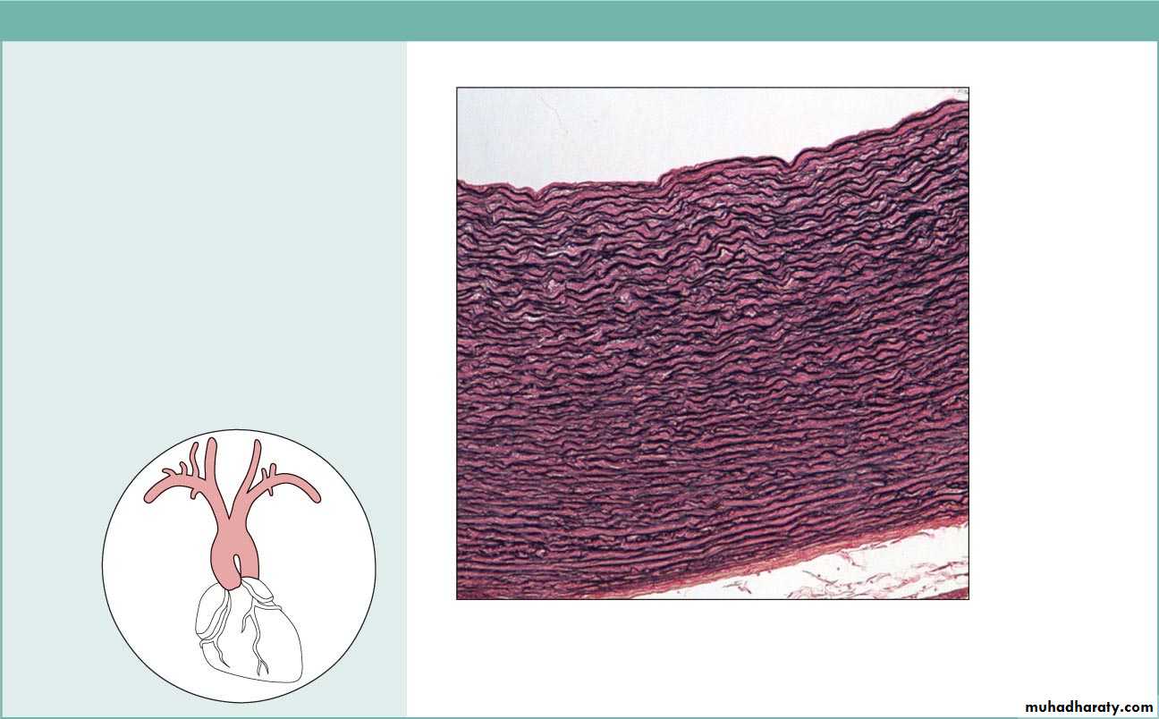
(f) Connective tissue proper: dense connective tissue, elastic
Description: Dense regularconnective tissue containing a high
proportion of elastic fibers.
Function: Allows recoil of tissue
following stretching; maintains
pulsatile flow of blood through
arteries; aids passive recoil of lungs
following inspiration.
Location: Walls of large arteries;
within certain ligaments associated
with the vertebral column; within the
walls of the bronchial tubes.
Elastic fibers
Aorta
Heart
Photomicrograph: Elastic connective tissue in
the wall of the aorta (250x).
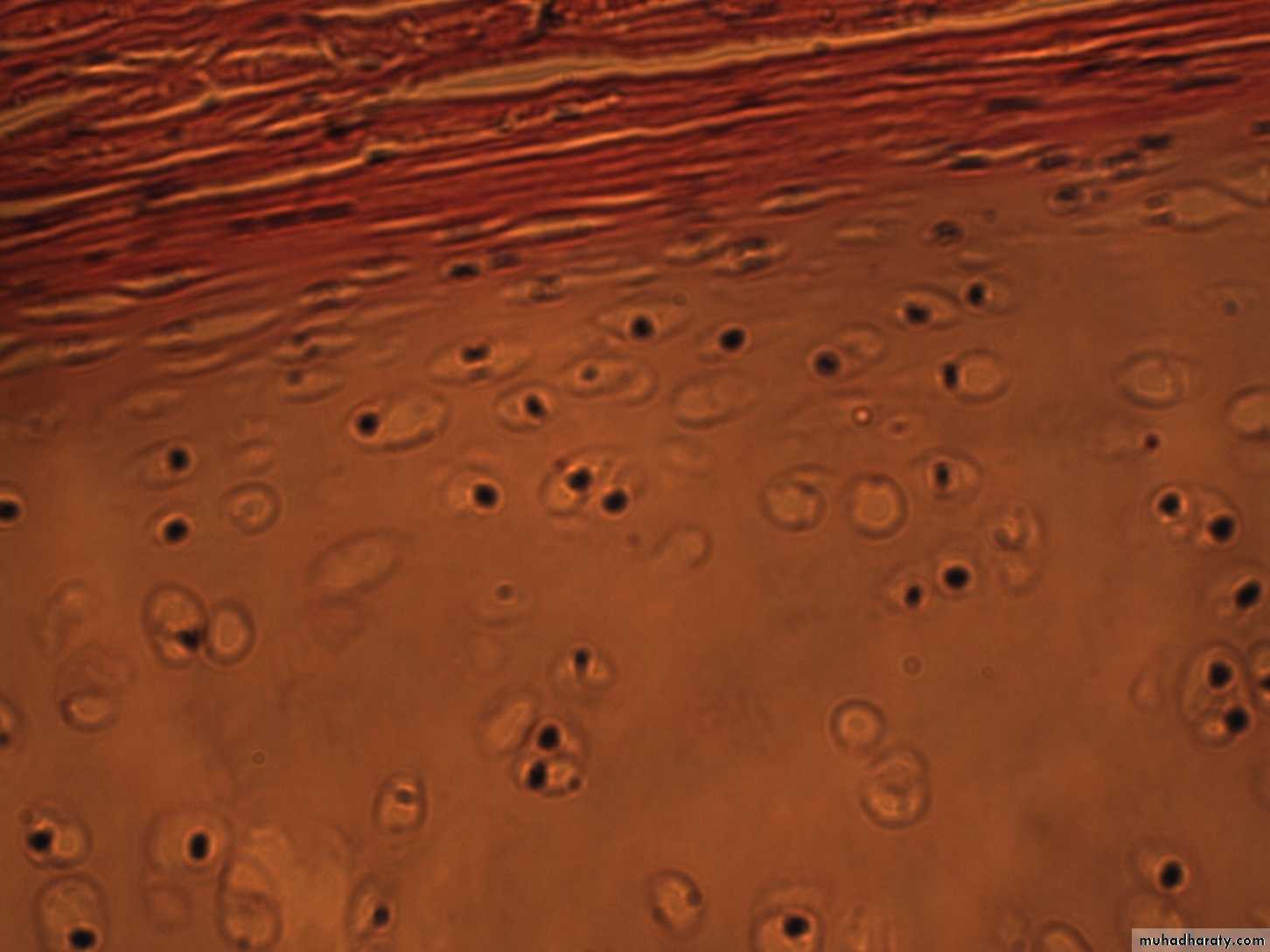
Figure 4.8g Connective tissues.
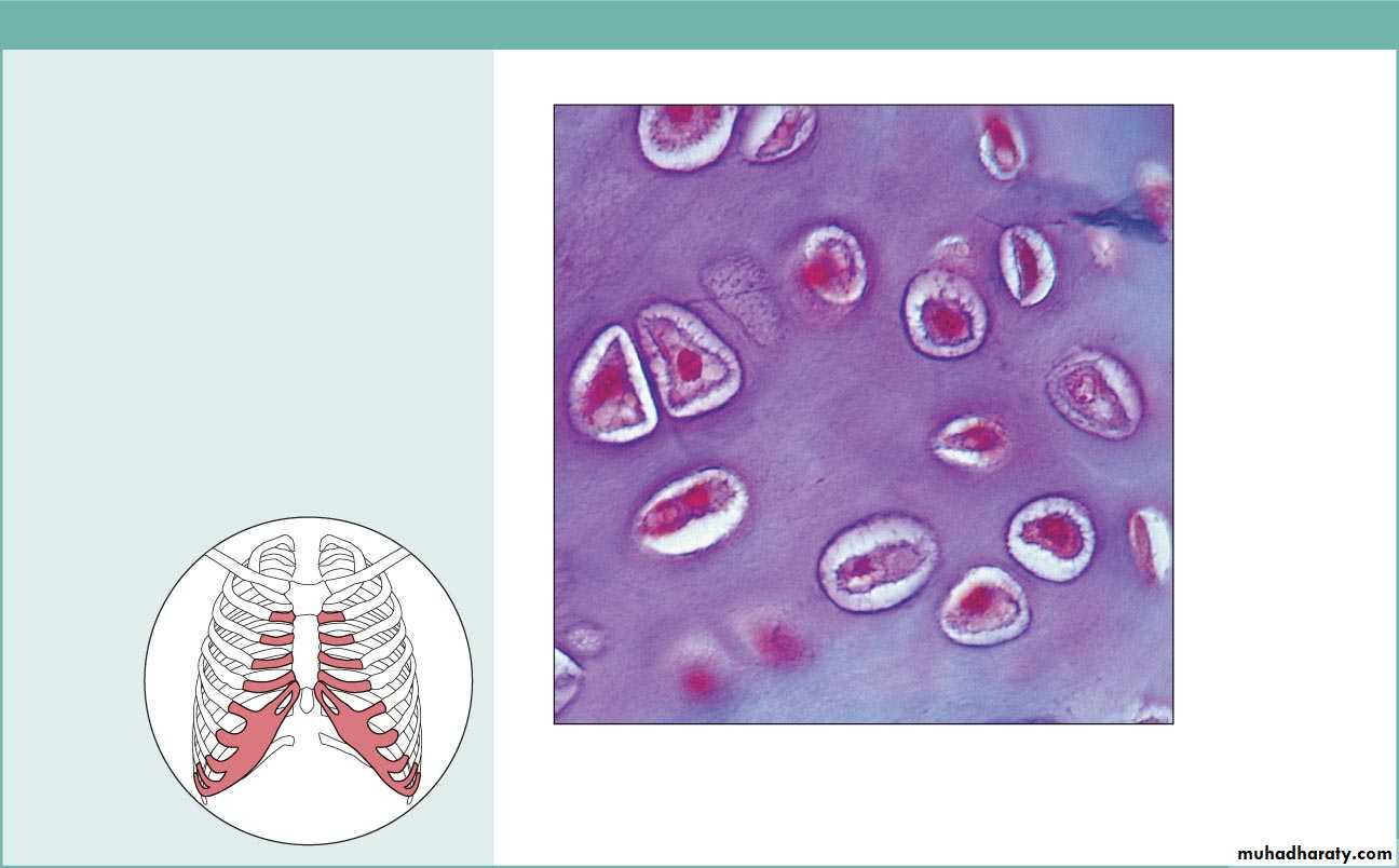
(g) Cartilage: hyaline
Description: Amorphous but firmmatrix; collagen fibers form an
imperceptible network; chondroblasts
produce the matrix and when mature
(chondrocytes) lie in lacunae.
Function: Supports and reinforces;
has resilient cushioning properties;
resists compressive stress.
Location: Forms most of the
embryonic skeleton; covers the ends
of long bones in joint cavities; forms
costal cartilages of the ribs; cartilages
of the nose, trachea, and larynx.
Photomicrograph: Hyaline cartilage from the
trachea (750x).
Costal
cartilages
Chondrocyte
in lacuna
Matrix
Figure 4.8h Connective tissues.
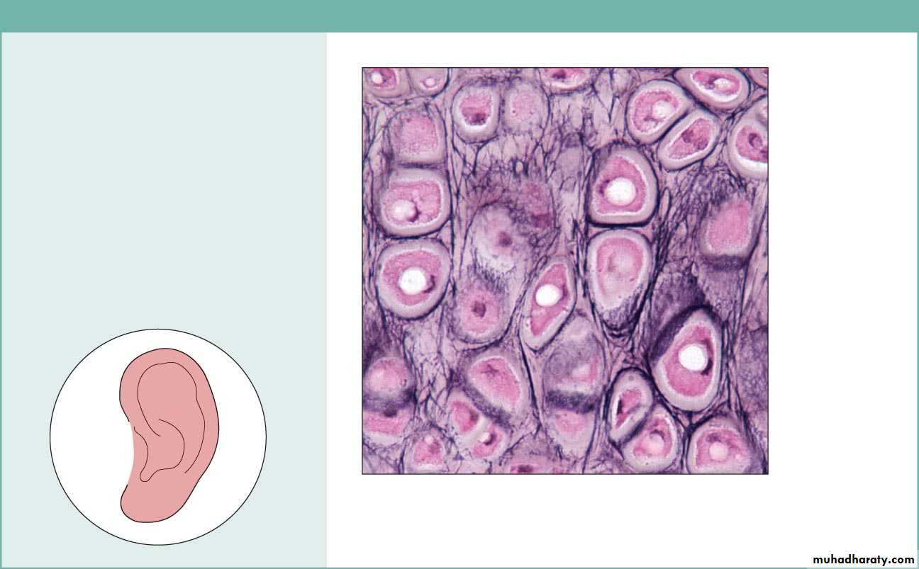
(h) Cartilage: elastic
Description: Similar to hyalinecartilage, but more elastic fibers
in matrix.
Function: Maintains the shape
of a structure while allowing
great flexibility.
Location: Supports the external
ear (pinna); epiglottis.
Photomicrograph: Elastic cartilage from
the human ear pinna; forms the flexible
skeleton of the ear (800x).
Chondrocyte
in lacuna
Matrix
Figure 4.8i Connective tissues.

(i) Cartilage: fibrocartilage
Description: Matrix similar tobut less firm than that in hyaline
cartilage; thick collagen fibers
predominate.
Function: Tensile strength
with the ability to absorb
compressive shock.
Location: Intervertebral discs;
pubic symphysis; discs of knee
joint.
Photomicrograph: Fibrocartilage of an
intervertebral disc (125x). Special staining
produced the blue color seen.
Intervertebral
discs
Chondrocytes
in lacunae
Collagen
fiber
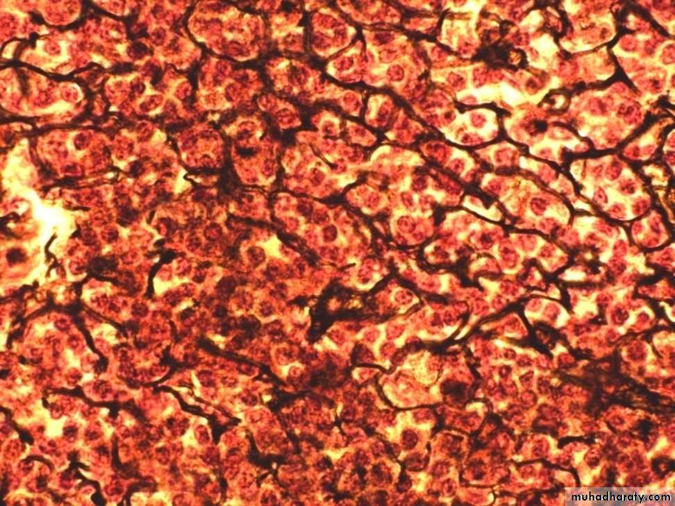
Figure 4.8j Connective tissues.
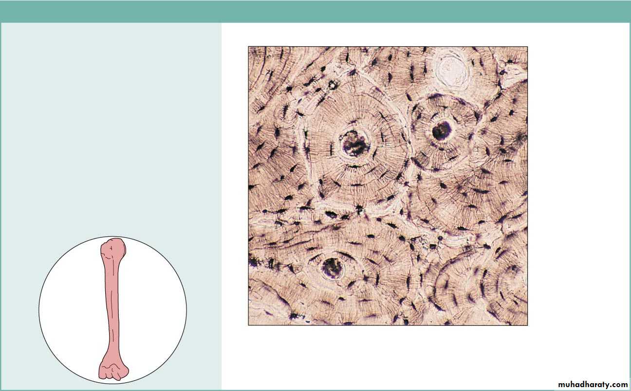
(j) Others: bone (osseous tissue)
Description: Hard, calcifiedmatrix containing many collagen
fibers; osteocytes lie in lacunae.
Very well vascularized.
Function: Bone supports and
protects (by enclosing);
provides levers for the muscles
to act on; stores calcium and
other minerals and fat; marrow
inside bones is the site for blood
cell formation (hematopoiesis).
Location: Bones
Photomicrograph: Cross-sectional view
of bone (125x).
Lacunae
LamellaCentral
canal