Dr. Monia M.N. Kandil
(1)
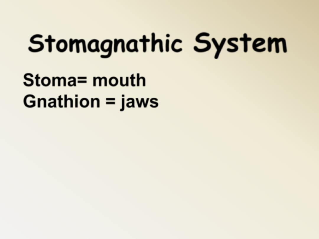
Stomagnathic
System
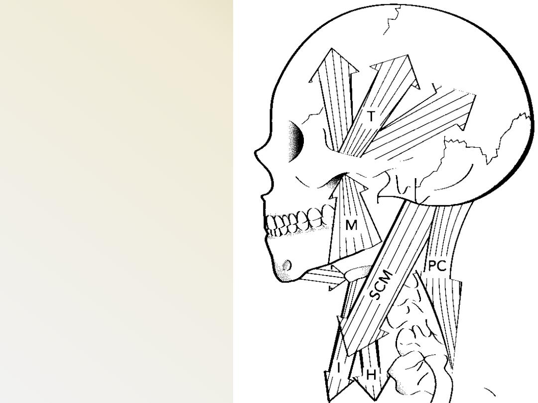
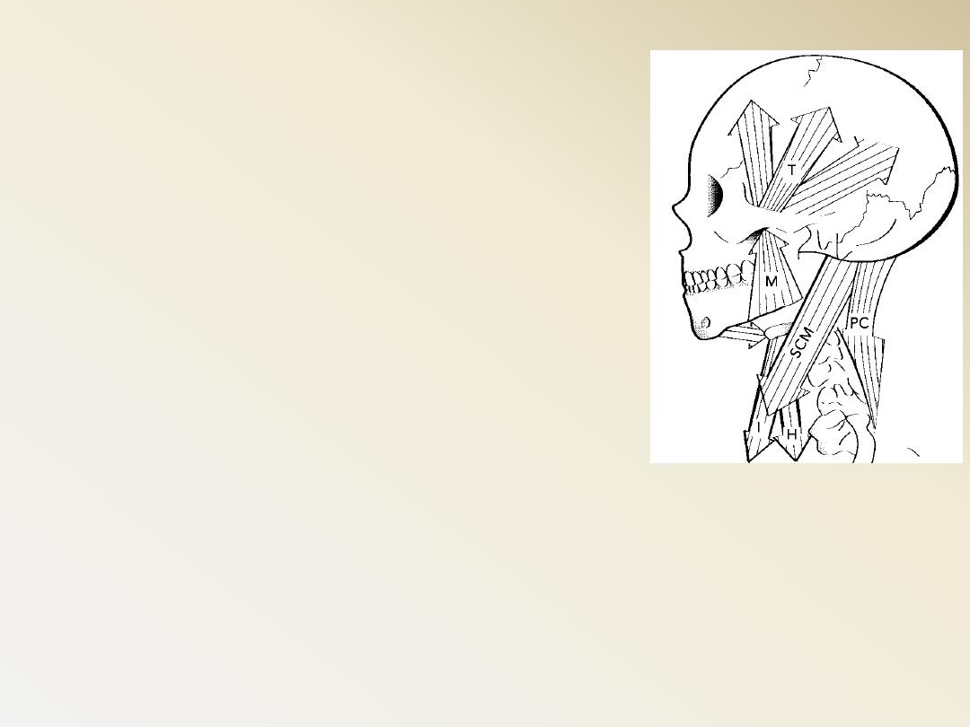
The movement of the jaw is intiated by
a complex set of muscles’ actions, which
are in turn controlled by the body's
local and central nervous system
Stoma= mouth
Gnathion = jaws
The stomatognathic
system
= Masticatory system =
• Teeth
• Periodontium
• Jaws
• TMJ
• Associated muscles +
tongue & ms of the soft
palate
• Investing tissues
• Neural control
The stomatognathic system
• There is a
complex, dynamic
balance
between the way that the
teeth come together, the muscle
that work the jaws, the joints, and
their relationship to the posture of the head & neck. A
change in any part of the system affects all the others
and change the balance
( Masticatory system)
The stomatognathic system
Is the functional unit of the body primary
responsible for chewing, speaking and
swallowing, as well as parafunctional
actions.
Disturbance of any part could disturb the
whole odontostomatognathic system and
subsequently the body as well.
When opposing teeth are in contact and
mandibular movements are made, the
direction of the movement is controlled
by the neuromuscular system as limited
by the movement
The stomatognathic system
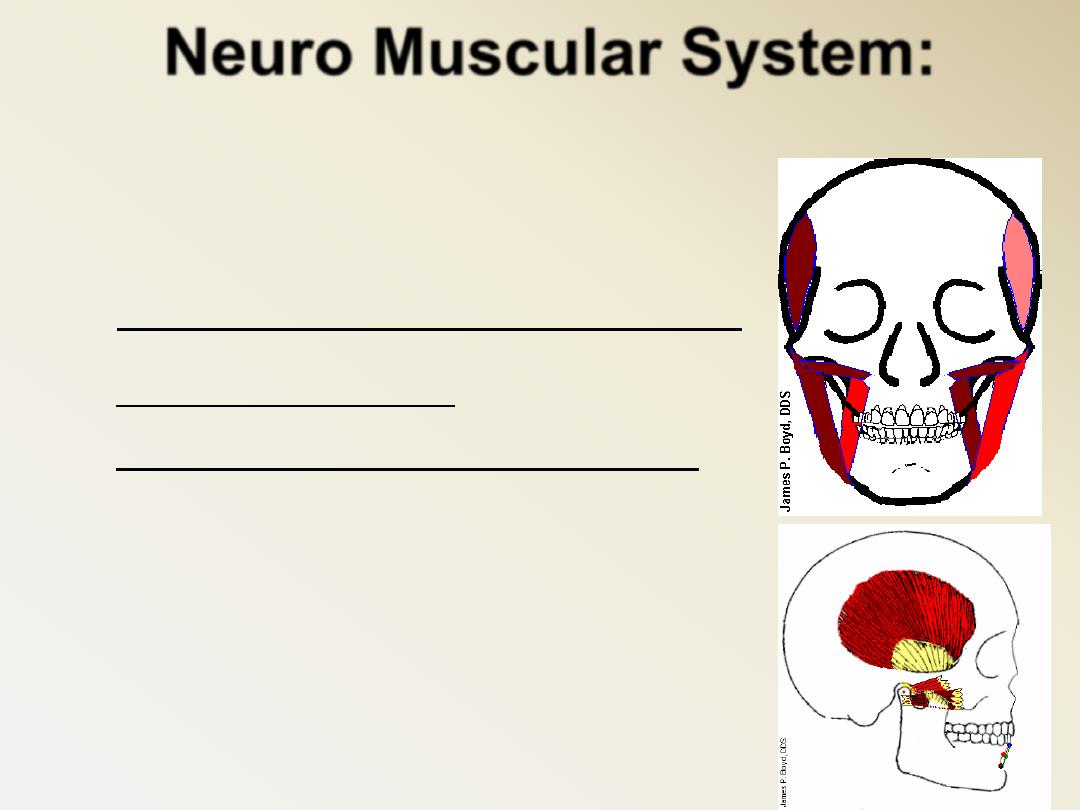
Muscles of Mastication:
Neuro Muscular System:
Tempromandibular
Sphenomandibular
Stylomandibular
TMJ Capsule
Associated Ligaments
The direction of mand. movement is controlled
by the neuromuscular system
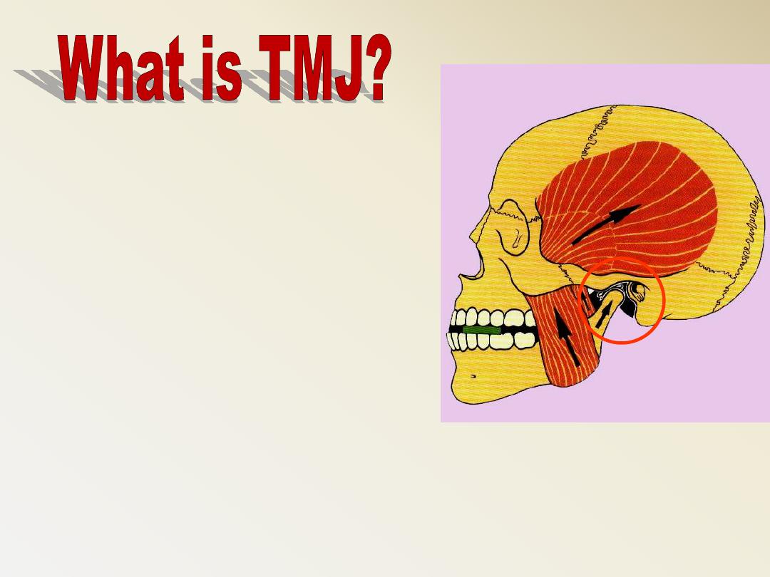
The letters TMJ are short
for of 'temporo-mandibular
joint', which is the joint
connecting your lower jaw
and your skull.
The movement in this joint lets you open and
close your mouth and chew from side to side.
Temporalis
Masseter
It has 4 anatomical parts
:
1- condyle
2- Articular fossa
3- Articular disc
4- Articular capsule
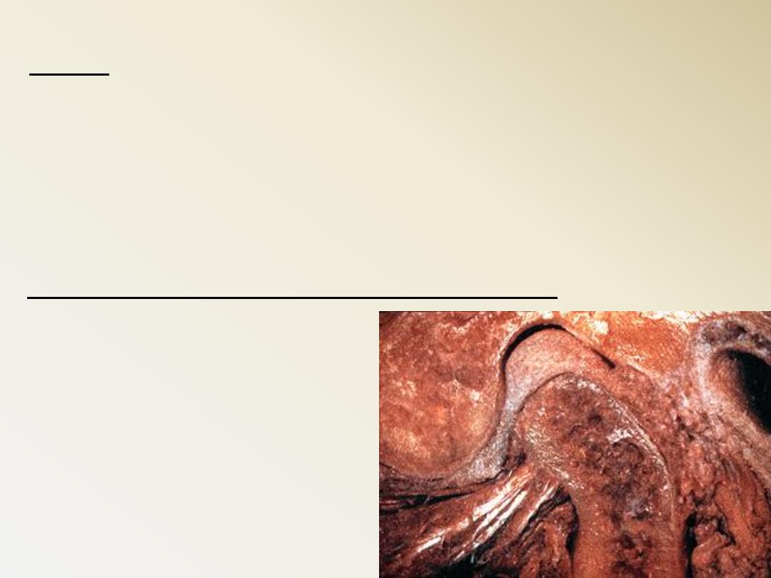
TMJ:
is a bilateral joints permits the mandible
to move as a unit with two functional
patterns:
- Hinge (inferior portion)
- Translation (superior portion)
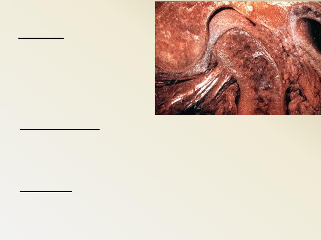
Condyle: The rounded
articular surface at
the end of the
mandible (lower jaw).
Glenoid fossa: A deep concavity in the temporal
bone a the root of the zygomatic arch that
receives the condyle of the mandible.
Tubercle: A slight elevation from the surface
of the bone giving attachment to a muscle or
ligament.
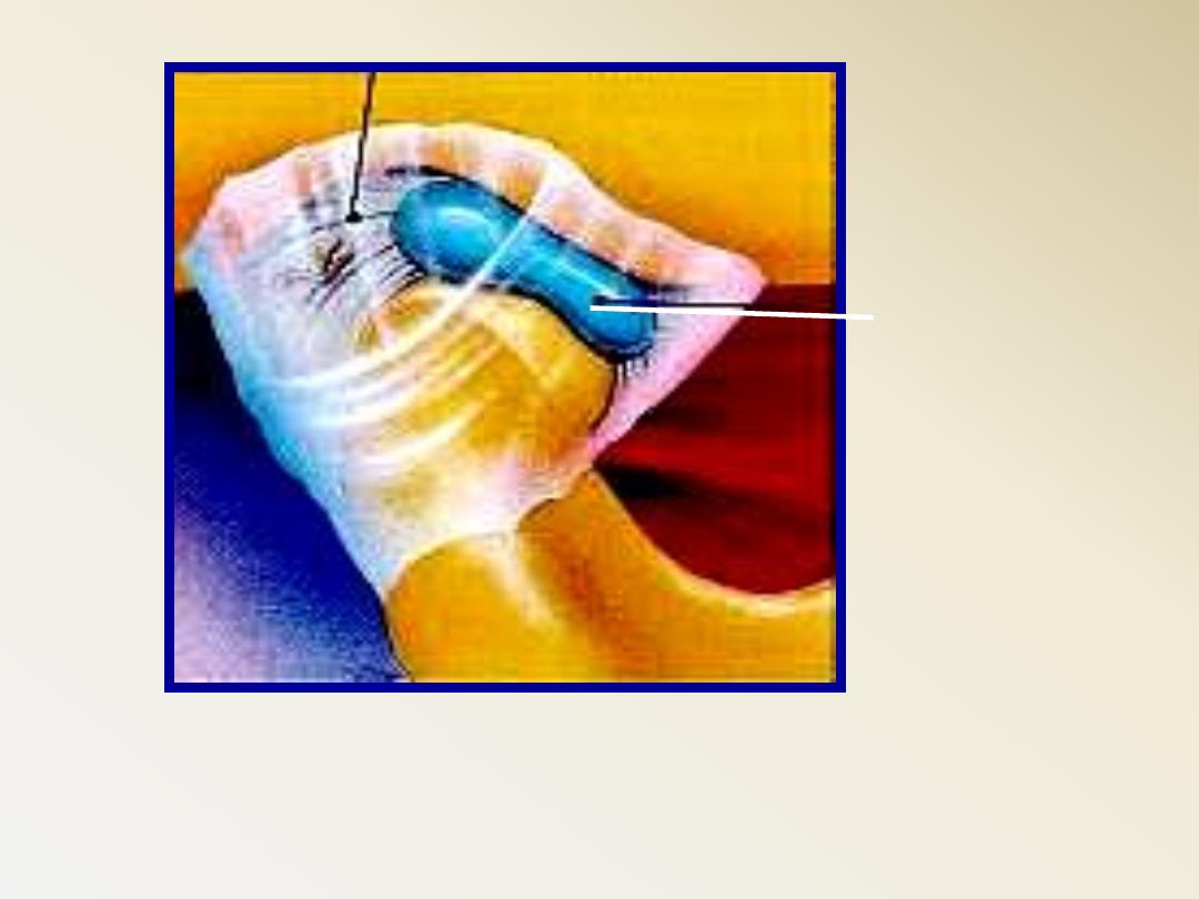
Biconcave
articular
disc
C.T. capsule
Dense fibrous connective tissue
Lacks blood vessels and nerves
Able to tolerate forces without damage or pain being produced
Provides protection to condyle and fossa during movements
The Synovial fluid:
Consist of small amounts of a clear,
straw-colored viscous fluid. It is an
infiltrate of the blood diffused out from
the rich capillary network of the
Synovial membrane.
Function:
1- Lubrication
2- Nutrition.
3- Clear the tissue
debris.
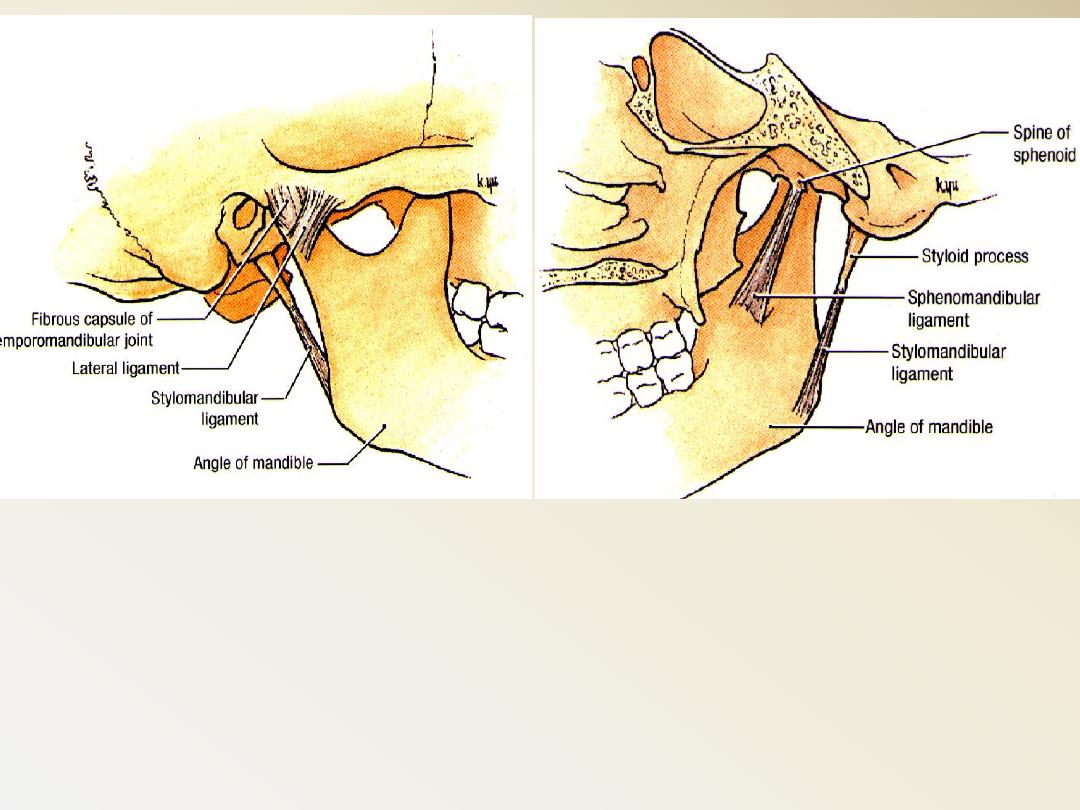
TMJ LIGAMENTS AND CAPSULE
1-Collateral(discal)
2-Capsular
3-Tempromandibular
4-Sphenomandibular
5-Stylomandibular
Introduction
Muscles of mastication develop from
the mesoderm of the first pharyngeal
arch.
They are innervated by the
Mandibular division of the trigeminal
nerve (cranial nerve V)
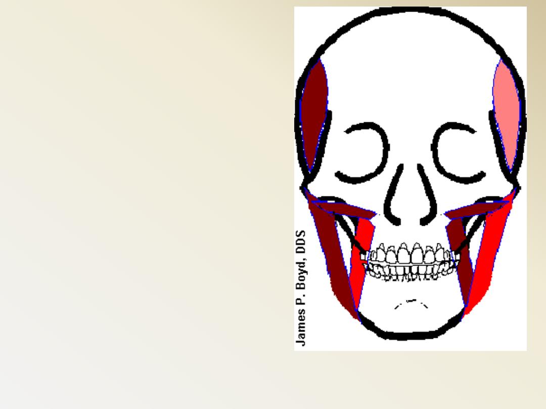
Muscles of Mastication
They are functionally classified as:
Jaw elevators
Masseter
Temporalis
Medial pterygoid
Upper head of lateral pterygoid
Jaw depressors
Lower head of lateral pterygoid
Anterior digastric
Geniohyoid
Mylohyoid
Muscles of Mastication
Other related Muscles
Orbicularis oris anterior oral
seal
Buccinator and Tongue Help
to keep the bolus of food on the
occ. Surface of teeth
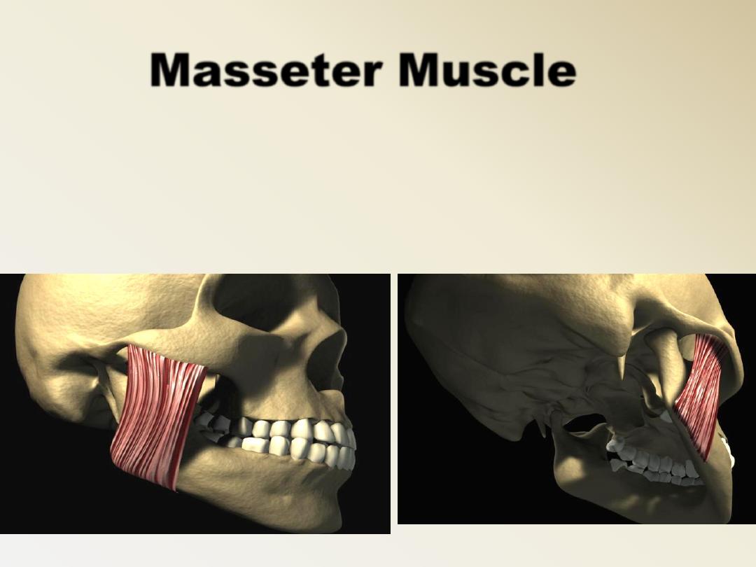
Masseter Muscle
It has 3 layers
Origin: border of the zygomatic arch
Insertion: lateral surface of the ramus
Action:
–
Elevation (deep fibers)
–
Protraction (superficial fibers)
Nerve supply:
–
Anterior division of mandibular nerve
(masseteric nerve)
Blood supply:
–
Transverse facial artery
Masseter Muscle
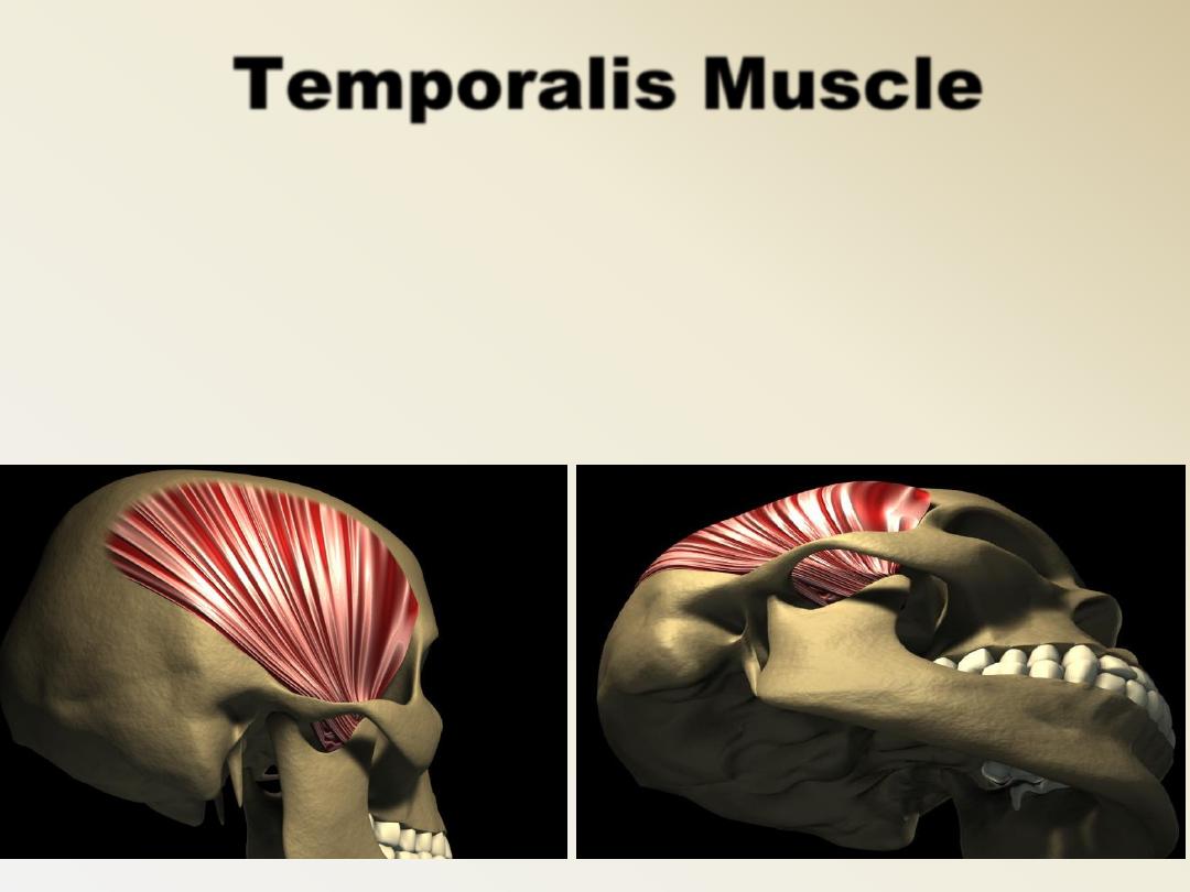
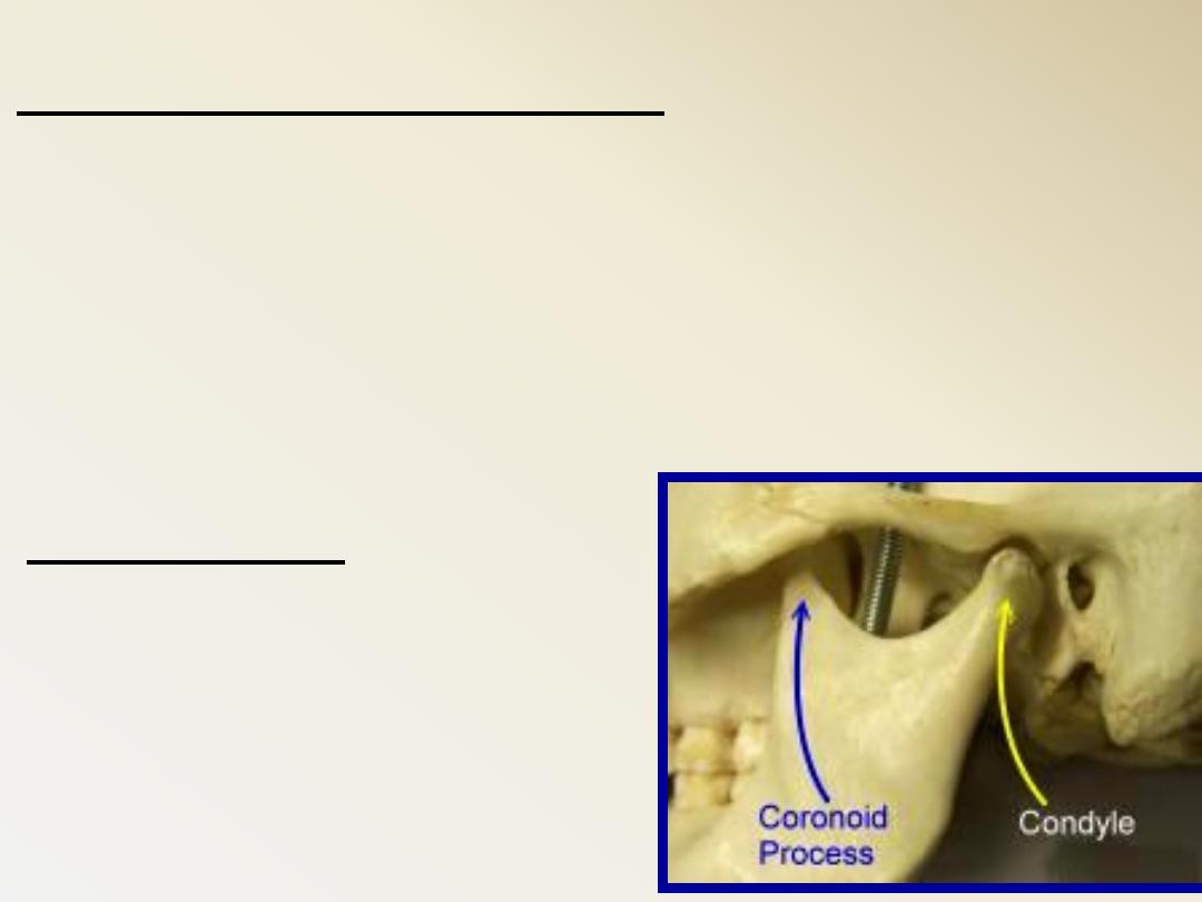
Temporalis Muscle
Has 2 heads:
•
Deep head (anterior, middle and
posterioe fibers)
•
Superficial head (much smaller)
Origin: Temporal fossa, Temporal fascia
–
In an area bound by the inferior temporal line
above and the infra temporal crest below
Insertion:
–
Cronoid process in its medial aspect (apex,
anterior and posterior border)
–
Anterior border of the ramus.
Temporalis Muscle
Action:
–
Elevation (anterior fibers)
–
Protraction (posterior fibers)
Nerve supply:
–
Anterior division of the mandibular
nerve
(2 deep temporal nerves)
Temporalis Muscle
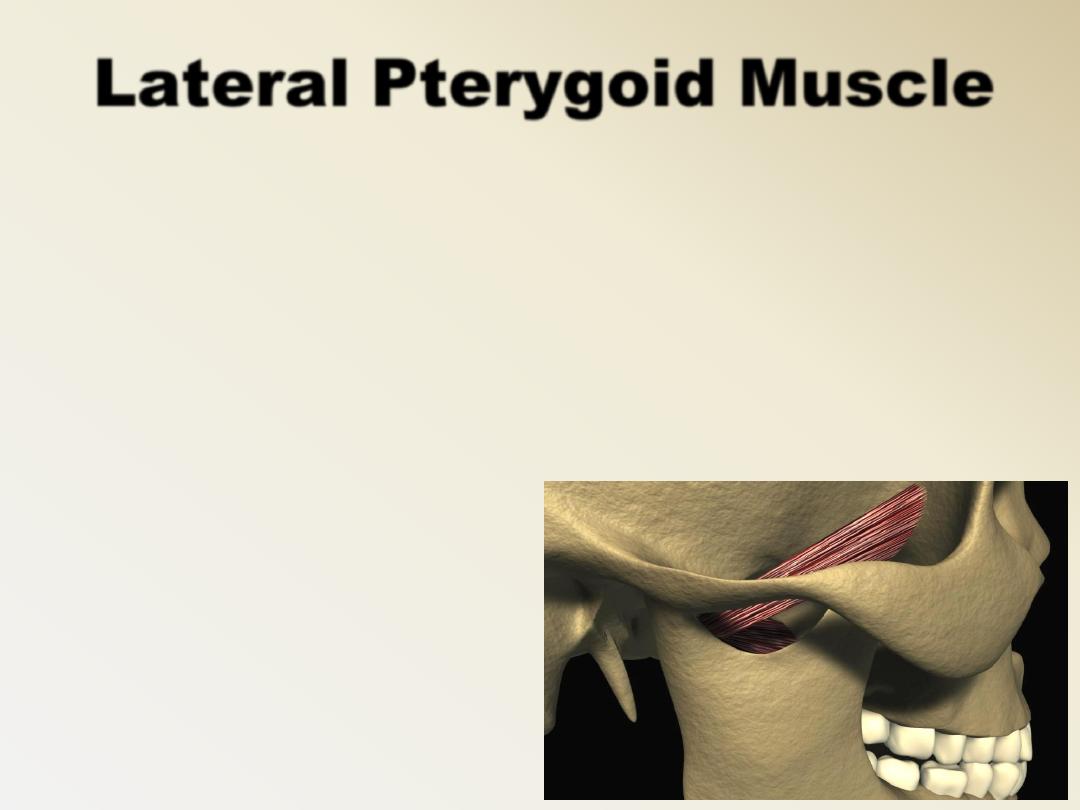
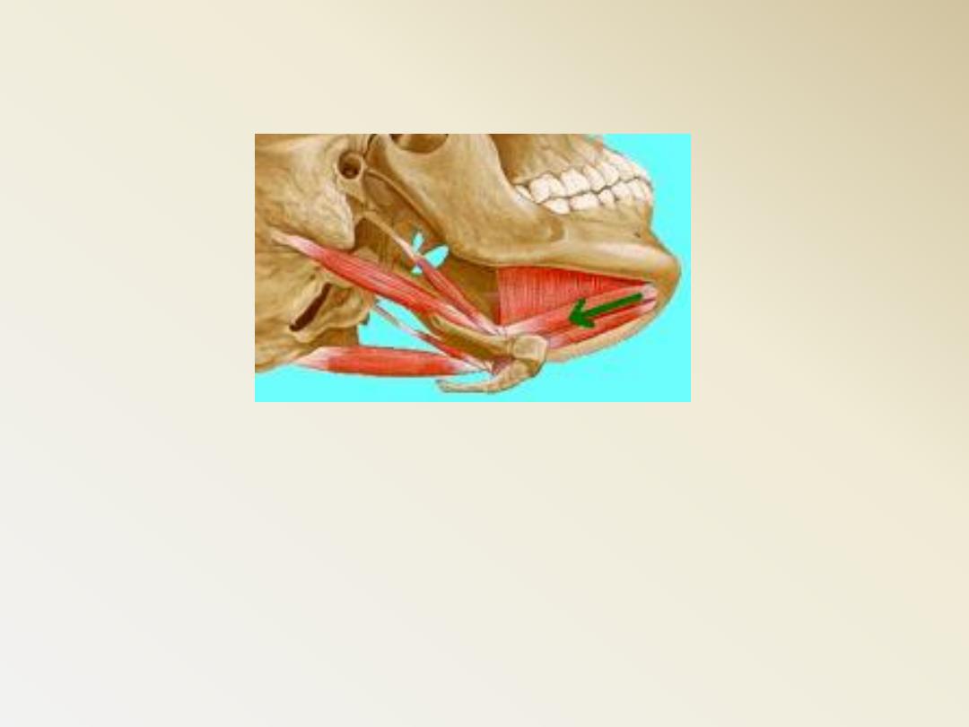
Lateral Pterygoid Muscle
Has 2 heads:
Upper head:
–
Origin: infratemporal surface & crest of the greater
wing of sphenoid
–
Insertion: enters the TMJ & inserted into:
a) Pterygoid fovea of the neck of the mandible
b) Articular disc
c) capsule of TMJ
(anterior aspect)
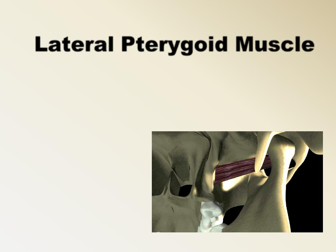
Lateral Pterygoid Muscle
•
Lower head:
–
Origin: Lateral surface of the lateral pterygoid plate
–
Insertion: as upper head
Action:
1.
Both muscles produce
depression of the mandible.
2.
Lat. & Med pterygoid on 1 side protrude the
mandible to the opposite side.
3.
Lat & Med pterygoid on the 2 sides cause
side to side movement
Nerve supply:
Anterior division of mandibular nerve (nerve to
lateral Pterygoid)
Lateral Pterygoid Muscle
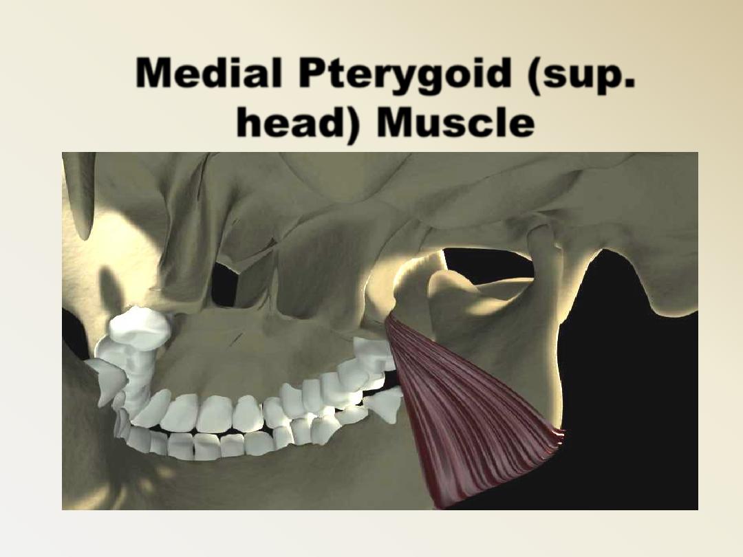
Has 2 heads
Superficial head:
–
Origin: a) maxillary tuberosity
b) neighboring part of palatine
bone.
_ Insertion: Medial surface of the angle
& ramus below the mandibular
foramen.
Medial Pterygoid (sup.
head) Muscle
Medial Pterygoid (sup.
head) Muscle
Medial Pterygoid (deep
head) Muscle
Deep head:
–
Origin: Medial surface of the lateral
Pterygoid plate.
–
Insertion: as upper head.
Action: 1) both muscles elevate the
mandible.
2 & 3 as in lateral Pterygoid.
Nerve supply: Trunk of the mandibular
nerve (nerve to medial
pterygoid muscle)
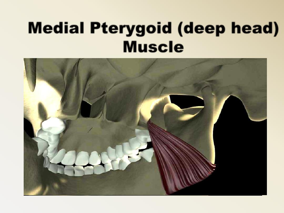
Medial Pterygoid (deep head)
Muscle
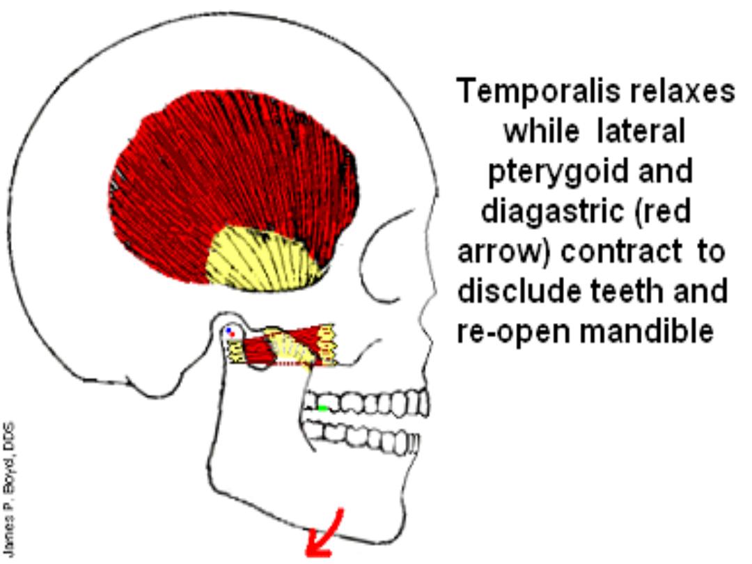
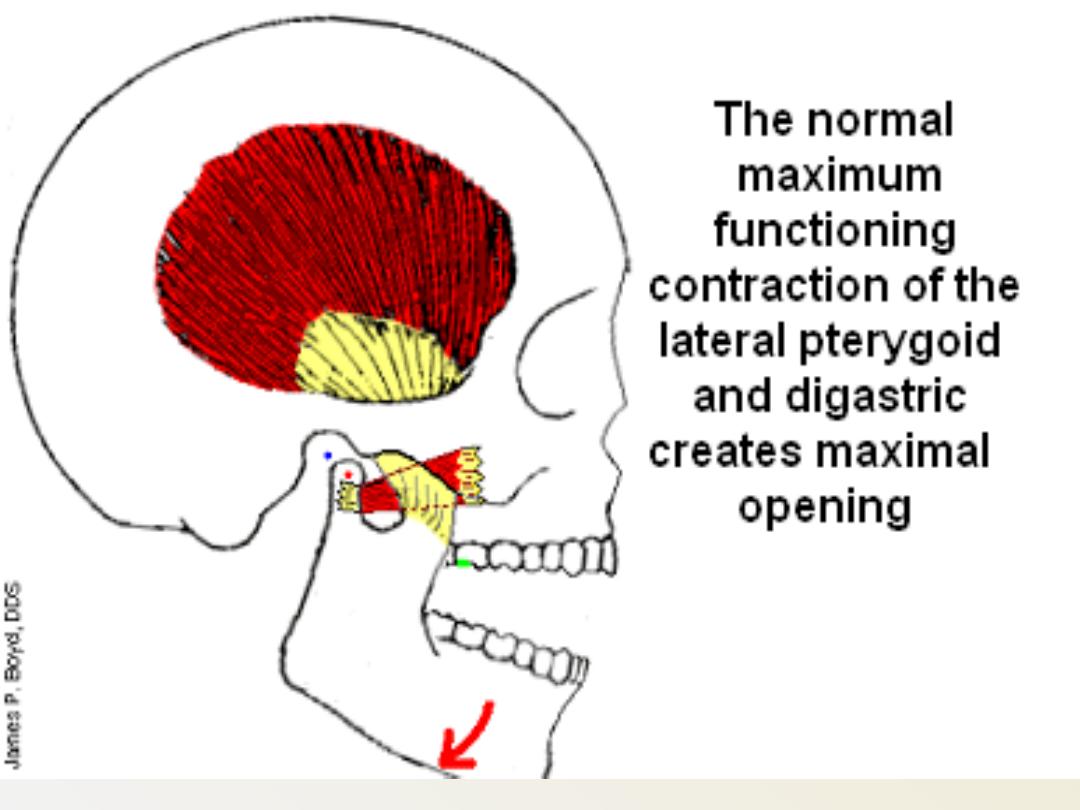
The Lat. Pterygoid
M. advance the
condyles, thereby
opening the mouth
(depressing the
mandible), with the
assistance of the
Digastric M.
Digastric muscles
is not a muscle of
mastication but it play an important role in
mandibular function
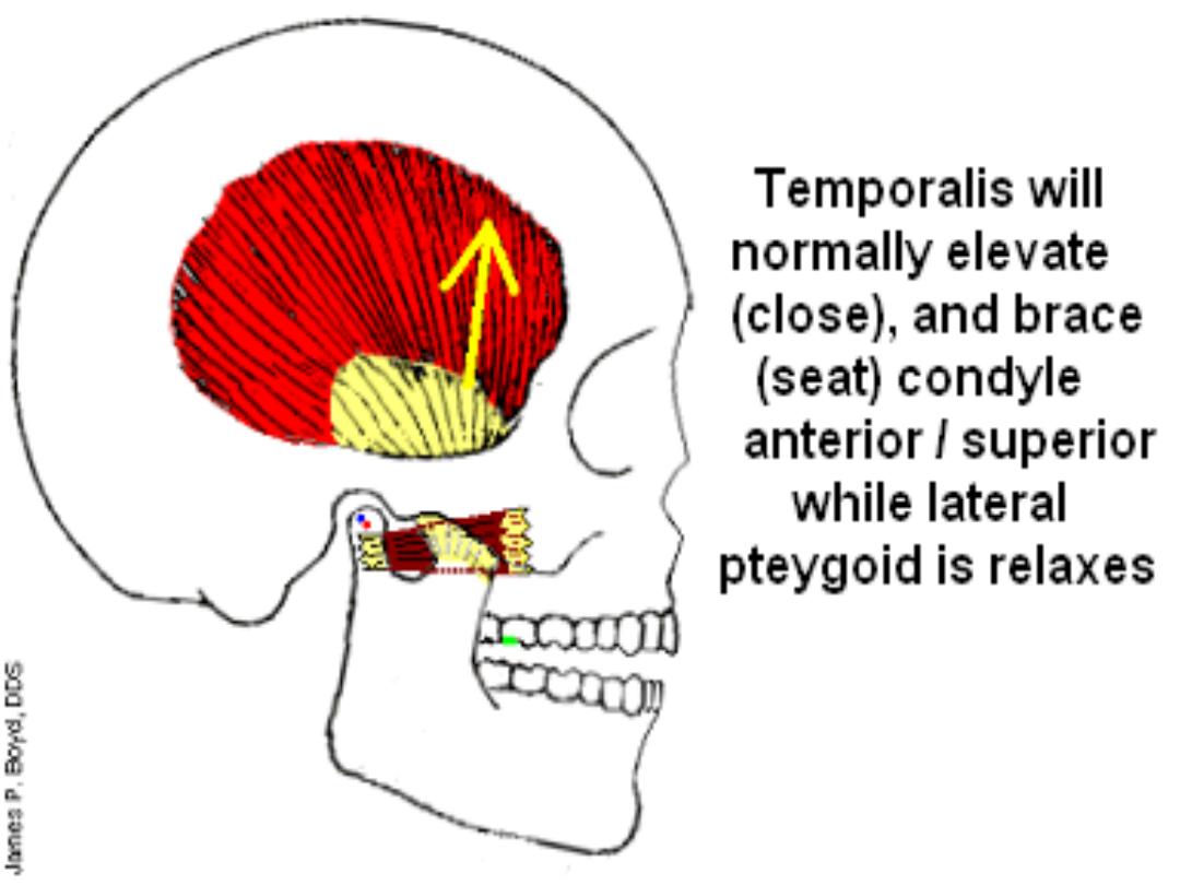
In normal chewing
function, while
initiating closing the
mand., there is
a shift
slightly to the side of
the bolus
, due to
the orientation of the
masseter and medial
pterygoid
Ms.
Occlusion Terminology
&
Definitions
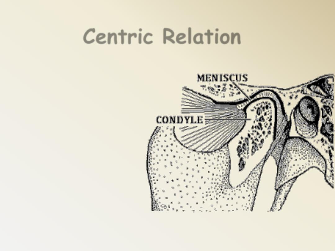
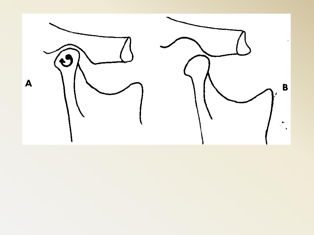
Centric Relation
It is bone to
bone relationship
independent
on
tooth contacts
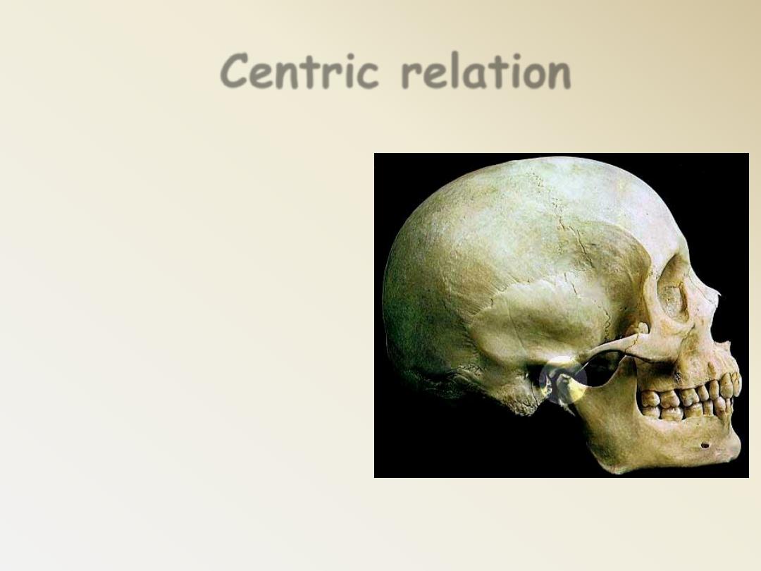
Centric relation
It is the most
retruded unstrained
position of the
condyle in the glenoid
fossa from which
lateral movements
can be performed.
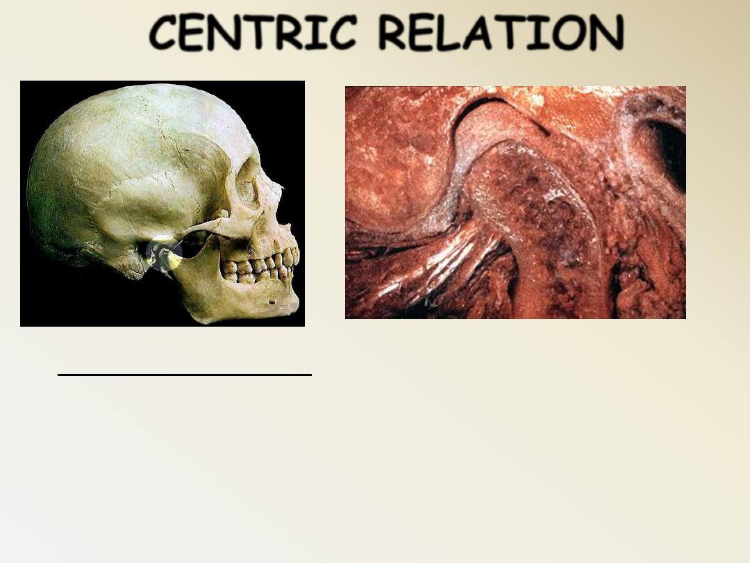
Others define as: The relation of the
mandible to the maxillae, at a given vertical
dimension, when the condyles occupy the
most supero-anterior position in their
mandibular fossae axis.
CENTRIC RELATION
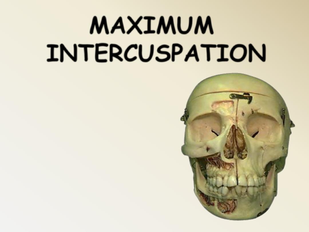
MAXIMUM
INTERCUSPATION
The complete
intercuspation of
the opposing teeth
independent
of condylar position.
Occlude close.
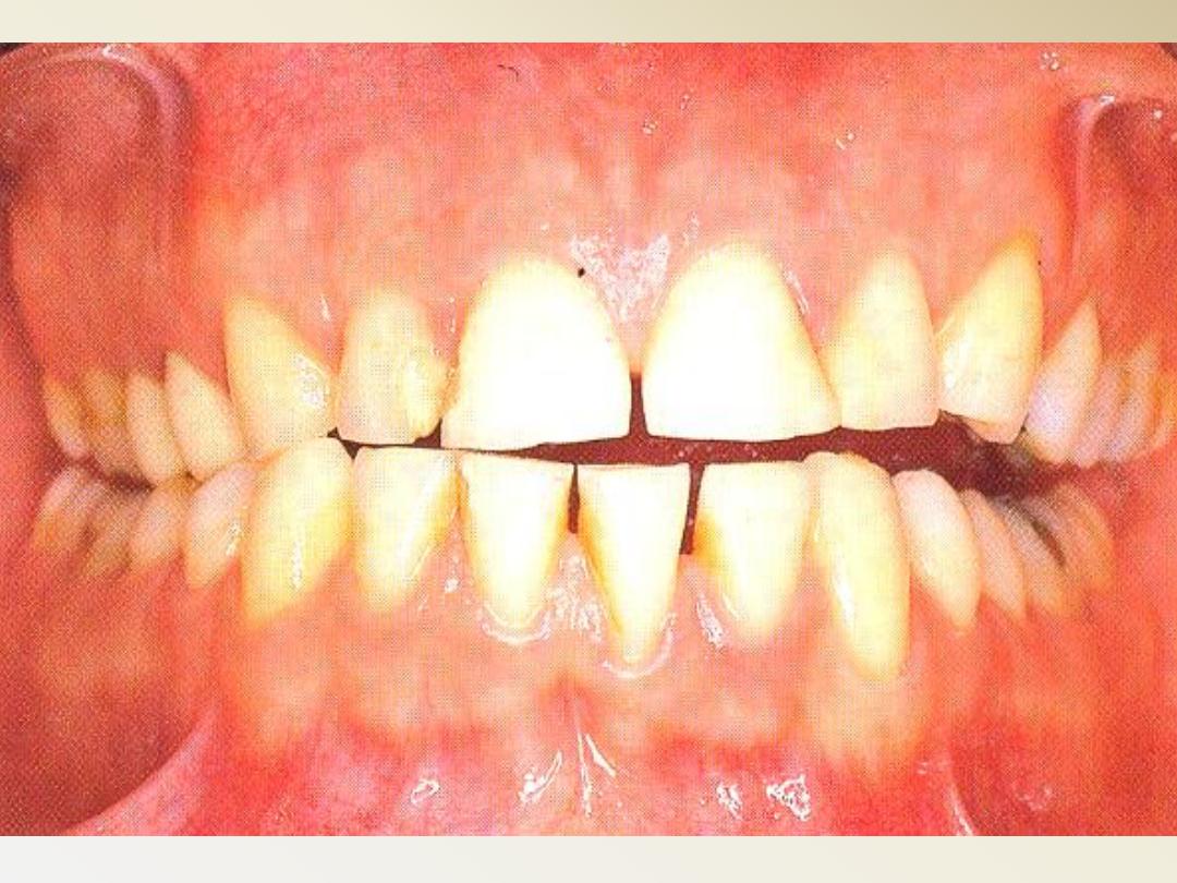
Occlusion is the act or process of closure
It is the
STATIC relationship between the
incising or masticating teeth analogues.
It is an important factor that influences stability
and retention of complete dentures.
So Occlusion is the closure of the maxillary and
mandibular teeth throughout the range of
functional and nonfunctional movements of the
mandible

CENTRIC
OCCLUSION
The occlusion of opposing teeth when
the mandible is in centric relation

Any occlusion other than centric
relation
Eccentric Occlusion
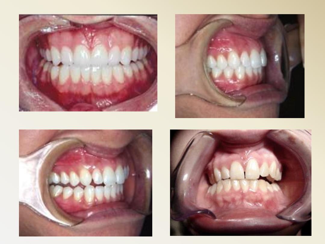
The dynamic movements of
the teeth in relation to
each other
Articulation:
VERTICAL
DIMENSION OF
OCCLUSION
The degree of separation between
the maxillae and the mandible
when the teeth are in occlusion.
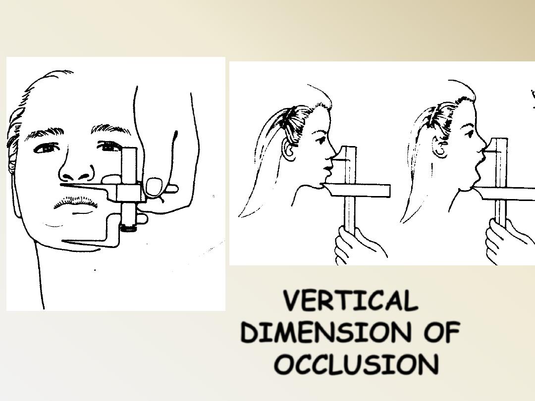
VERTICAL
DIMENSION OF
OCCLUSION
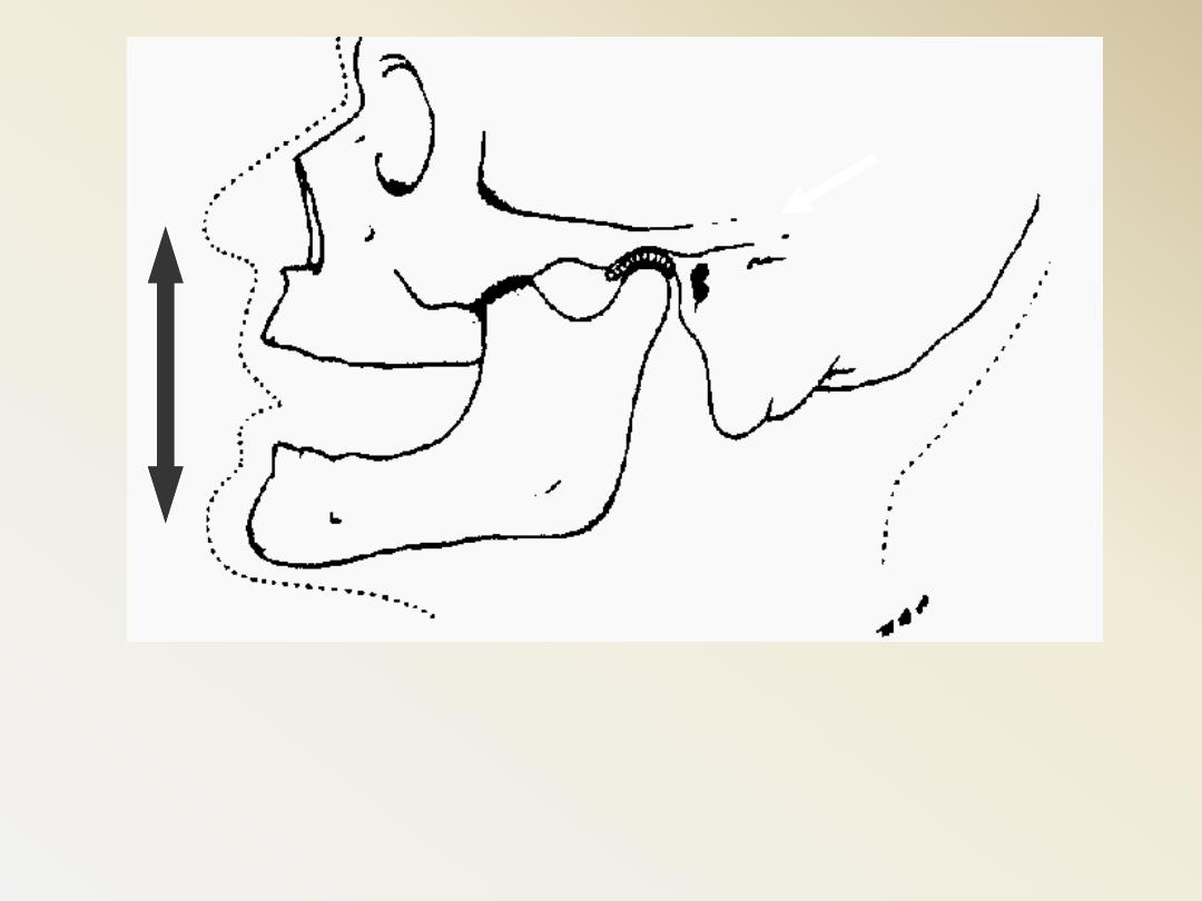
Interocclusal distance
(Free way space)
The distance between the
occluding surfaces of the
maxillary and mandibular
teeth when the mandible is in
a specified relaxed position, it
ranges from 2-4 mm.
In the edentulous patients, use the most
retruded position of the condyle in its
fossa (centric relation)
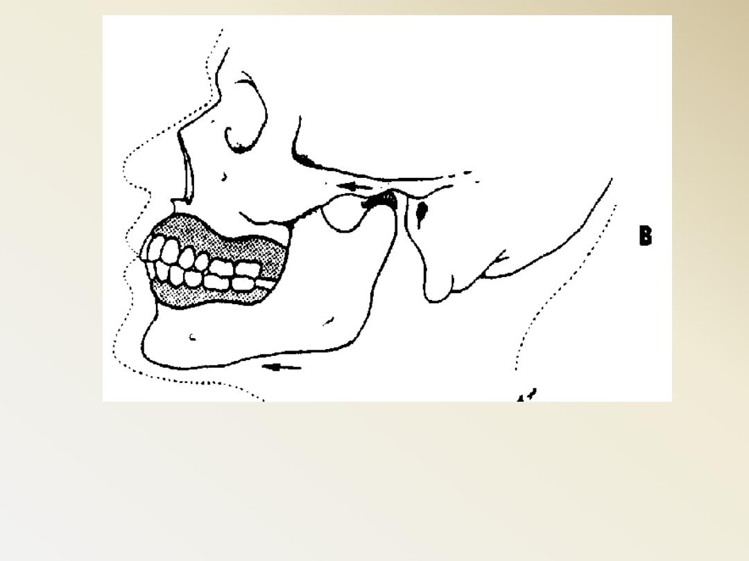
Centric Occlusion can be
made to coincide with CR
the occlusal surface of the teeth could
be altered to
allow freedom of tooth
movement
in harmony with the rotation
of condyle. (from hinge position to
habitual intercuspal position).
long centric or Freedom in centric
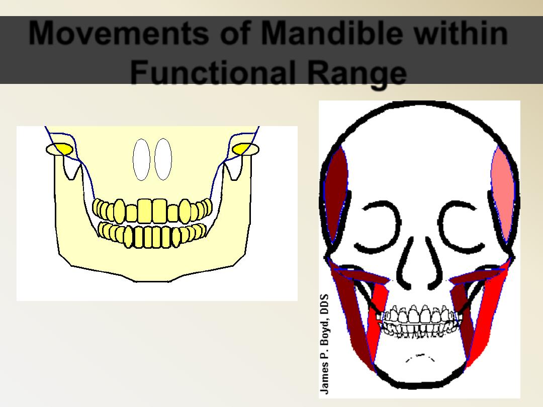
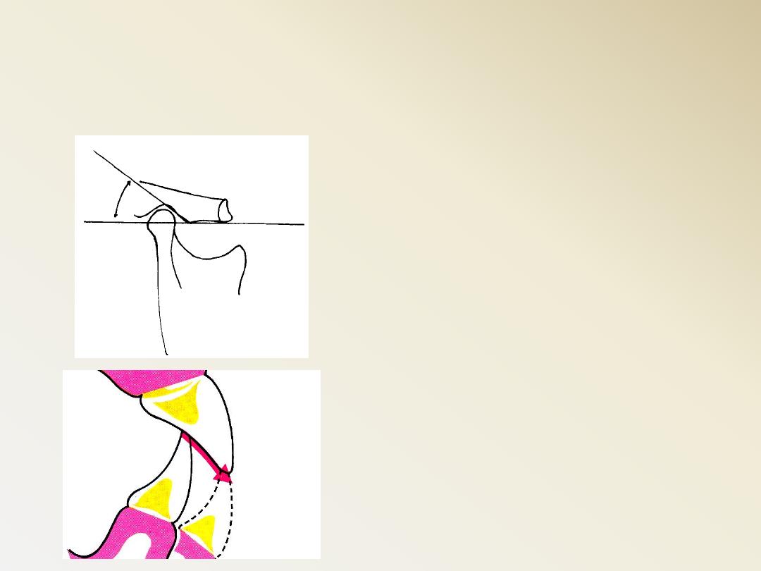
Movements of Mandible within
Functional Range
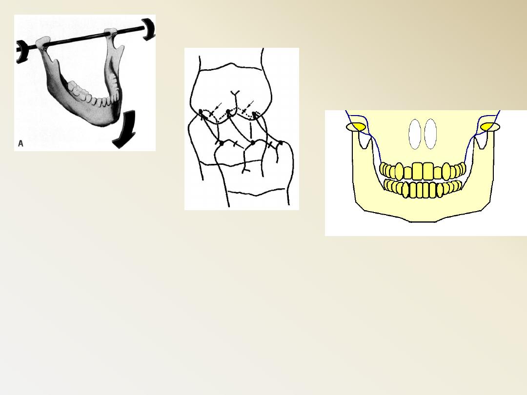
The closure of the mandible does not
occur in a straight upward movement but
rather in a curve
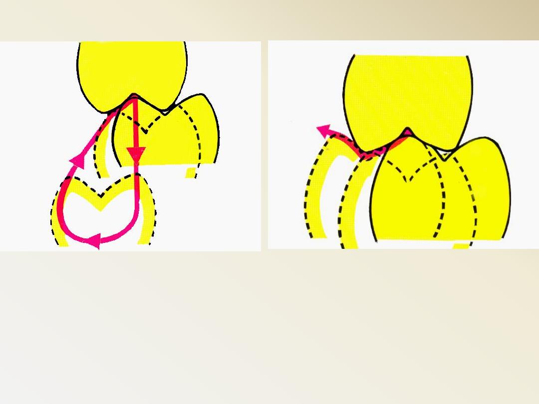
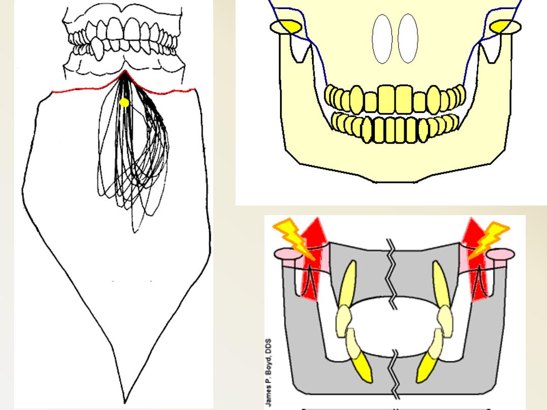
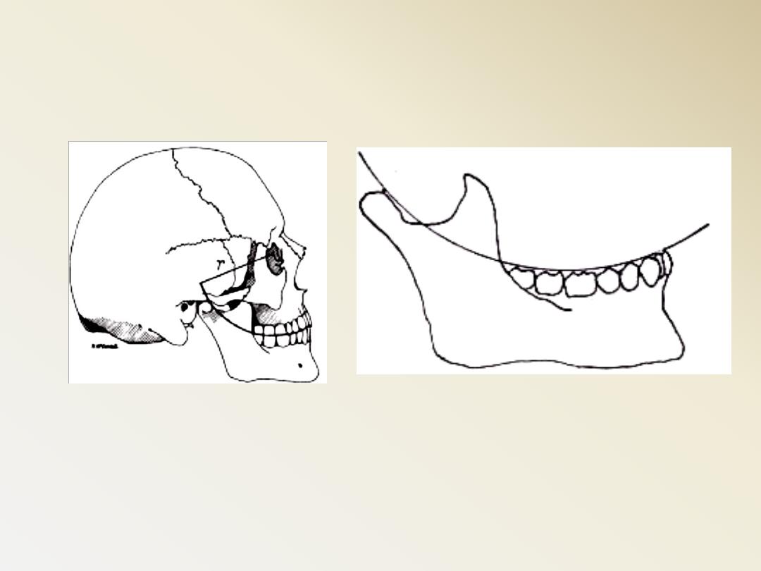
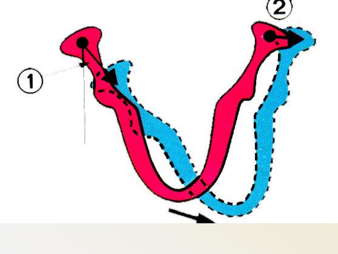
Masticatory cycle
(Balanced Articulation)
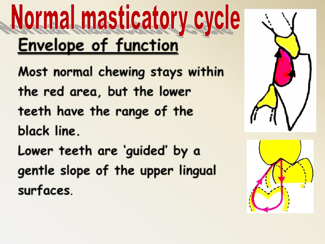
Envelope of function
Most normal chewing stays within
the red area, but the lower
teeth have the range of the
black line.
Lower teeth are ‘guided’ by a
gentle slope of the upper lingual
surfaces.
Teeth harmonious with bone / craniofacial
structures
Anterior Guidance
in harmony with
the border
movements of the
Envelope of
Function.
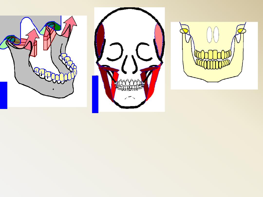
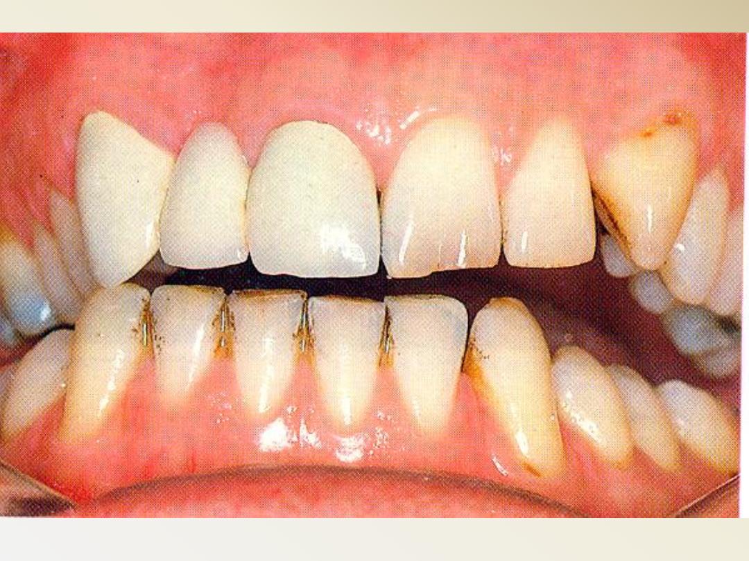
In normal chewing function, the mandible
opens, and then, while initiating closing, there
is a shift slightly to the side of the bolus,
due to the orientation of the masseter and
medial pterygoid.
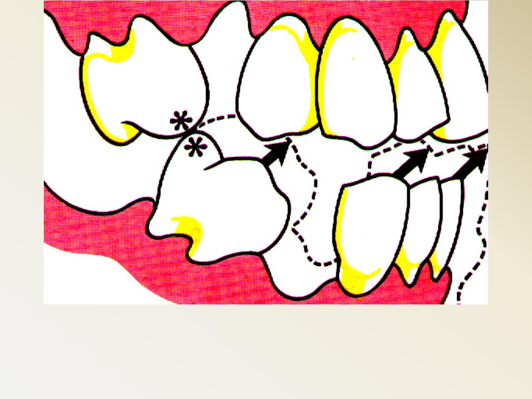
Change the pattern of mandibular closure
as a result of premature contact
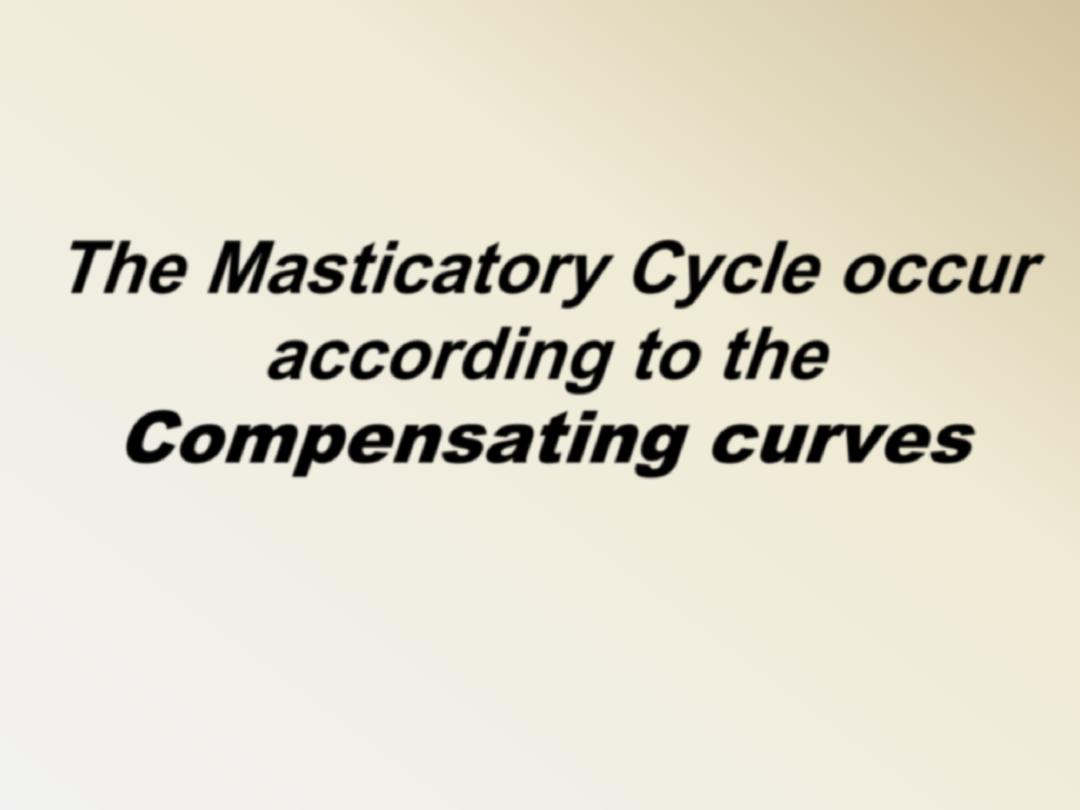
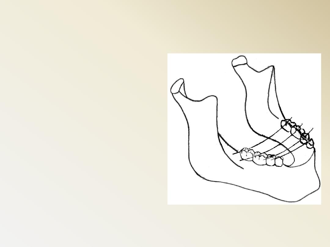
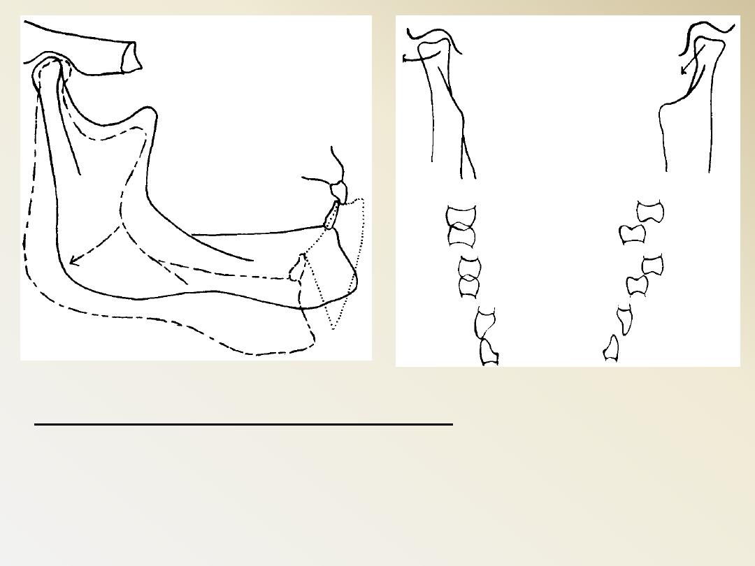
The Masticatory Cycle occur
according to the
Compensating curves
Spee’s curve
The anatomic curvature of the occlusal alignment
of the lower teeth beginning at the tip of the
lower cuspid and following the buccal cusps of the
natural bicuspids and molars continuing to the
anterior border of the ramus
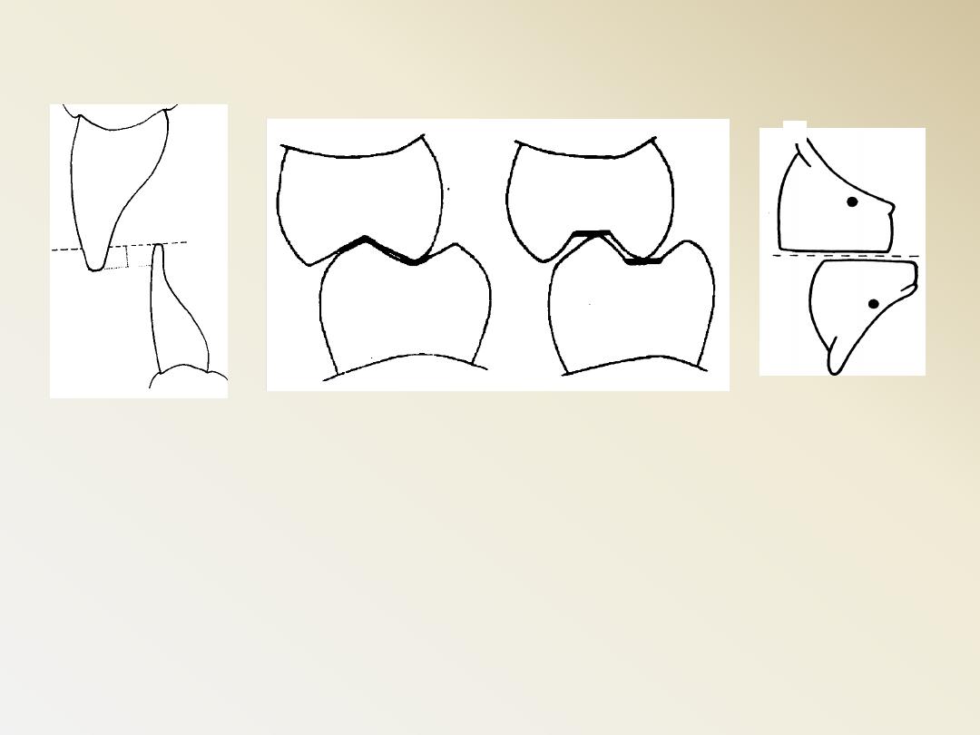
Compensating curves
The buccal cusps of the
lower posterior teeth
are slightly higher than
the lingual cusps, and a
line drawn through the
buccal and lingual
cusps of the teeth on
the other side forms a
lateral curve called the
curve of Wilson
Curve of Wilson
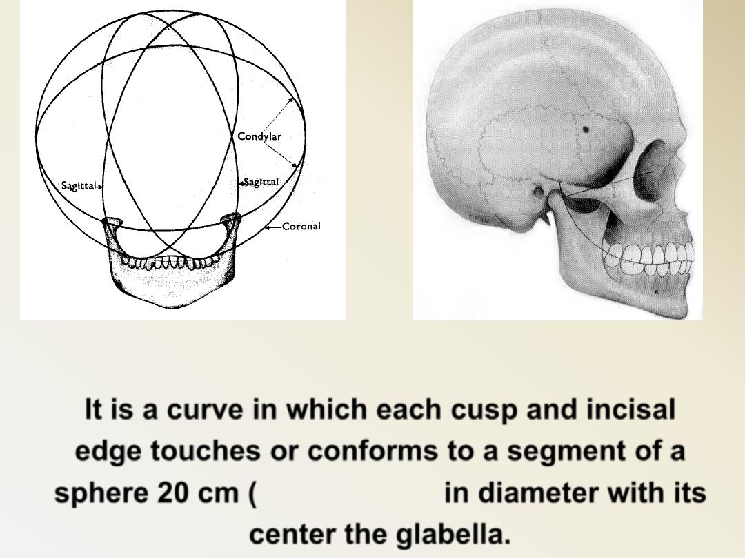
Monson’s curve
It is a curve in which each cusp and incisal
edge touches or conforms to a segment of a
sphere 20 cm (eight inches) in diameter with its
center the glabella.

The compensating curve of
the artificial occlusion
corresponds to a
combination of these
curves in natural teeth. It
is considered one of the
most important factors in
establishing balanced
occlusion
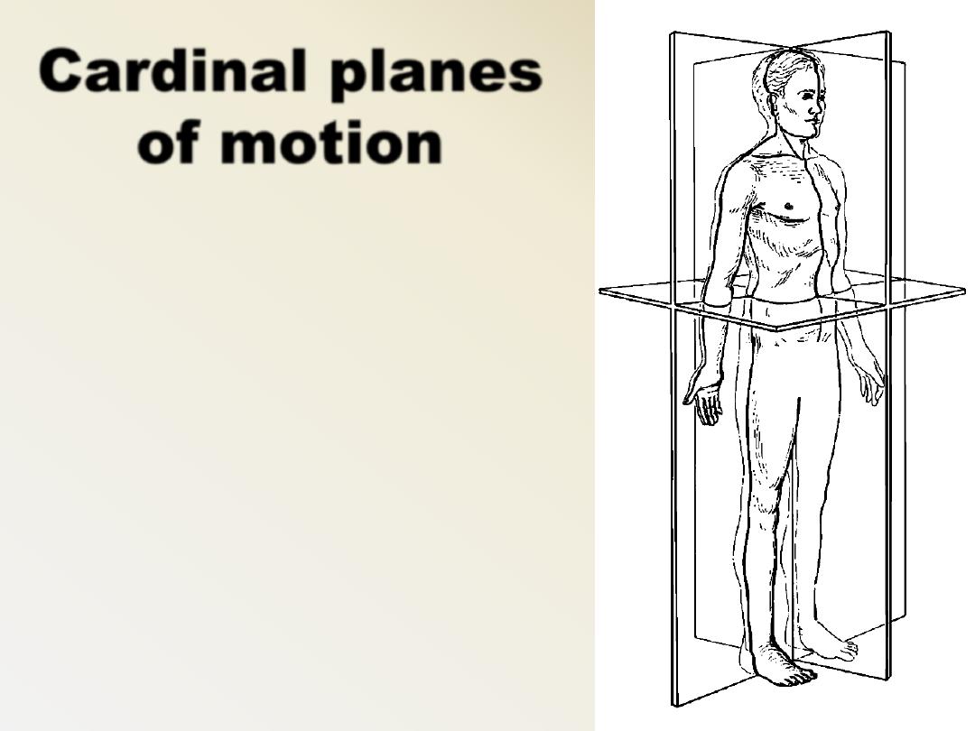
3 basic or traditional
planes
in relation to the
body, not in relation
to the earth
Anteroposterior or
Sagittal Plane
Lateral or Frontal Plane
Transverse or
Horizontal Plane
Cardinal planes
of motion
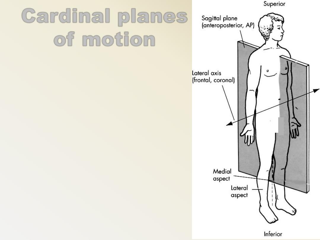
Cardinal planes
of motion
Anteroposterior Plane
–
divides body into equal,
bilateral segments
–
It bisects body into 2 equal
symmetrical halves or a
right & left half
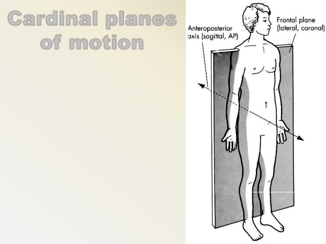
Cardinal planes
of motion
Lateral Plane
–
divides the body into (front)
anterior & (back) posterior
halves
–
Ex. Jumping Jacks
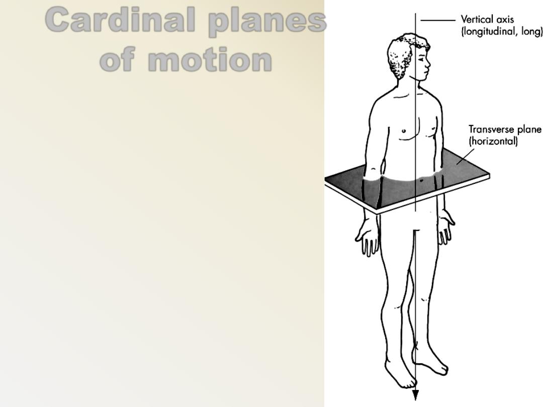
Cardinal planes
of motion
Horizontal Plane
–
divides body into (top)
superior & (bottom) inferior
halves when the individual is
in anatomic position
–
Ex.Spinal rotation to left or
right
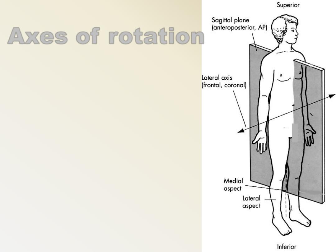
Axes of rotation
Lateral axis
–
Has same orientation as
frontal plane of motion &
runs from side to side at a
right angle to sagittal plane
of motion
–
Runs medial / lateral
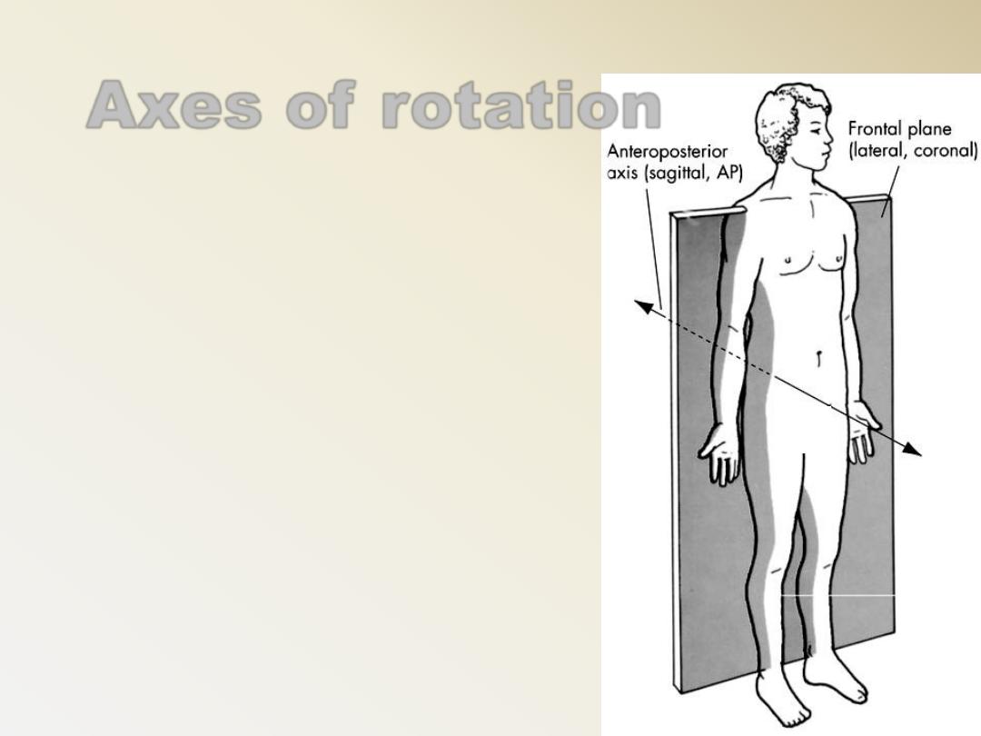
Axes of rotation
Anteroposterior axis
–
Has same orientation as sagittal
plane of motion & runs from front
to back at a right angle to frontal
plane of motion
–
Runs anterior / posterior
Axes of rotation
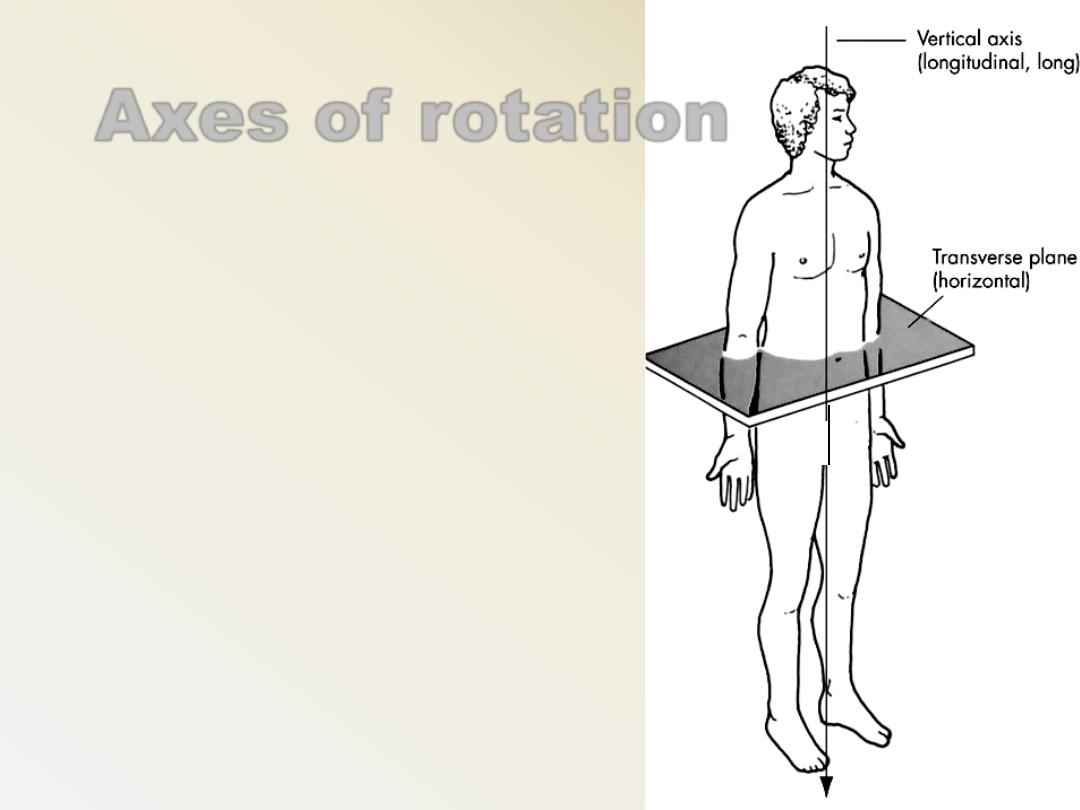
Vertical axis
–
Runs straight down through
top of head & is at a right
angle to transverse plane of
motion
–
Runs superior/ inferior
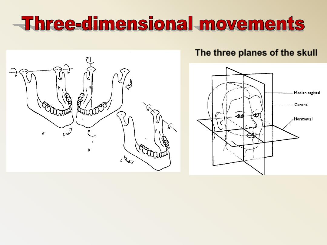
The three planes of the skull
For movement to occur in a plane, it must turn or
rotate about an axis as previously mentioned
The axes are named in relation to their orientation

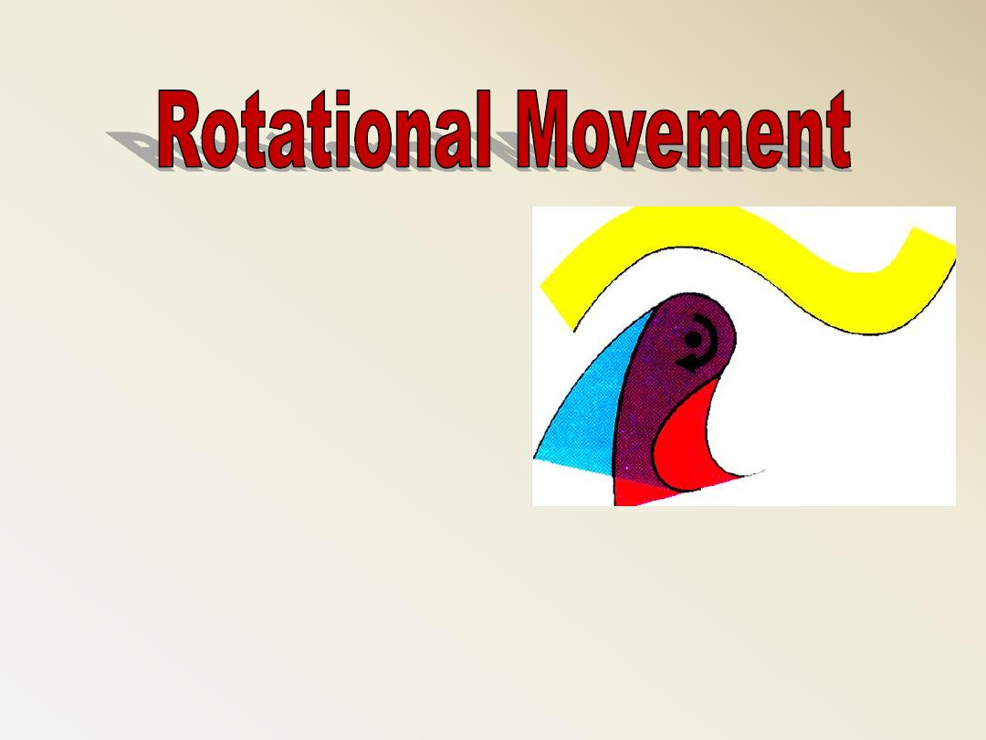
Rotation
occurs when the
mandible makes a hinged
movement.
Translation
occurs when the
mandible moves into a
protrusive or lateral
position, or a combination
of the two
• It Is a Simple Hinge
Movement
Occurs during the early
opening and late closing
movement of the
mandible
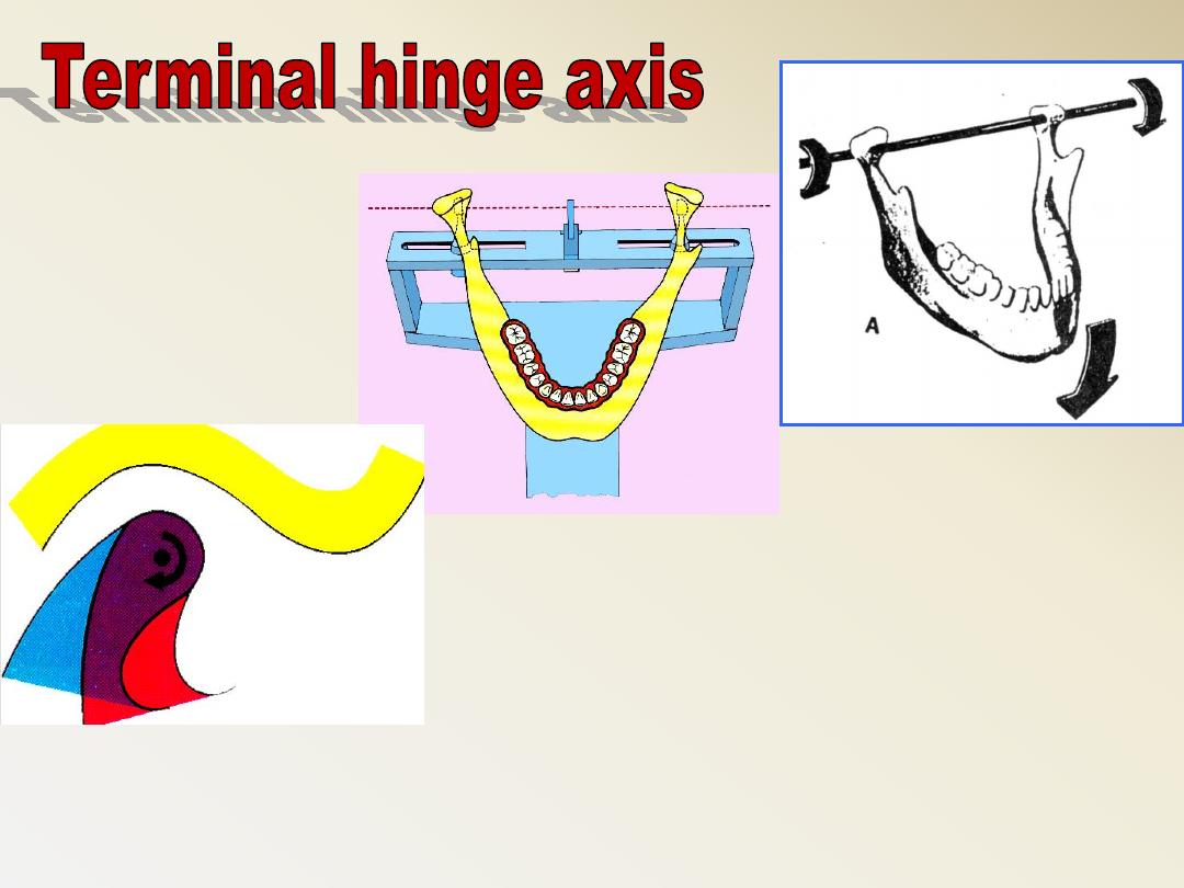
It is an imaginary line around which the condyles rotate
during early opening and late closing.
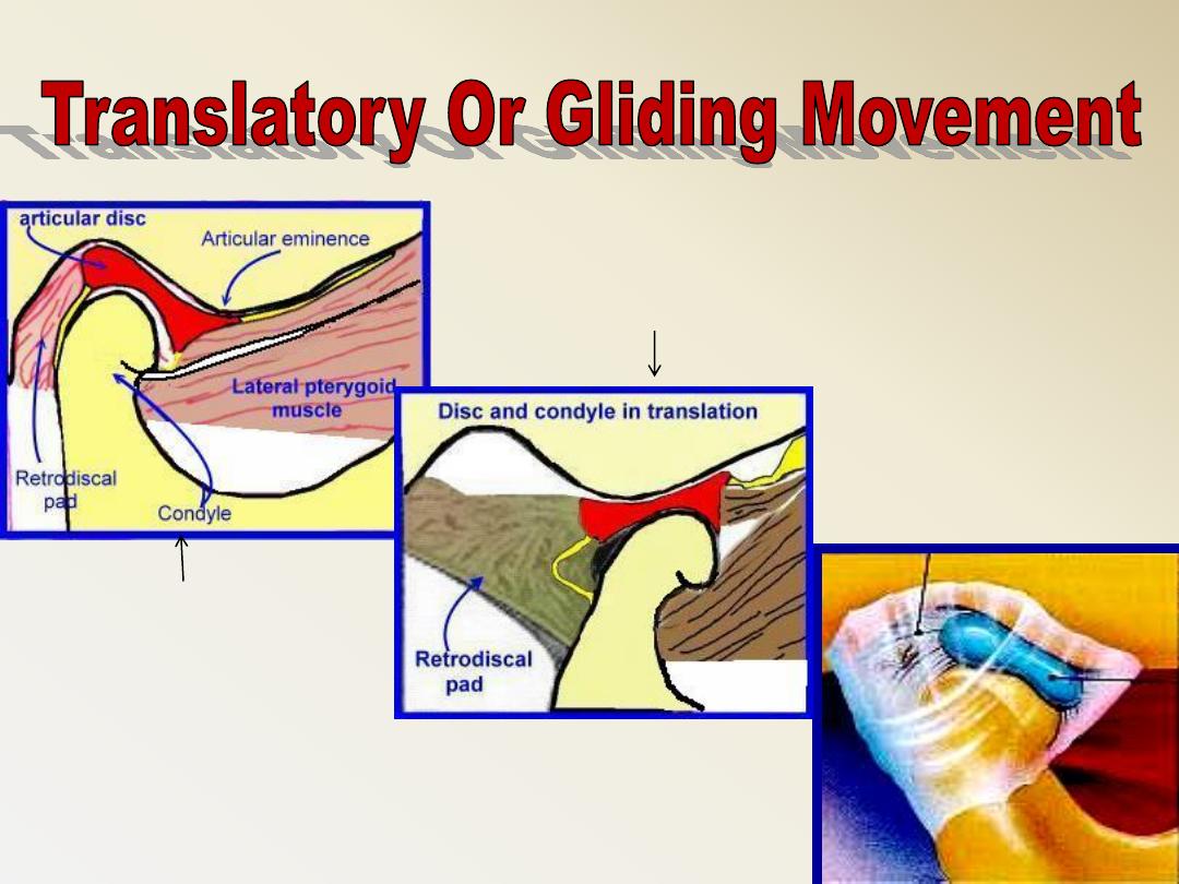
Translation movements
Rotation
movements
Translatory movement of the mandible takes place in the
upper compartment of the T.M.J between the superior
surface of the articular disc as it moves with the condyle.
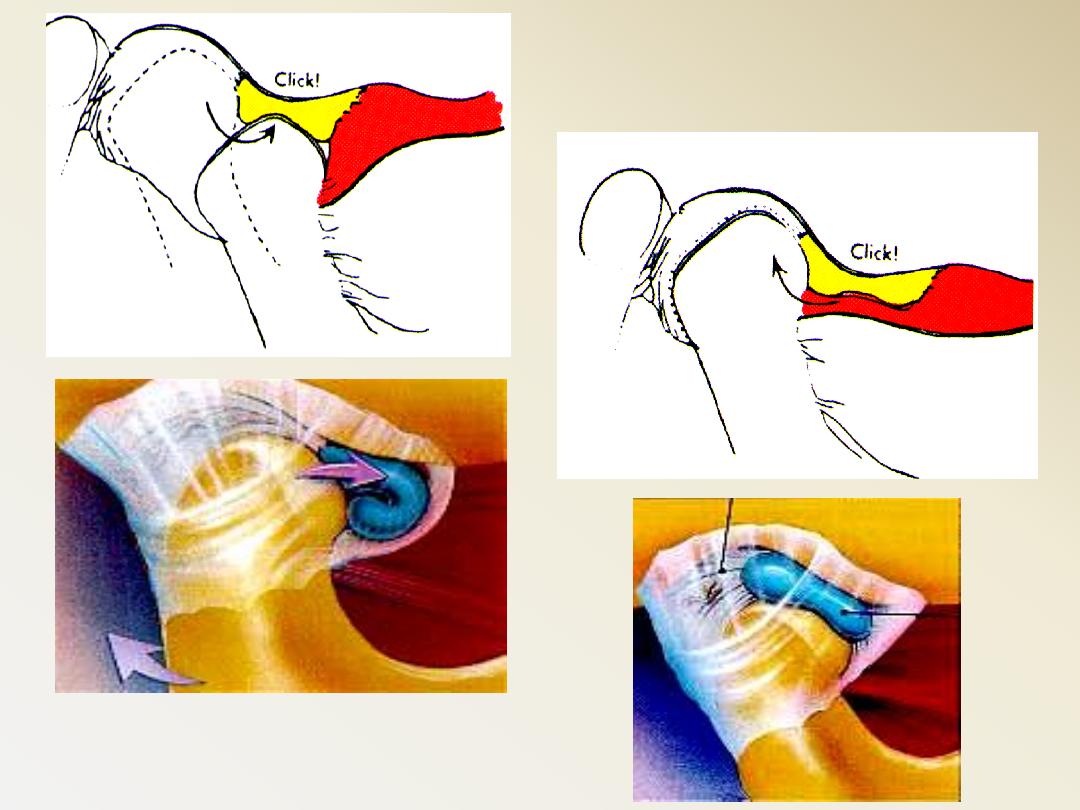
Reciprocal Click
Dislocation
Articular disc as it moves
with the condyle
A- Rotation occurs when the head of the
condyle rotates around an imaginary axis
B. Translation is the bodily movement of the
head of the condyle
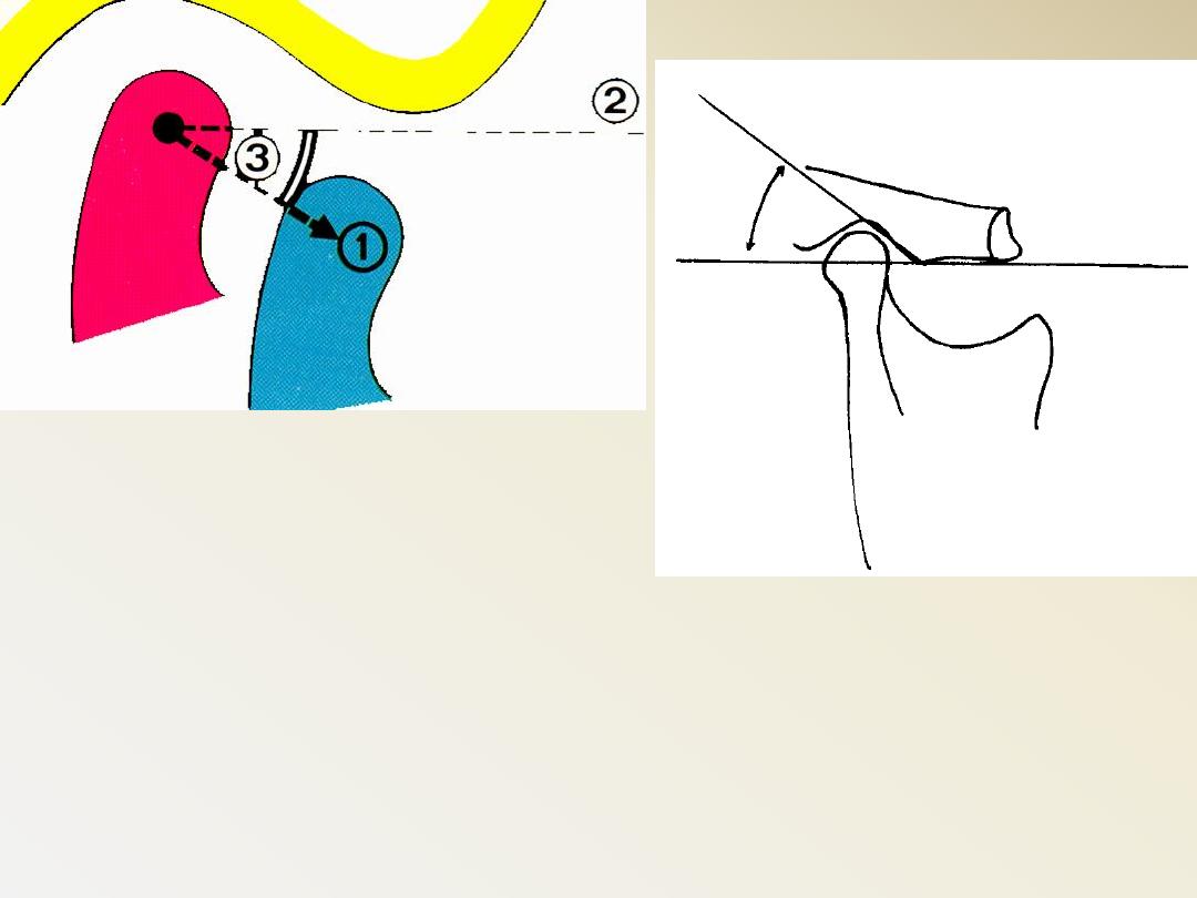
The angle formed by the steepness of the
articulator surface that is related to a
horizontal plane is called the sagittal
inclination
(
Condylar inclination)
1. The shape of the glenoid
fossa.
2. The variation of the
thickness of the articular
disc in its different parts.
3. The relation of the
condyle to the disc
during movement.
4. The extent of mandibular
protrusion
The inclination of the condylar paths varies in
different individuals and from side to side in
the same person… It depends upon:
The condylar guidance
:
refers to the path
of the condyle follows in the TMJ when the
mandible moves into protrusive or lateral
movements
Bennet
angle
Bennet
movement