Human Anatomy
Temporal fossaTemporal fossa
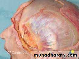
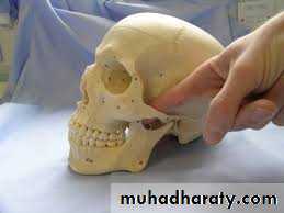
The temporal fossa is the region on the side of the head ( the spaces on side of calvaria) above the external ear canal, which is covered by the temporalis muscle. The side of the head anterior and superior to the ear is commonly called the temple the skin, fascia , and portions of the extrinsic muscle of the ear in this region overlie the deeper fan-shaped temporalis muscles that attached to the bones of the temporal fossa . superiorly, this fossa is bounded by the superior temporal line , whereas its inferior boundary is the zygomatic arch, even though the temporalis muscle extends inferiorly below this arch into the infratemporal fossa .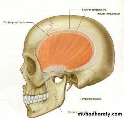
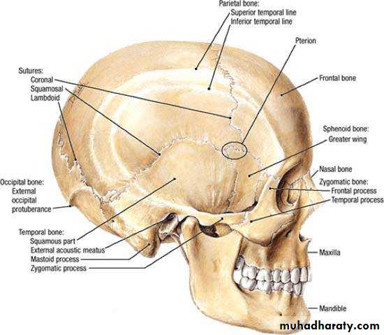
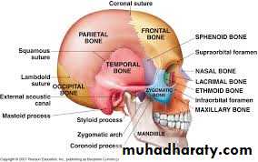
The floor of the temporal fossa is formed by the bones of the side of the head-portions of the frontal , sphenoid, temporal and parietal bones .
The inferior and superior temporal lines begin at the zygomatic process of the frontal bone and arch posteriorly over the parietal bone before descending to the temporal bone and blending into the zygomatic process of temporal bone.
The tough fascia covers the temporal muscle is called temporalis fascia attaching superiorly to the superior temporal line . Inferiorly , the fascia splits into two layers which attach to the lateral and medial surfaces of the zygomatic arch .
Boundaries
a- Postero superiorly : superior temporal lineb- Inferiorly : infratemporal crest .
c- Anteriorly : frontal process of zygomatic bone .
d- Laterally : zygomatic arch .
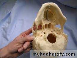
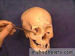
e- Floor : formed by 4 bones : frontal ,parietal , temporal, and sphenoid forming pterion which is thinnest part of the lateral wall of the skull where the anteroinferior corner of the parietal bone articulates with the greater wing of the sphenoid . Clinically , the pterion is an important area because it overlies the anterior division of the middle meningeal artery and vein .
Contents :
Temporalis muscle , deep temporal nerves and vessels , auriculotemporal nerve , superficial temporal vessels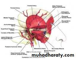
Infratemporal fossa
The infratemporal fossa is an irregularly shaped space deep and inferior to the zygomatic arch , deep to the ramus of the mandible and posterior to the maxilla (Deep lateral region of face ) .
The pterygomaxillary fissure is a vertical fissure that lies within the fossa between the pterygoid process of the sphenoid bone and back of the maxilla. It leads medially into the pterygopalatine fossa. The inferior orbital fissure is a horizontal fissure between the greater wing of the sphenoid bone and the maxilla. It leads forward into the orbit. Superiorly , the fossa is limited by the infratemporal surface of the greater wing of the sphenoid bone and the very anteroinferior-most portion of the squamous part of temporal bone .
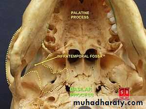
The boundaries:
Laterally : the ramus of the mandible.Medially: lateral pterygoid plate .
Anteriorly : the posterior aspect of the maxilla.
Posteriorly : the tympanic plate and the mastoid and styloid processes of the temporal bone.
Superiorly : the inferior ( infratemporal) surface of the greater wing of the sphenoid.
Inferiorly: where the medial pterygoid muscle attaches to the mandible near its angle.
Communications:
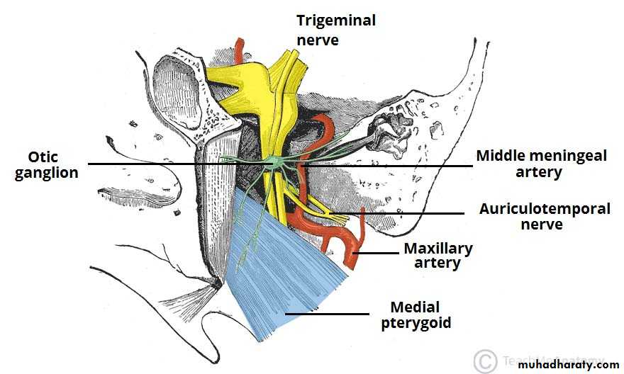
The infratemporal fossa communicates with the temporal fossa as the temporalis muscle descends from its origin in the temporal fossa to be inserted onto the coronoid process of the mandible . nerves and vessels supplying the temporalis muscle pass from the infratemporal fossa to the temporal fossa to pierce the deep surface of this muscle. Two foramina open onto its roof on the medial aspect of the infratemporal region of the greater wing of the sphenoid :1. The larger of the two , the foramen ovale, transmits the mandibular division of the trigeminal nerve exiting from the cranial vault and the accessory meningeal artery proceeding to the cranium .
2. The smaller foramen , the foramen spinosum, lies between the foramen ovale and the spine of the sphenoid. It transmits the middle meningeal artery and the recurrent meningeal nerve from the fossa into the cranium.
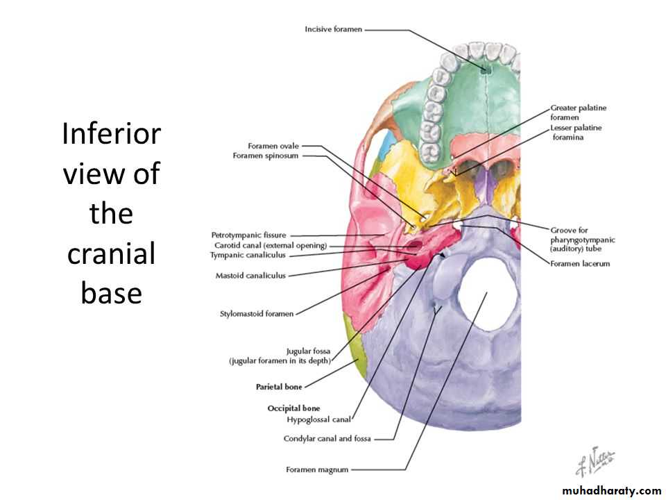
The fossa communicates with the orbit at its most superoanterior aspect via the inferior orbital fissure between the maxilla and the greater wing of the sphenoid . through this fissure pass the maxillary division of the trigeminal nerve, on its way to the floor of the orbit , as well as the zygomatic branch which arises from it.
The cleft between the maxilla and the lateral pterygiod plate is the pterygomaxillary fissure communicating with the pterygopalatine fossa medially . it is through this fissure that the maxillary artery distribute in to the fossa, eventually to reach the nasal cavity via the sphenopalatine foramen
Foramina opened in the infratemporal fossa: (summary)
• Foramen spinosum: for middle meningeal artery into middle cranial fossa.
• Foramen ovale: for mandibular nerve ( CN V3) and accessory meningeal artery.
• Pterygomaxillary fissure: medial cleft leading into pterygopalatine fossa; for terminal part of maxillary artery .
• Inferior orbital fissure: leads anteriorly into orbit; for zygomatic and infraorbital branches of maxillary nerve ( CN V2) , infraorbital artery , and communication between pterygoid plexus and inferior ophthalmic vein
Contents
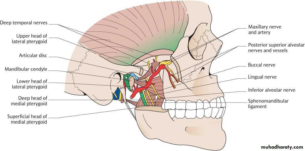
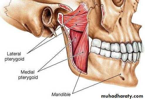
1. Muscles of mastication ( masseter and most of temporalis lie outside of infratemporal fossa)• Lower portion of temporalis muscle: passes medial to zygomatic arch to insert on coronoid process and anterior border of ramus of mandible
• Lateral pterygoid muscle: from lateral pterygoid plate and greater wing of sphenoid to neck of mandible and articular disc of TMJ
• Medial pteryygoid muscle: from medial surface lateral pterygoid plate and tuberosity of maxilla to medial surface of ramus and angle of mandible .
2. Mandibular nerve ( CN V3) and its branches, chorda tympani, and otic ganglion .
3. Maxillary artery.4. Pterygoid plexus .
Muscles of mastication
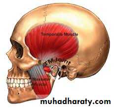
Temporalis
1. Origin from temporal fascia and temporal fossa from temporal lines to infratemporal crest.
2. Insertion: coronoid process and anterior border of ramus of mandible .
3. Action : closes jaw, posteroinfrior part retracts jaw.
4. Innervation: anterior and posterior deep temporal branches of mandibular nerve ( CN V3), which curve around infratemporal crest to pass beneath temporalis
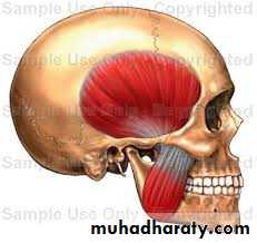
Masseter
1. Orginea. Superficial part: anterior 2/3 of lower border of zygomatic arch
b. Deep part: posterior and medial side of zygomatic arch
2. Insertion
a. Superficial part : angle and lower lateral surface of ramus of mandible
b. Deep part : upper lateral surface of ramus
3. Action : closes the jaw
4. Innervation : masseteric nerve from mandibular nerve ( CN V3), which passes over mandibular notch to enter muscle
Medial pterygoid ( internal pterygoid )
1. Origin : medial surface of lateral pterygoid plate of sphenoid , pyramidal process of palatine and tuberosity of maxilla .2. Insertion : lower and posterior of angle medial surface of ramus of mandible
3. Action : closes jaw with bilateral contraction; helps grinding movements with1-sided contraction ( moving jaw side to side).
4. Innervation: nerve to medial pterygoid from mandibular nerve ( CN v3 ), which also send brabches to innervate tensor tympani and veli palatine .
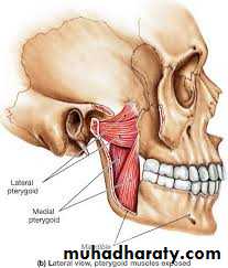
Lateral pterygiod ( external pterygoid )
1. Origina. Superior head : from inferior surface of greater wing of sphenoid .
b. Inferior head : from lateral surface of lateral pterygoid plate .
2. Insertion
a. Superior head : articular disc of TMJ
b. Inferior head : pterygoid fovea on neck of mandibular condyle
3. Action : protrudes mandible, opening mouth by drawing mandible and articular disc forward onto articular tubercle; unilateral contraction moves mandible from side to side, assisting in grinding motion ( note : anterior belly of digastric, geniohyoid, mylohyoid and platysma help in opening mouth )
4. Innervation: nerve to lateral pterygoid from mandibular nerve ( CN V3) .
Clinical significabce
The temporalis muscle can be used as a flap for various deformities . the primary indications for the temporalis muscle flap are for intraoral, cranial base, and orbital reconstructions . the use of split temporalis muscle as a sling for the lower eyelid and lip in facial paralysis is another common indication : some dynamic movement is possible through the V3 branch of the trigeminal nerve .(also masseter muscle can be used in patient with facial paralysis ) . less common indication are for palate and maxillary reconstruction .Trauma to the temporal region
The bone of calvarium is thinnest in the temporal fossa. Strong blows to the side head may cause a depressed fracture, in which a fragment of bone is depressed inward to compress or injure the brain. At the pterion , the middle meningeal artery is easily ruptured following such an injury CAUSING EXTRA DURAL HEMATOMA that compress the brain and could be fatal if untreated.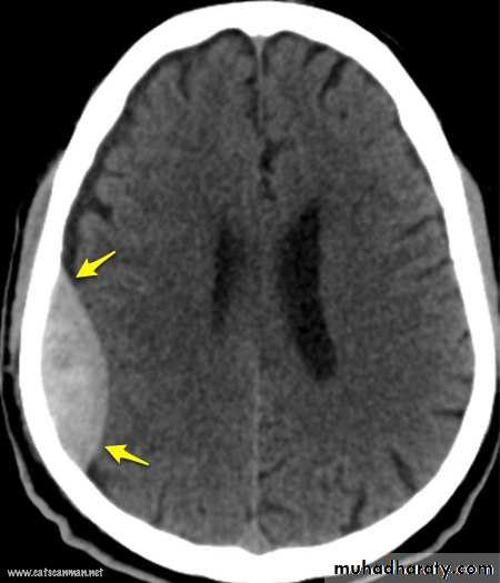
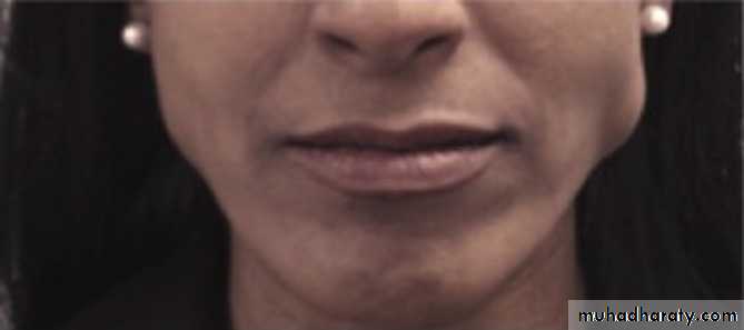
Benign masseteric hypertrophy is a relatively uncommon condition that can occur unilaterally or bilaterally . pain may be a symptom, but most frequently a clinician is consulted for cosmetic reasons . although it is tempting to point to malocclusion, bruxism , clenching or based on awareness of the condition, clinical and radiographic finding, and exclusion of more serious pathology such as benign and malignant parotid disease

Thank
You
for
Listening