Interpretation of Dental Caries
Interpretation of Dental Caries
To detect dental caries, careful clinical examination and careful interpretation are both necessary. A dental examination for caries cannot be considered complete without dental images.Dental images enable the dental professional to identify carious lesions that are not visible clinically. In addition, dental images allow the dental professional to evaluate the extent and severity of carious lesions.
RADIOGRAPHIC TECHNIQUES
A. Posterior bitewing views.Premolar and molar view with a kVp of 75 - 85 kVp.
The most important view for caries.
Remember anterior B.W. views can be used for Perio.
B. Periapical views.
Remember in Radiology a PA is a Postero-Anterior view. The parallel technique must be used to see caries best.
C. Panoramic view.
Often not sufficient detail to see incipient caries but is Often the best view to see occlusal caries.
FREQUENCY OF RADIOGRAPHIC PROCEDURES.
The frequency that radiographs are required can be determined only clinically and by taking a history. Such factors as oral hygiene, fluoride exposure, diet, history of caries and age are important.
Radiographs for caries should be taken at 1 - 3 year intervals in adults depending on the above factors. In young children one can consider taking radiographs every 6 - 12 months on caries-prone children.

Approximately 40-50 % demineralization is required for radiographic detection of a lesion. As seen in the occlusal view, above right, the thickness of the tooth buccolingually masks the carious lesion when it is small.
*The actual depth of penetration of a carious lesion is actually deeper than it appears on the radiograph.
Proximal caries susceptible zone
caries0
1- Buccolingual thickness of tooth, The thicker the tooth, the more difficult it is to see the extent of the caries.
2- Limitations of two-dimensional film, The extent of carious involvement can not be seen in a buccolingual (cheek to tongue) direction.
Factors affecting appearance of caries on radiographs:
3- X-ray beam angle (horizontal or vertical), This is especially important when trying to identify recurrent caries, since changes in angulation may cause the superimposition of the existing restoration with the carious lesion.
4- Exposure factors,. Caries detection is improved with a lower kVp setting, which provides a higher contrast. If the overall density of the film is too light or too dark, the diagnostic potential of the film is limited.
Factors affecting appearance of caries on radiographs:
CLASSIFICATION OF CARIES ONDENTAL IMAGES
The appearance of caries on dental images can be classified according to the location of the caries on the tooth.Caries that involves:
1-interproximal, 2-occlusal, 3-buccal&lingual, and 4-root surfaces may be seen on a dental image.
In addition, recurrent and rampant caries may also be viewed on dental images.
1- Interproximal Caries
The term interproximal means “between two adjacent surfaces.”Caries found between two teeth is termed interproximal caries.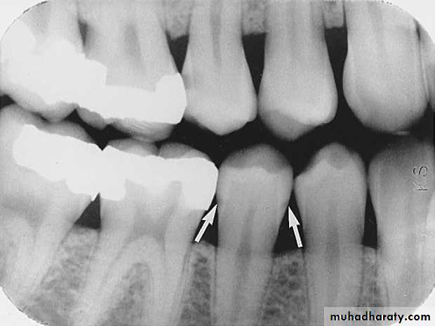
Interproximal Caries
On a dental image, interproximal caries is typically seen at or just below (apical to) the contact point.Interproximal caries can be classified according to the depth of penetration of the lesion through enamel and dentin. Interproximal carious lesions can be classified as incipient, moderate, advanced, and severe.
I
M = Moderate (Stage II)
I = Incipient (Stage I)
A = Advanced (Stage III)
S = Severe (Stage IV)
Caries Classification
S
A
M
A
Interproximal Caries
(Incipient)I
Up to half the thickness of enamel
Usually not restored unless patient has high level of caries activity (high risk). Treat with fluoride.
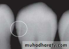
Incipient Interproximal Caries
An incipient carious lesion on the distal surface of the mandibular second premolar.
M
Interproximal Caries
(Moderate)
More than halfway through the enamel (up to DEJ)
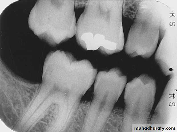
Moderate Interproximal Caries
A moderate carious lesion on the distal surface of the mandibular second premolar
Moderate Interproximal Caries
A moderate carious lesion on the distal surface of the mandibular second premolar
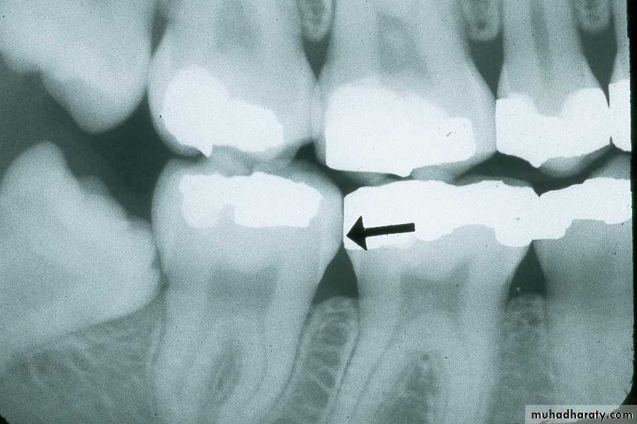
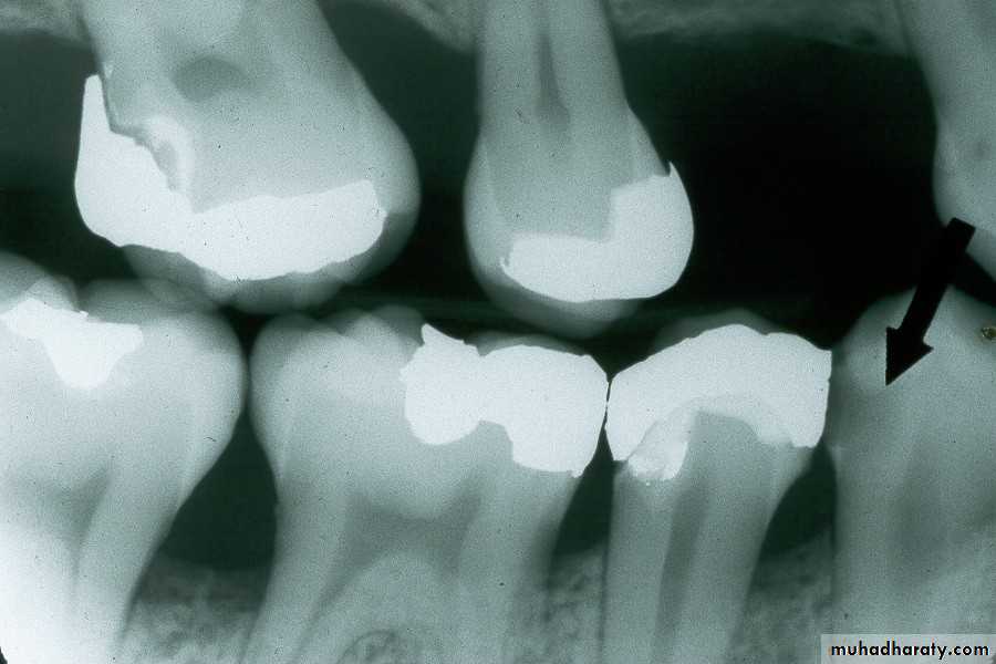
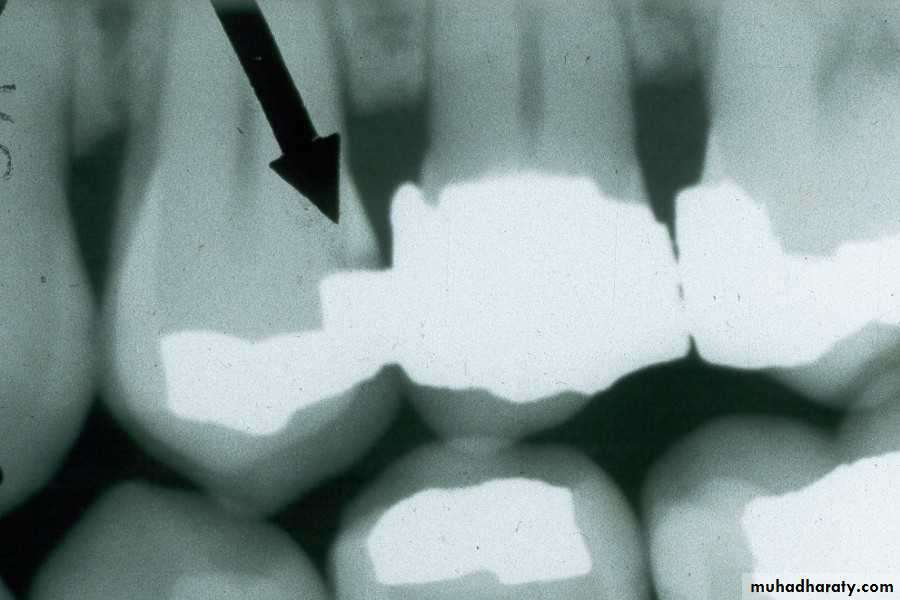
Interproximal Caries (Advanced)
extends to or through the DEJ and into dentin but does not extend through dentin more than half the distance toward the pulpAdvanced Interproximal Caries
An advanced carious lesion, which extends through the dentino-enamel junction (DEJ) and into dentin, seen on the distal surface of the mandibular first molar.
Advanced Interproximal Caries
Advanced lesion identified by arrows.
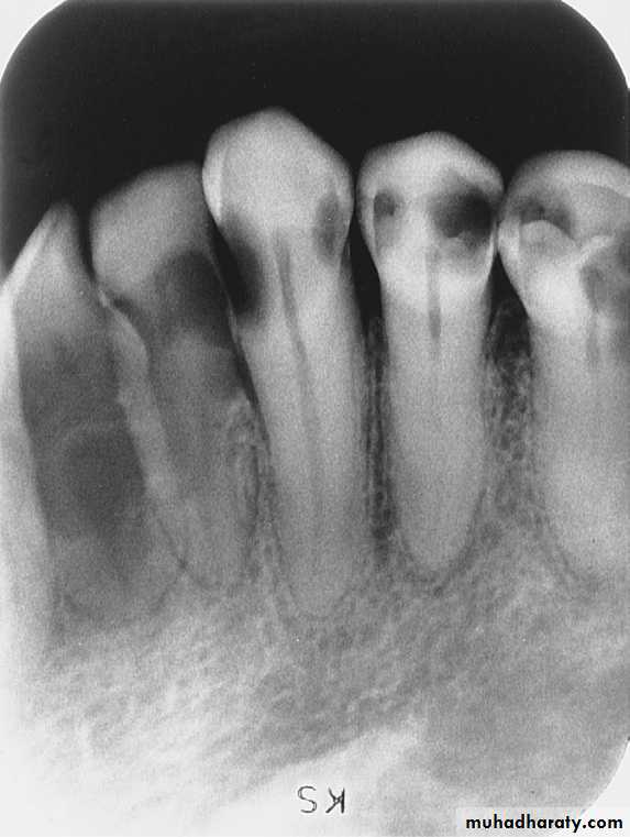
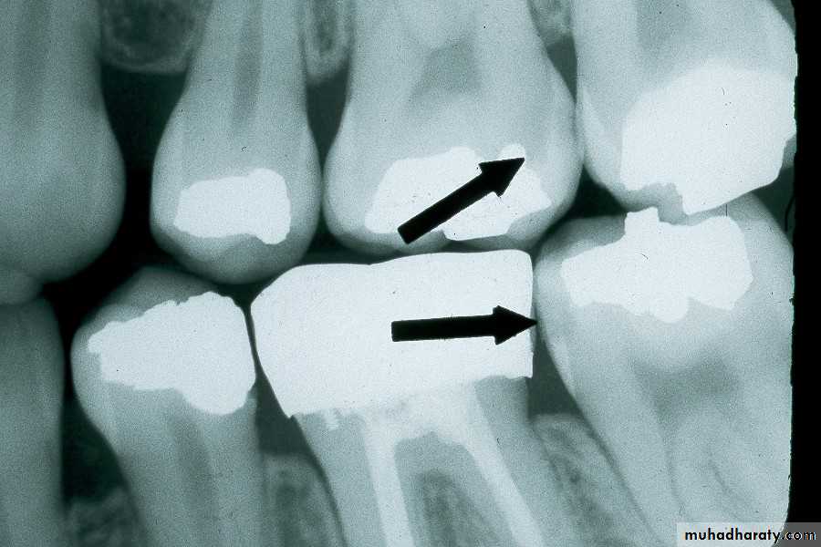
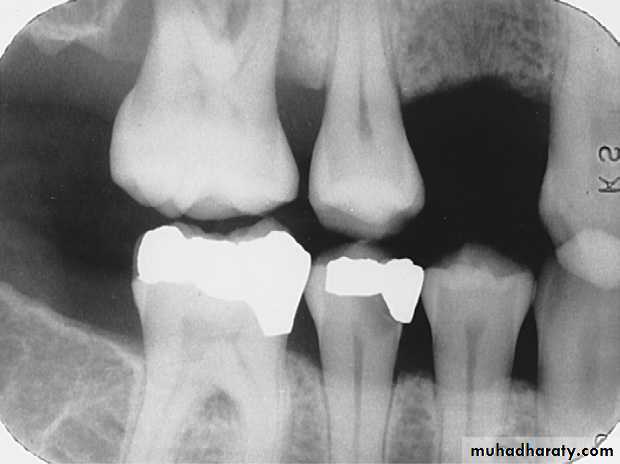
Interproximal Caries (Severe)
extends through enamel, through dentin, and more than half the distance toward the pulpSevere Interproximal Caries
A severe carious lesion on the distal surface of the mandibular first molar.
Severe lesion
2- Occlusal CariesThat involves the chewing surfaces of posterior teeth is termed occlusal caries.
Must have penetrated into dentin. Diagnosed from clinical exam. May be seen as thin radiolucent line or cup-shaped zone underlying occlusal enamel, but difficult to see on radiographs unless lesion is large.
A thorough clinical examination with the mirror, explorer, and light is the method of choice for the detection of occlusal caries.
Occlusal carious lesions can be classified as incipient, moderate, or severe.
Occlusal Caries (Incipient)
cannot be seen on a dental image and must be detected clinically with an explorer.
Occlusal Caries (Moderate)
extends into dentin and appears as a very thin radiolucent line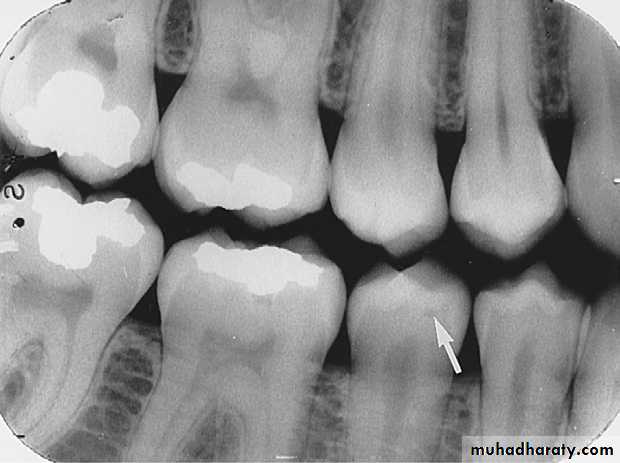
Appears as a tiny radiolucency just below the dentino-enamel junction (DEJ) on the mandibular second premolar.
Occlusal Caries( Severe )
extends into dentin and appears as a large radiolucency.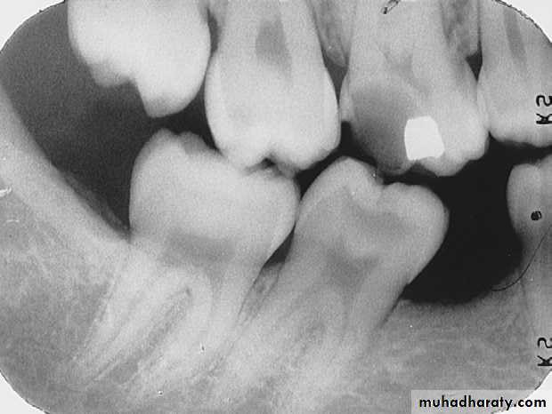
A severe occlusal carious lesion seen as a large radiolucency in dentin on the mandibular first molar
3- Buccal and Lingual Caries
As the names suggest, buccal caries involves the buccal tooth surface, whereas lingual caries involves the lingual tooth surface. Because of the superimposition of the densities of normal tooth structure, buccal and lingual caries are difficult to detect on a dental image and are best detected clinically.When viewed on a dental image, caries that involves the buccal or lingual surface appears as a small, circular radiolucent area.
Buccal and Lingual Caries
Buccal caries seen as a small, circular radiolucency on
the mandibular second molar.- Saucer-like cratering on the roots of the teeth, involving the cementum. Usually found on older individuals with prominent recession and/or periodontitis. May have xerostomia due to medications.
4- Root surface Caries
- Involves only the roots of teeth. The cementum and dentin located just below the cervical region of the tooth are involved. No involvement of enamel occurs.
Root surface Caries
Root caries appearing as a crater-shaped radiolucency
just below the cemento-enamel junction (CEJ) on the mandibular second premolar.Found around the margins of existing restorations. May be due to;
1- unusual susceptibility to caries,2- poor oral hygiene,
3- failure to remove all of the caries during cavity preparation,
4- a defective restoration
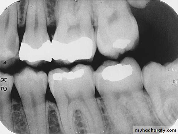
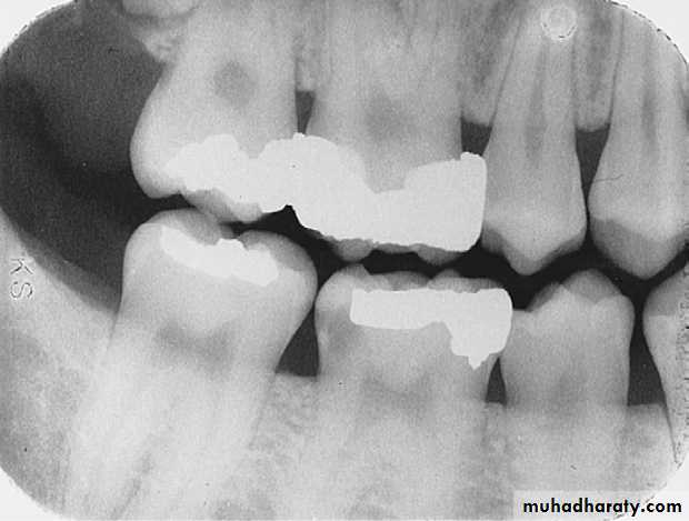
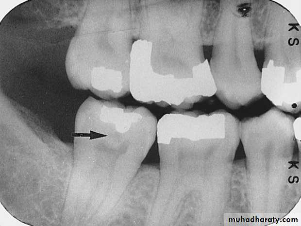
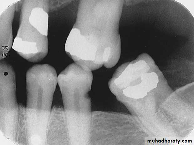
Recurrent Caries
Recurrent Caries
Recurrent caries seen as a radiolucency below a two surface amalgam restoration on the mandibular second premolar.
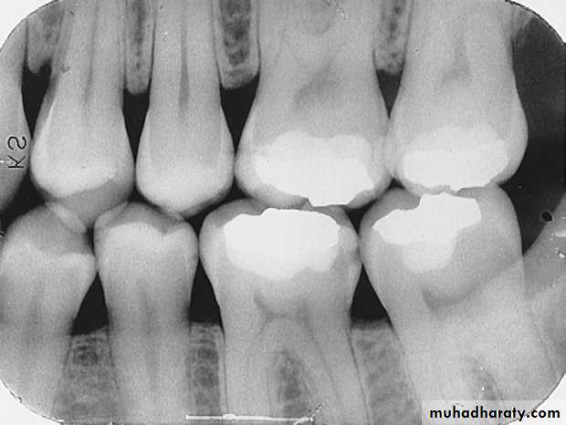
Recurrent caries on upper first premolar
Rampant CariesRampant caries is advanced and severe caries that affects numerous teeth
Rampant caries is typically seen in children with poor dietary habits or in adults with decreased salivary flow.