An Entrance to Periodontology
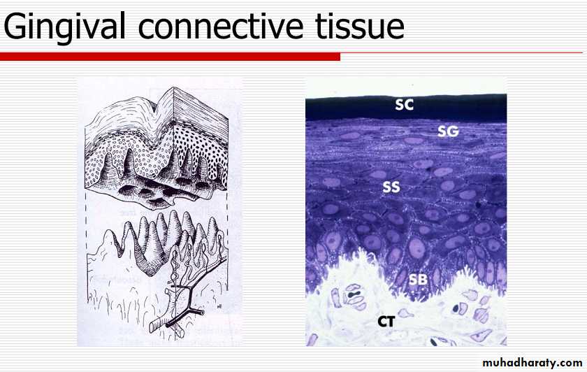
Gingival Connective Tissue The major components of the gingival connective tissue
are collagen fibers (about 60% by volume), fibroblasts
(5%), vessels, nerves, and matrix (about 35%).
It is known as the lamina propria and consists of two
layers:
1. a papillary layer subjacent to the epithelium, which consists of papillary projections between the epithelial rete pegs, a reticular layer with the periosteum of the alveolar bone.
2. The ground substance fills the space between fibers and
cells, is amorphous, and has a high content of water.
Gingival Connective Tissue
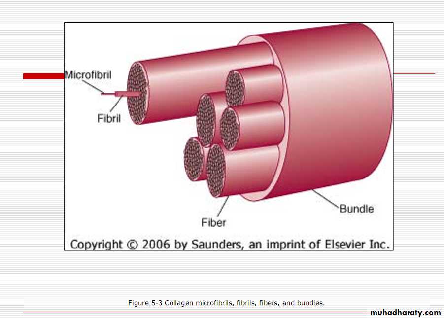
The three types of connective tissue fibers are
• collagen• reticular
• elastic
Gingival Connective Tissue
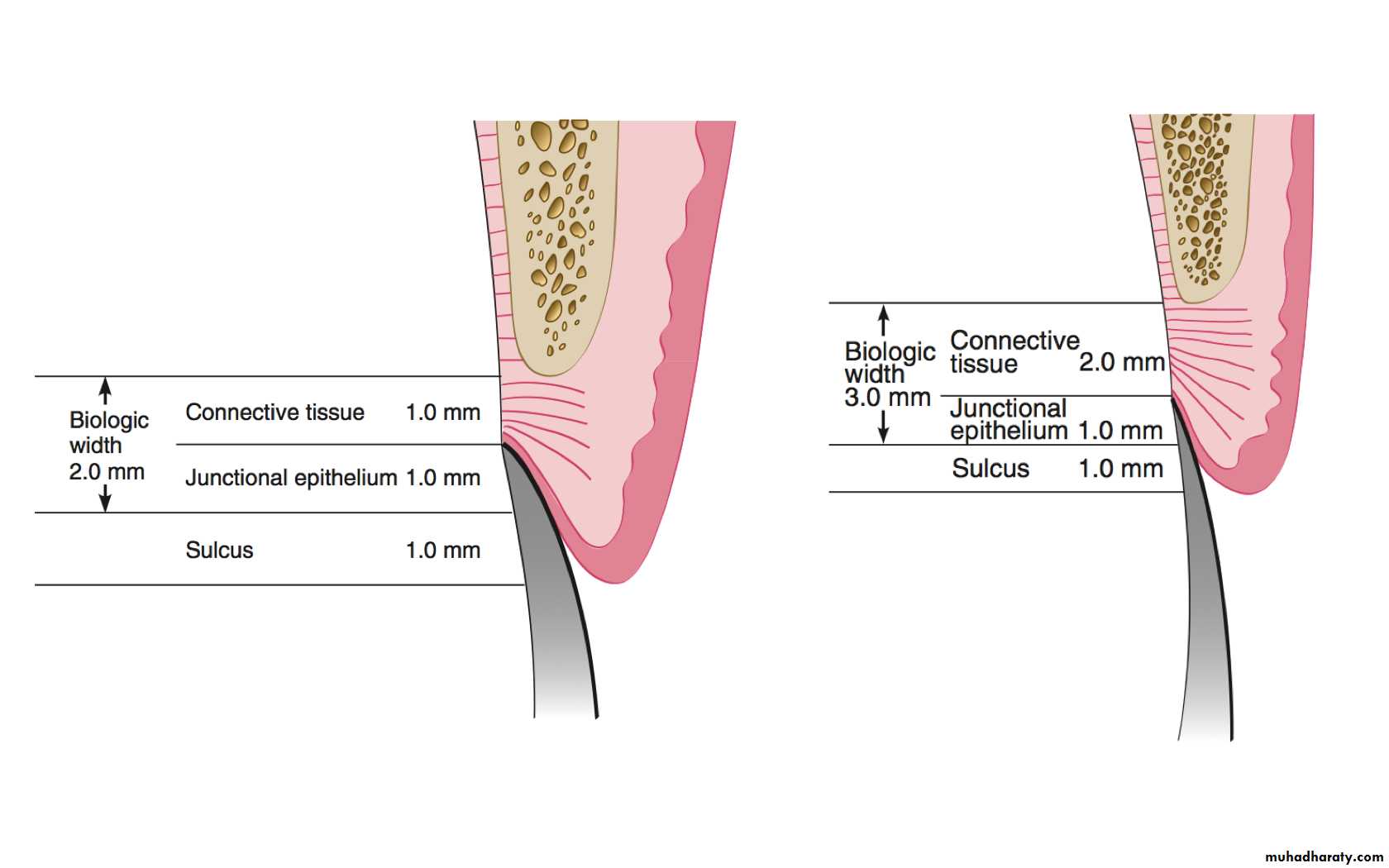
Collagen type I forms the bulk of the lamina propria and provides the tensile strength to the gingival tissue.Therefore, densely packed collagen bundles that are anchored into the acellular extrinsic fiber cementum just below the terminal point of the junctional epithelium form the connective tissue attachment.
The stability of this attachment is a key factor in limiting the migration of junctional epithelium.
The gingival fibers
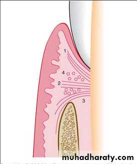
1. Gingivodental GroupThe gingivodental fibers are those on the facial, lingual, and interproximal surfaces.
They are embedded in the cementum just beneath the epithelium at the base of the gingival sulcus.
They also extend externally to the periosteum of the facial and lingual alveolar bones, terminating in the attached gingiva or blending with the periosteum of the bone.
Interproximally, the gingivodental fibers extend toward the crest of the interdental gingiva.
Diagram of the gingivodental fibers extending from the cementum (1) to the crest of the gingiva, (2) to the outer surface, and (3) external to the periosteum of the labial plate. Circular fibers (4) are shown in cross-section.
The gingival fibers
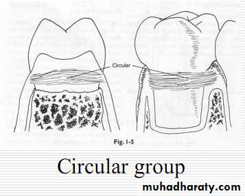
2. Circular GroupThe circular fibers course through the connective tissue of the marginal and interdental gingiva and encircle the tooth in ringlike fashion.
The gingival fibers
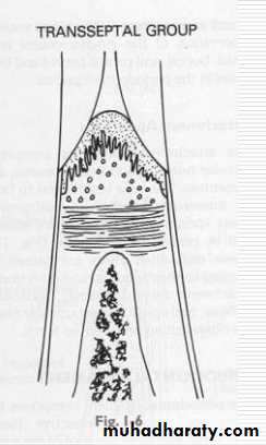
3. Transseptal Group
Located interproximally, the transseptal fibers form horizontal bundles that extend between the cementum of approximating teeth into which they are embedded. They lie in the area between the epithelium at the base of the gingival sulcus and the crest of the interdental bone and are sometimes classified with the principal fibers of the periodontal ligament.
The functions of Gingival Fibers
• 1. To brace the marginal gingiva firmly against the• tooth.
• 2. To provide the rigidity necessary to withstand the
• forces of mastication without being deflected away
• from the tooth surface.
• To unite the free marginal gingiva with the cementum of the root and the adjacent attached gingiva.
Gingival cells
• Fibroblasts• Macrophages
• mast cells
• Osteoblasts
• Cementoblasts
• Osteoclasts
• Odontoclasts
• polymorphonuclear leucocytes, lymphocytes and plasma cells
• undifferentiated ectomesenchymal cells
Repair of Gingival Connective Tissue
It has high turnover rate, good healing and regenerative capacity, with little evidence of scarring after surgical procedures.
However, the reparative capacity of gingival connective tissue is not as great as that of the periodontal ligament or the epithelial tissue.
CORRELATION OF CLINICAL AND MICROSCOPIC FEATURES
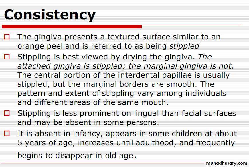
Consistency
The papillary layer of the connective tissue projects into the elevations, and the elevated and depressed areas are covered by stratified squamous epithelium.The surface texture of the gingiva is also related to the presence and degree of epithelial keratinization.
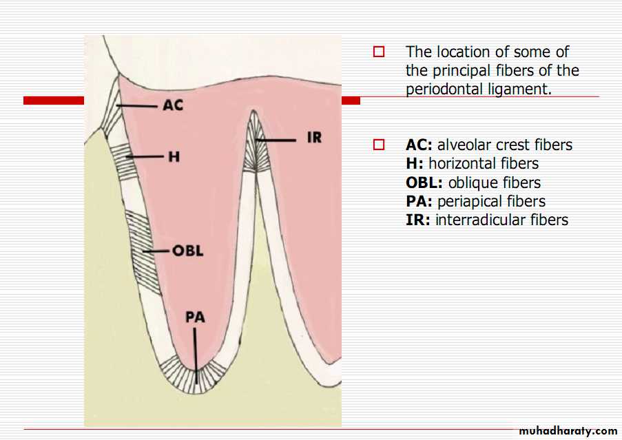
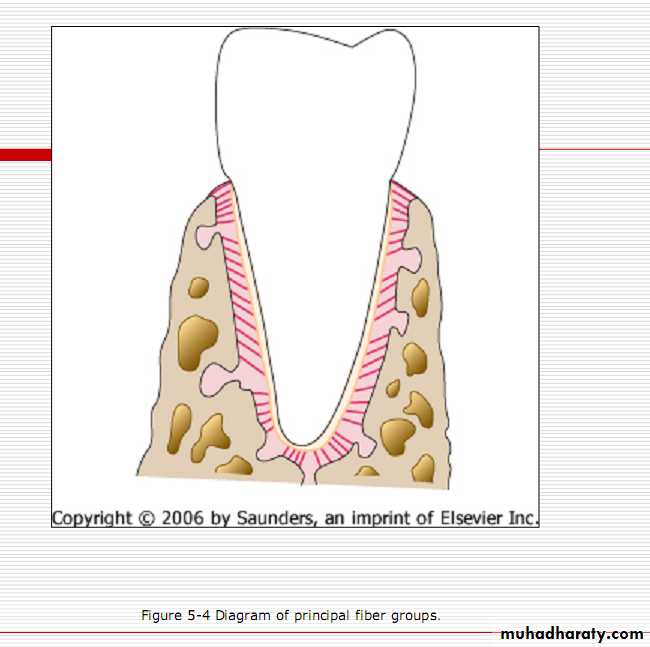
Periodontal ligament
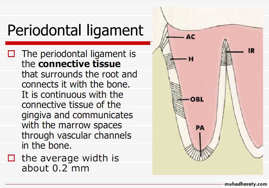
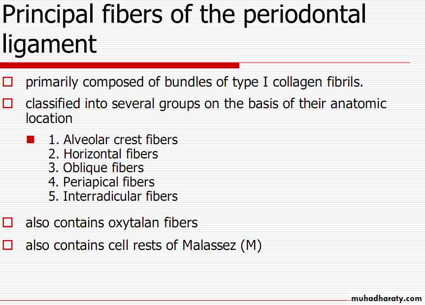
1. Alveolar crest group
Alveolar crest fibers extend obliquely from the cementum just beneath the junctional epithelium to the alveolar crest.Fibers also run from the cementum over the alveolar crest and to the fibrous layer of the periosteum covering the alveolar bone.
The alveolar crest fibers prevent the extrusion of the tooth and resist lateral tooth movements. The incision of these fibers during periodontal surgery does not increase tooth mobility unless significant attachment loss has occurred.
2. Horizontal group
Horizontal fibers extend at right angles to the long axis of the tooth from the cementum to the alveolar bone.3. Oblique group
Oblique fibers, the largest group in the periodontal ligament, extend from the cementum in a coronal direction obliquely to the bone. They bear the brunt of vertical masticatory stresses and transform them into tension on the alveolar bone.
4. Apical group.
The apical fibers radiate in a rather irregular manner from the cementum to the bone at the apical region of the socket. They do not occur on incompletely formed roots.5. Interradicular group
The interradicular fibers fan out from the cementum to the bone in the furcation areas of multirooted teeth. Interradicular fibers are only found between the roots of multi-rooted teeth.