Dr. Wafaa Khalil
جراحة فم \ خامس اسناند.وفاء م(7)
28-11-2016
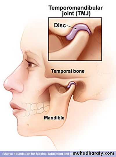
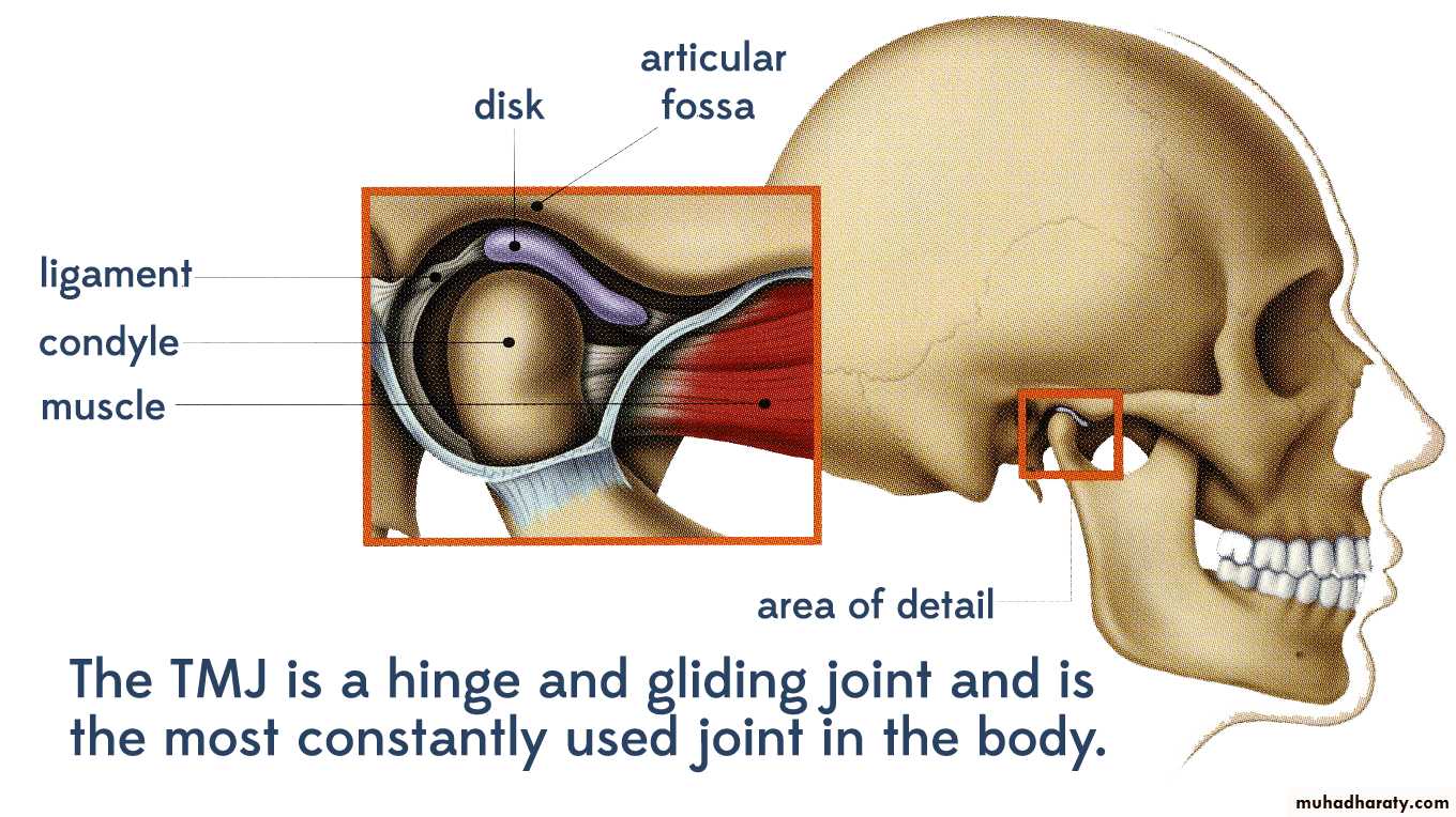
Anatomy of TMJ
• Temporomandibular Joint• consists of mandible suspended from temporal bone via ligaments and muscules, including stylomandibular and sphenomandibular ligaments
• a true synovial joint capable of gliding, hinging, sliding and slight rotation
• mandible and temporal bone separated by meniscus (disc)
Anatomy of TMJ
• Condylar process of mandible articulates with glenoid fossa of temporal bone• anterior: anterior eminance of TMJ
• posterior: EAC
• lateral: zygomatic arch
• medial: styloid process
• Anatomy of TMJ
• Condylar process, continued• lined by fibrous tissues, primarily hyaline cartilage
• this is the primary growth center of the mandible
• damage leads to facial maldevelopment, including both the mandible and the maxilla
• Coronoid process
• insertion for portions of temporalis and masseter
Anatomy of TMJ
• Meniscus (disc)• synovial fluid above and below disc
• “shock absorber”
• internal derangement in 50% of all people
• anteriorly and medially most common
• jaw “pops”
• held in place by medial and lateral capsular ligaments and retrodisc pad
• Disease of TMJ
• Developmental• Inflammatory / infectious condition
• Degenerative / autoimmune
• Cystic lesion
• Traumatic injury
• Neoplasm
• Miscellaneous
• Developmental Disturbances
• Condylar agenesis• the absence of all or portions of condylar process, coronoid process, ramus or mandible
• other first and second arch anomalies seen
• early treatment maximizes condylar growth
• a costocondral graft may help with facial development
Disease of TMJ
• Hypoplasia
• Unilateral, Bilateral• Example “first arch syndrome” , Hemifacial microsomia ( 1st & 2nd arch syndrome or otomandibular dysostosis), Treacher Collins Syndrome (mandibulofacial dysostosis)
• usually developmental secondary to trauma or infection
• most common facial deformity is shortening of mandible
• jaw deviates towards affected side
• Flatness of the face on the unaffected side. (The side to which the ramus is short causes muscles to appear fuller; the muscles on the unaffected side are stretched so that side appears flatter.) Mandibular deviation causes malocclusion
• Treatment for child: costochondral graft
• Treatment for adult: shorten normal side or lengthen involved side
•
Disease of TMJ
• Hyperplasia
• Hemifacial hyper atrophy
• an idiopathic, progressive overgrowth of mandible
• deviation of jaw away from affected side
• presents in 2nd decade
• Treat by condylectomy
Disease of TMJ
• Infection
• ( specific, non specific infection)• Acute, chronic
• Viral, bacterial, fungal
• Origin
• Ear
• Direct injury
• Systemic spread
Disease of TMJ
Disease of TMJ
Degenerative / autoimmune- Joint Disorders are the second most common cause of temporomandibular pain after myofacial pain dysfunction syndrom
- Include internal derangements, degenerative joint disease, traumatic arthritis, ankylosis
• Joint Disorders, Continued
Disease of TMJ
Cardinal features are jaw popping (clicking) and pain
• 50% of the population has a jaw pop, which usually occurs with opening (between 10-20 mm)
• may elicit a history of “lock” jaw
• Internal Derangement
• the most common joint disorder• involves the abnormal repositioning of the disc
• disc location is usually anteromedial
• four types of derangements (see other screen)
Joint Disorders, Continued
Joint Disorders, Continued
• Type IA• popping over the joint without associated pain (50% of normal subjects)
• Type IB
• popping over the joint with pain
• due to chronic streching of capsular ligaments and tendons
Joint Disorders, Continued
• Type II• similar to type IB, but a history of “lock jaw” can be elicited
• closed lock vs open lock
• Type III
• a persistent lock, usually closed
• No click
• Rx of Internal Derangements
• Type I and II
• similar to myofascial disorders: NSAIDs, anxiolytics/relaxers, “oral” hygiene and appliances if necessary for four weeks
• progression of symptoms may require surgical intervention
• main goal is lysis of adhesion and repositioning of disc
• open vs arthroscopic
Rx of Internal Derangements
• Type III• usually requires general anesthesia to mobilize jaw
• agressive medical and physical therapy is initiated, including a bite appliance
• if no improvement after 3 weeks, surgery is indicated to lyse adhesions and/or reposition disc
Disease of TMJ
• Traumatic injury to the joint• Traumatic arthritis
• Acute subluxation
• Chronic subluxation
• Dislocation
• Acute, chronic
• Anterior, posterior, medial , lateral
• Tearing of the disk
• Damage to the condyle
• # condyle
• Infection
• Still’s disease (Systemic-Onset Juvenile Rheumatoid Arthritis) is a disorder characterized by inflammation with high fever spikes, fatigue, salmon-colored rash and/or arthritis. Though there have been several theories regarding the cause(s) of Still's disease, the cause is not yet known. Many symptoms of Still's disease are often treatable with anti-inflammatory drugs)
• Ankylosis of the Temporomandibular join
Disease of TMJ
• Traumatic injury to the joint, continued
• Traumatic Injuries
• Fractures of the condyle and subcondyle are common• unilateral fracture involves deviation of jaw towards affected side with or without open bite
• Treat: Maxillo-mandibular fixation (MMF) with early mobilization
• bilateral fracture usually has anterior open bite
• often requires open reduction with internal fixation (ORIF) of one side with MMF
• Dislocation of the TMJ
Traumatic Injuries• Acute dislocation
• new onset Type III derangement, surgery of the mouth
• treatment is reduction under anesthesia manually (downward,backward,upward)
• Chronic dislocation
• usually secondary to abnormally lax tendons
• Rx: sclerosing agents like (alcohol, sodium tetradecyl sulfate,...ect.) capsulorraphy, myotomy of lateral pterygoid
• Limitation of Mouth opening
• Extra capsular causes
• # zygoma
• Coronoid bone hyperplasia ( enlargement)
• Scar tissue due to burn of the skin of the face
• Neurogenic causes
• Myogenic causes ( scar tissue in the muscles of mastication, myositis ossificans due to injury)
• Psychogenic causes (hysterical condition)
• Submucous fibrosis
• Inferior Dental block
• Haematoma
• Low grade infection
• Wisdom tooth removal (oedema , swelling and trismus)
• Tetanus
• Intra capsular causes
• Ankylosis of the TMJ either: Fibrous Ankylosis
• Bonny ankylosis
Disease of TMJ
• Ankylosis of the TMJ
• Defn: the obliteration of the joint space with abnormal bony morphology• etiologies include prolonged MMF, infection, trauma.
• False ankylosis: an extracapsular condition from an abnormally large coronoid process, zygomatic arch or scar tissue
• Ankylosis of the TMJ, Continued
• Treatment
• Child: a costochondral graft to help establish a growth plate
• Adult: prosthetic replacement
• the new joint should be established at highest point on ramus for maximal mandibular height
• an interpositional material is needed to prevent fusion
• Arthritis of the TMJ
• The most frequent pathologic change of the TMJ• Most are asymptomatic
• Rheumatoid arthritis
• usually seen in other joints prior to TMJ
• when present, both joints usually affected
• early radiographic changes include joint space narrowing without bony changes
• Arthritis of the TMJ, Continued
• late radiographic changes may involve complete obliteration of space with bony involvement and even ankylosis• end stage disease results in anterior open bite
• Juvenile RA may progress to destruction of the growth plate, requiring costochondral graft
• Treatment
• NSAIDs, penicillamine, gold
• Surgery limited to severe JRA and ankylosis
• Degenerative Arthritis
• “wear and tear” of the joints
• most asymptomatic
• Primary Degenerative arthritis
• “wear and tear” - usually in older people
• asymptomatic or mild symptoms
• Secondary Degenerative arthritis
• due to trauma, infection and bruxism
• symptoms severe
• radiographic findings include osteophytes an derosion of the condylar surface
Arthritis of the TMJ, Continued
• Treatment is initially similar to myofascial disorders, including NSAIDs, benzos and “oral” hygiene. Bite appliance may be necessary
• After 3-6 months, surgery is considered
• lysis of adhesions, osteophyte removal
• condylar shave. Resorption of the condyle is a known complication
Arthritis of the TMJ, Continued
• Muscular Disorders (Myofascial Pain Disorders) are the most common cause of TMJ pain
• It refers to a group of muscle disorders characterized by diffuse facial pain and limited mouth opening.• the cause of MPD is often multifactorial. Biologic, behavioral, environmental, social,emotional.
• More in female
Myofascial Pain Dysfunction Disorders
Signs and symptoms
• unilateral dull, aching pain worse with use (gum, candy, bruxism)
• Associated with sleep disturbances, otalgia, headache, burning tongue and cervical pain.
• Tooth clenching or grinding and bruxism,
• Limited and painful mouth opening.
- Sore teeth and frequently complain of jaw tiredness and fatigue when eating.
- Tenderness to palpation of the masticatory and
cervical muscles,
Myofascial Pain Dysfunction Disorders
Myofascial Pain Dysfunction Disorders
Treatment: most patients with MPD will improve with conservative treatment. TMJ surgerywill not resolve and will usually make it worse.
• The patient should be educated and reassured that the pain usually resolves with simple treatment and that a more serious condition does not exist
• Soft diet
• “oral” hygiene: no gum chewing, candy chewing, jaw clenching
• Bed rest, NSAIDs, muscle massage and physical therapy
• heat, muscle relaxants (benzos)
• Bite appliance (splint)
• psychological evaluation
• Disease of the TemporoMandibular Joint (TMJ)
• Osteophyte
• Pannus formation
Arthrocentesis (is the irrigation of the joint) for painful limited opening. TMJ arthroscopy is performed to lysis and lavage for the management of patients with painful limited mouth opening.
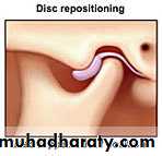
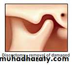
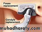
• Arthroplasty refers to all types of open surgery for TMJ, including disk repositioning, discectomy, and joint replacement
• Disk repositioning is used when the protective cartilage disk has slipped out of place inside the TMJ
• A discectomy is performed when the disk providing padding and protection to the TMJ has deteriorated or become damaged
• TMJ replacement
• Benign tumors and lesion
• Osteoma• Osteochondroma
• Chondroma
• Chondroblastoma
• Giant cell granuloma
• Giant cell tumor
• Neurofibroma
• Hemangioma
• Arteriovenous malformation
• Synovial chondromatosis
• Osteochondrosis dissecans
• Ganglion cyst
Disease of TMJ
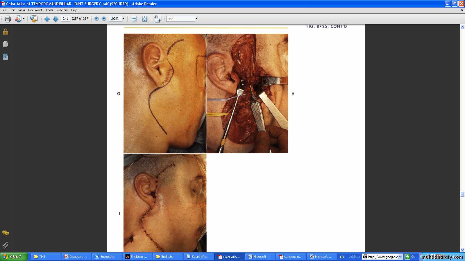
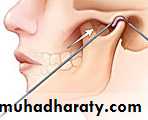
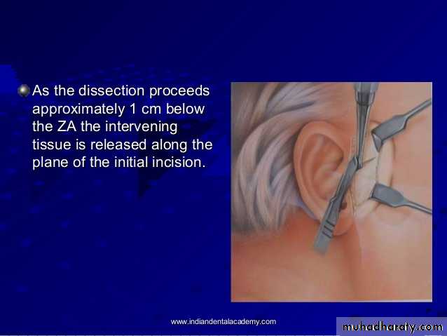
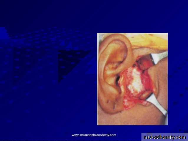
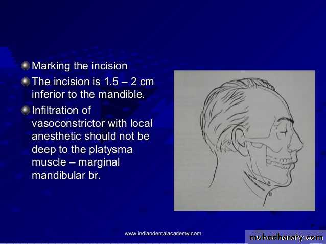
Surgical approaches to TMJ
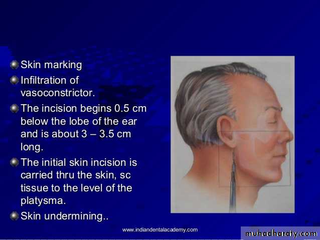
• Pre auricular approach
• Endaural approach
• Post auricular approach
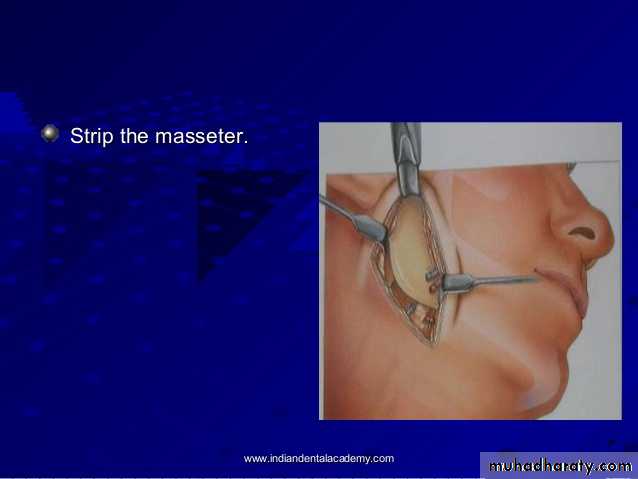
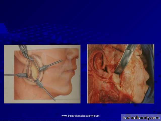
• Submandibular (Risdon’s ) approach
• Post ramal ( Hind’s ) approach
• Parotidectomy type of incision with temporal extension
• Hemicoronal approach
• Coronal or bicoronal approach
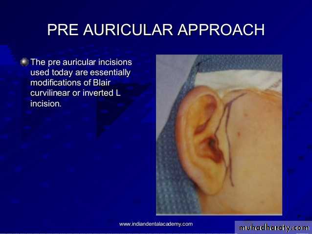
Preauricular approach
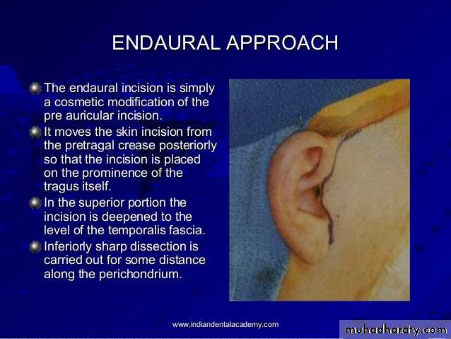
Endaural approach
Submandibular approach
Post ramal approach
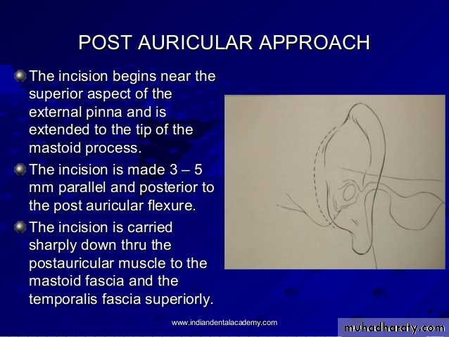
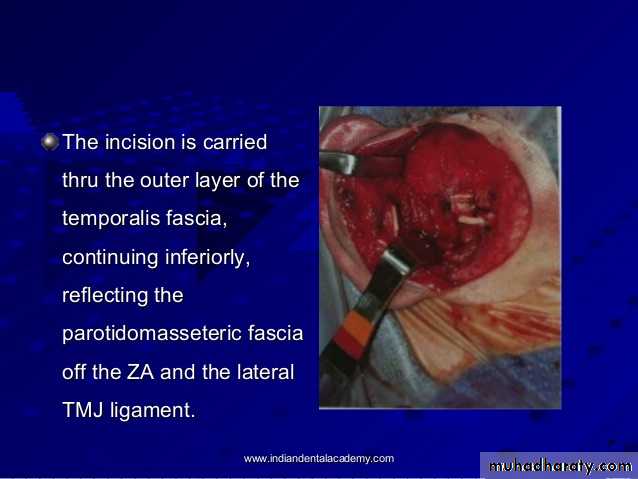
Post auricular approach