Interpretation of trauma, pulpal and periapical lesion
BY: Dr. Rebuar Fazil Khalid12/12/2016
1- radiographic changes caused by trauma
2- radiographic changes caused by resorption3- radiographic features of pulpal lesions:
pulpal sclerosis
pulpal obliteration
pulp stones
4- radiographic features of periapical lesions
periapical radiolucencies
periapical radiopacities
interpretation of trauma, pulpal and
periapical lesion1-Radiographic changes caused by trauma
Trauma: can be defined as an injury produced by an external force. trauma may affect the crown and roots of teeth as well as alveolar bone. trauma may result inA- Fractures of teeth and bone and
B-Injuries such as intrusion, extrusion, and avulsion.
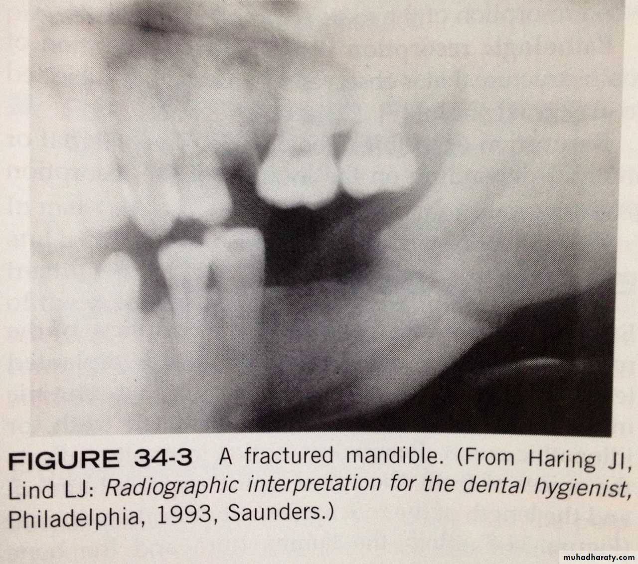
Fractures:
Fractures may affect the crowns and root of teeth or the bones of maxilla and mandible. whenever a fracture is evident or suspected, radiographic examination of the injured area is necessary.
Crown fractures:
Fractures that effect crown most often involve the anterior teeth. Most crown fractures result from an accident involving a fall or a motor vehicle.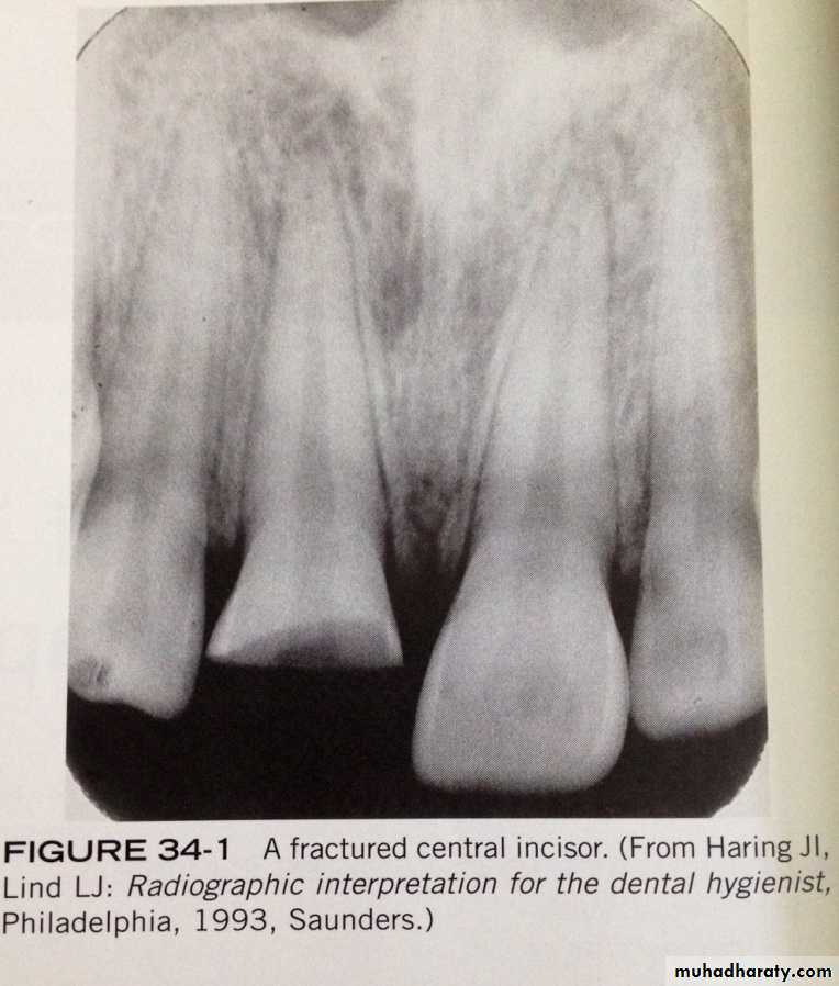
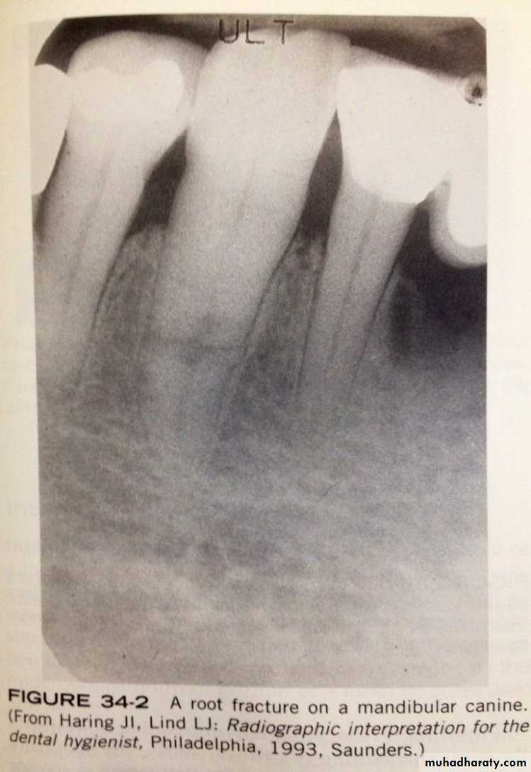
Root fractures:
- Root fractures are less common than crown fractures.- Root fractures occurs most often in the maxillary central incisor region.
-if the x-ray beam is parallel with the plane of the fracture, the root fracture appear as sharp radiolucent line on a periapical radiograph
Root fractures:
Jaw FracturesFractures of the mandible occur more often than fractures on any other bone of the face and frequently result from assaults, accidents, and sport injuries.
Injuries:
in addition to fractures, trauma may result in the displacement of teeth.- Tooth displacement includes:
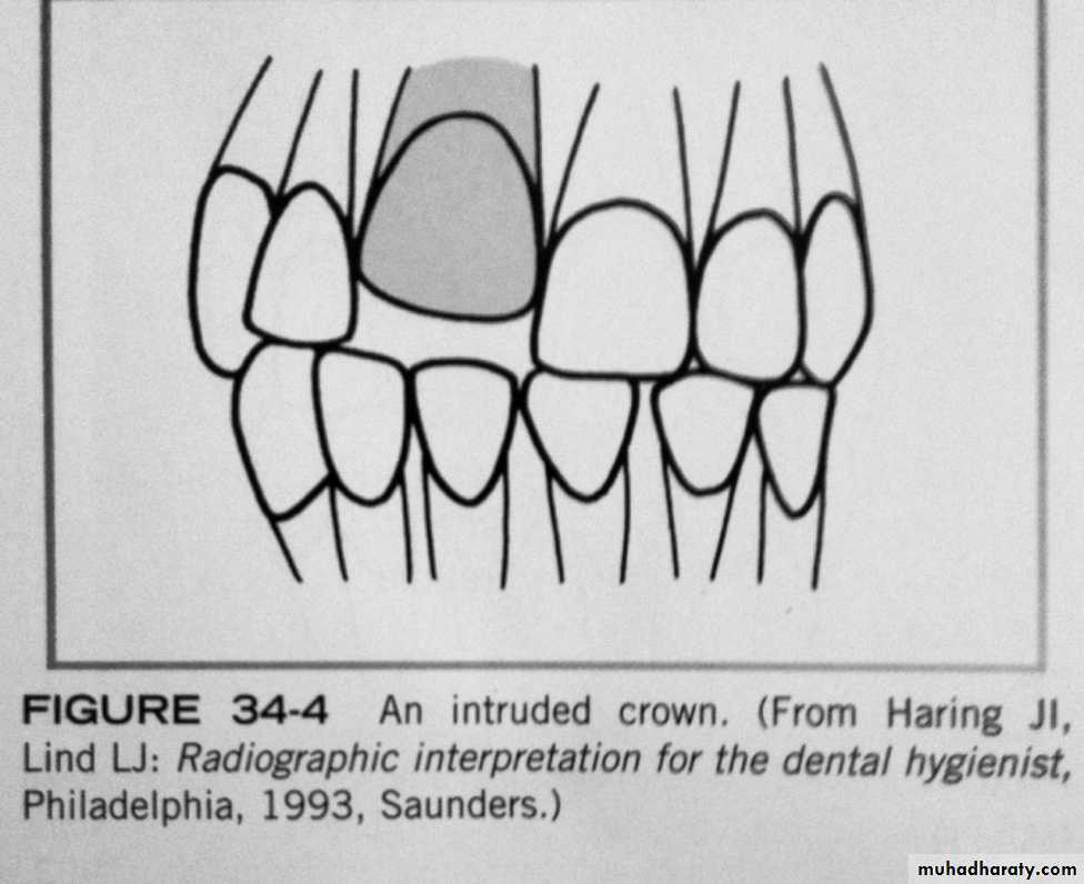
1- luxation(intrusion or extrusion)
2- avulsion.
Luxation
Luxation : is the abnormal displacement of teeth. either intrusion or extrusionIntrusion refers to the abnormal displacement of teeth in to bone.
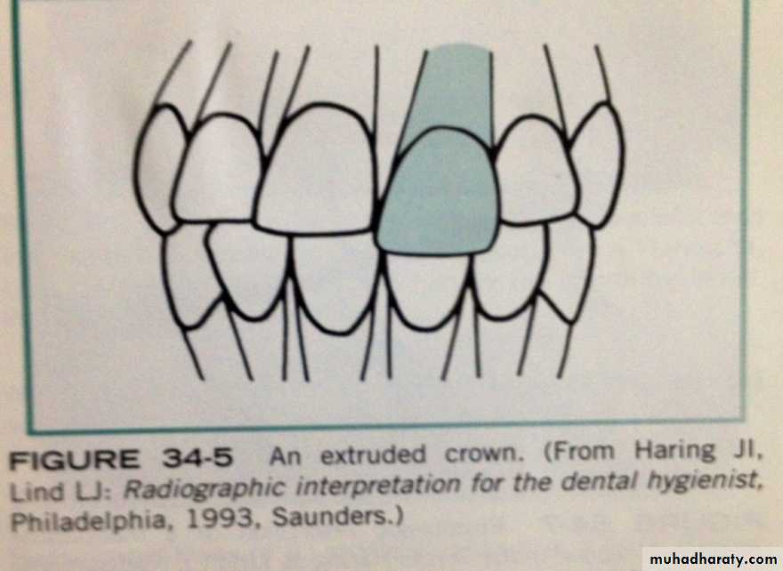
Extrusion
refers to the abnormal displacement of teeth out of bone. Teeth that have been luxated should be evaluated by a periapical radiograph and examined for:root and adjacent alveolar bone fractures, damage to the periodontal ligament, and pulpal problems.
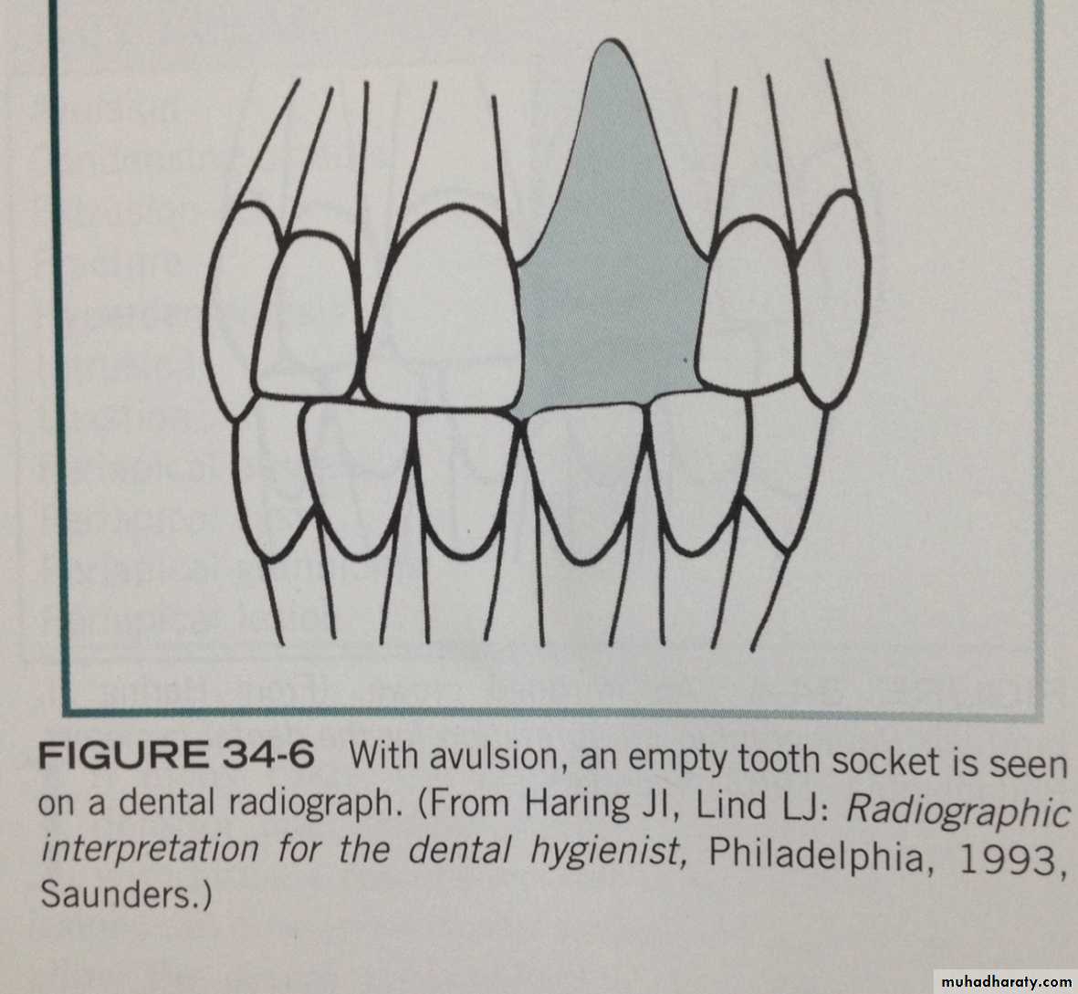
AVULSION
-is the complete displacement of a tooth from alveolar bone.
-An avusled tooth not seen in dental radiograph, instead important in the evaluation of the socket area and should be used to examine the region for splinted bone.
AVULSION
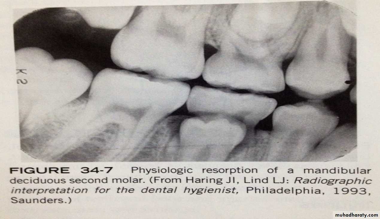
2-Radiographic changes caused by Resorption1-physiologic resorption:
is the process that seen with the normal shedding of primary teeth. the primary tooth is shed when resorption completed.
2-Pathologic resorption:
is a regressive alteration of tooth structure that is observed when a tooth is subjected to abnormal stimuli.either external resorption or internal resorption.
External Resorption
is seen a long periphery of the root surface and is often associated with abnormal mechanical force, trauma, chronic inflammation, reimplanted teeth, tumors, cysts and impacted teeth.
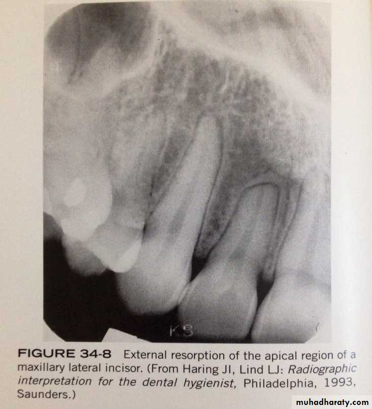
External Resorption
- external resorption most often affect the apices of teeth, the apical region appear blunted, and the length of the tooth appear shorter than normal.- its asymptomatic and there is no effective treatment to external resorption.
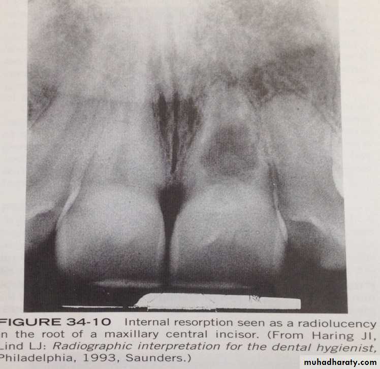
Internal resorption:
- occur with in crown or root of tooth and involves the pulp chamber , pulp canal. precipitating factor such as pulp capping, trauma, and pulp polyps.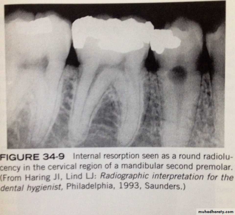
Internal resorption:
- internal resorption appear as around to ovoid radiolucency in the midcrown or midroot portion of a tooth.
- its asymptomatic, treatment is variable either endodnotic therapy or extraction.
3-RADIOGRAPHIC FEATURE OF PULPAL LESIONS:
1-pulpal sclerosis2- pulpal obliteration
3- pulp stones
Pulpal sclerosis:
- is a diffuse calcification of pulp chamber and pupl canals of teeth that result decreased pulp cavity. its associated with aging. appear on radiograph incidentally and have little clinical significance.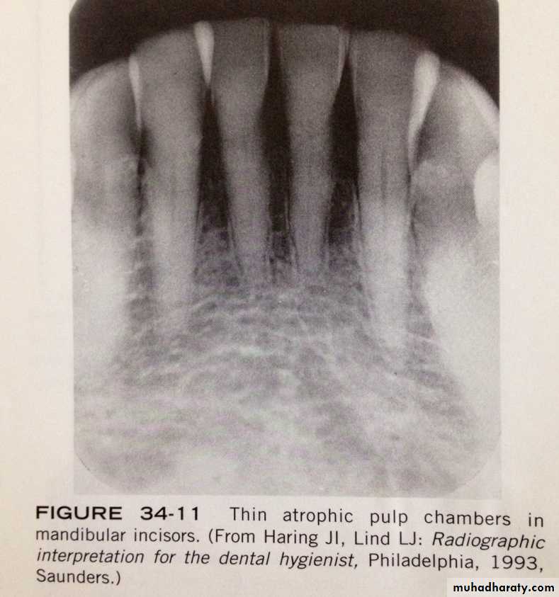
Pulpal obliteration:
some conditions( e.g: attrition, abrasion, caries, trauma) may act as irritant to the pulp and stimulate the production of secondary dentin, which results in obliteration of pulp. - radiographically: tooth with this condition does not appear to have pulp chamber or pulp canals and nonvital.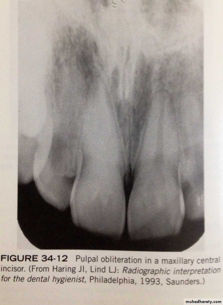
Pulpal obliteration:
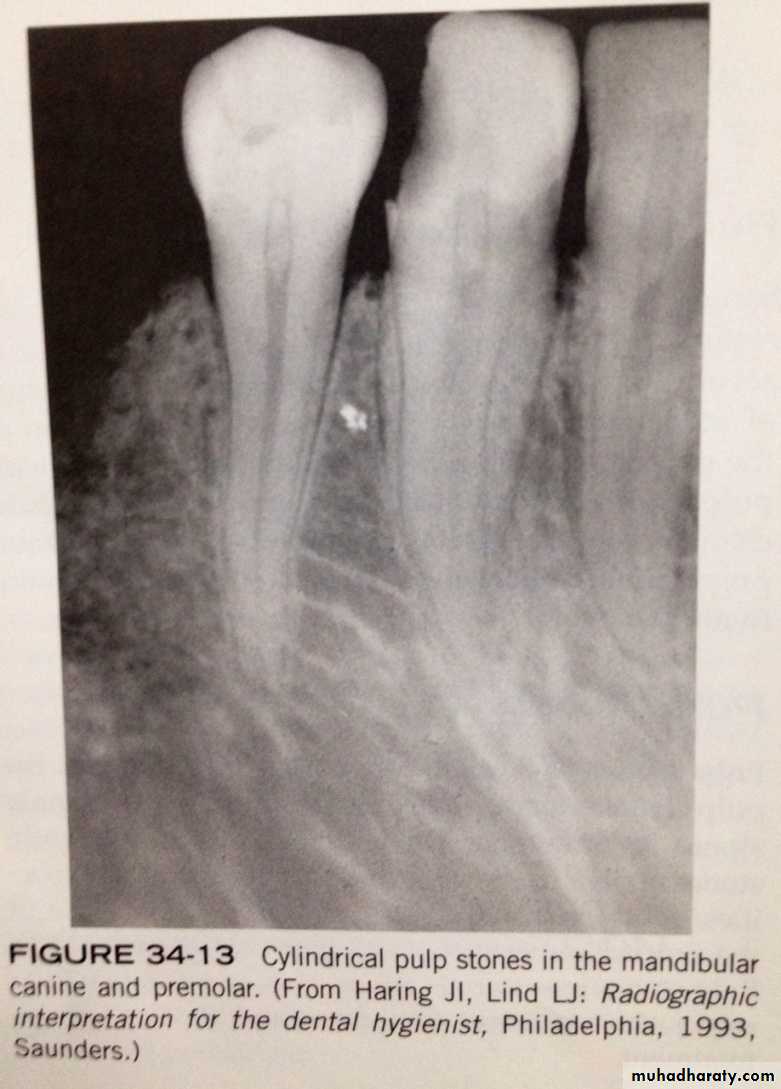
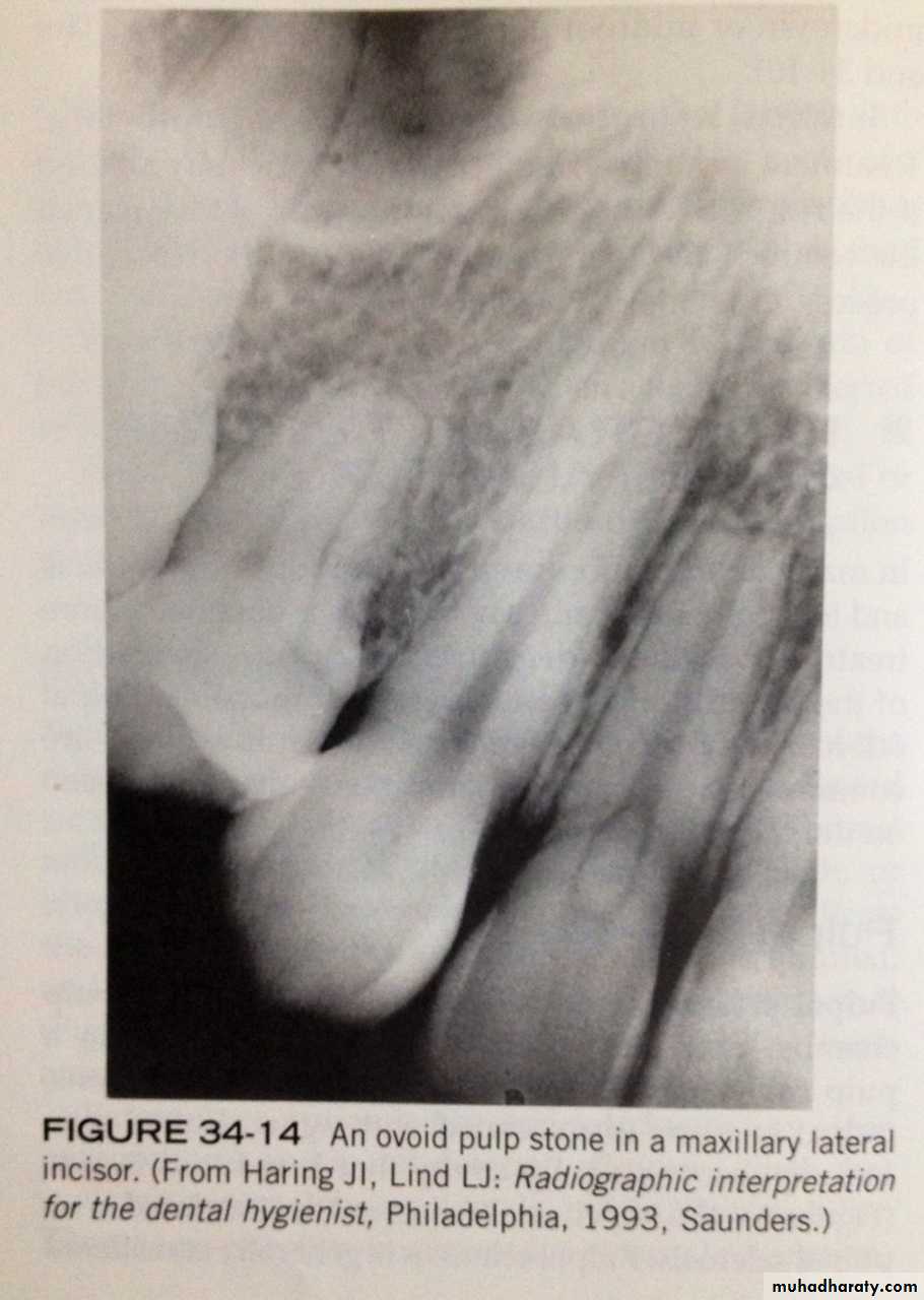
pulp stones- are calcifications that are found in pulp chamber or pulp canal. the cause unknown.
- On dental radiograph: pulp stone appear as round, ovoid or cylindrical radiopacities. do not cause symptoms and do not require treatment.
pulp stones
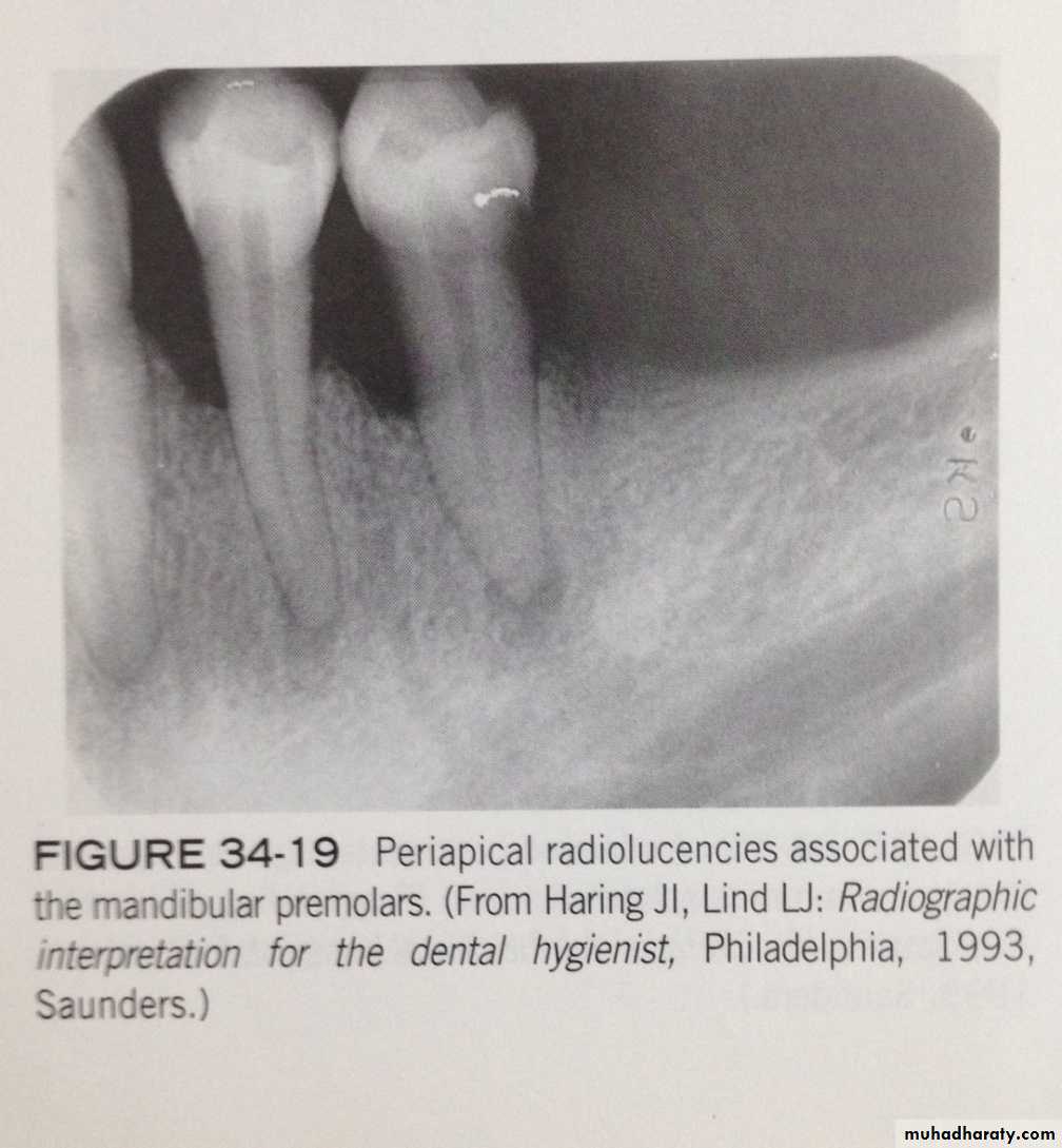
4-RADIOGRAPHIC FEATURES OF PERIAPICAL LESIONS:Periapical radiolucencies
periapical granulomas, cysts, and abscesses are common periapical radiolucencies that can be seen on dental radiographs. these lesions cannot be diagnosed by there radiographic appearences alone;instead diagnosis is based on the clinical features and radiographic and microscopic appearences. Because its impossible to distinguish between these three periapical lesions based on there radiographic appearence, the dental radiographer should refer to these lesions simply as "periapical radiolucencies".
PERIAPICAL GRANULOMA
A periapical granuloma is a localized mass of chronically inflammed granulation tissue at the apex of a non vital tooth.
The periapical granuloma results from pulpal death and necrosis.
is the most common sequlae of pulpitis. A periapical granuloma may give rise to a peiapical cyst or periapical abscess. A tooth with this condition is typically asymptomatic but has a previous history of prolonged sensitivity to heat or cold.
PERIAPICAL GRANULOMA
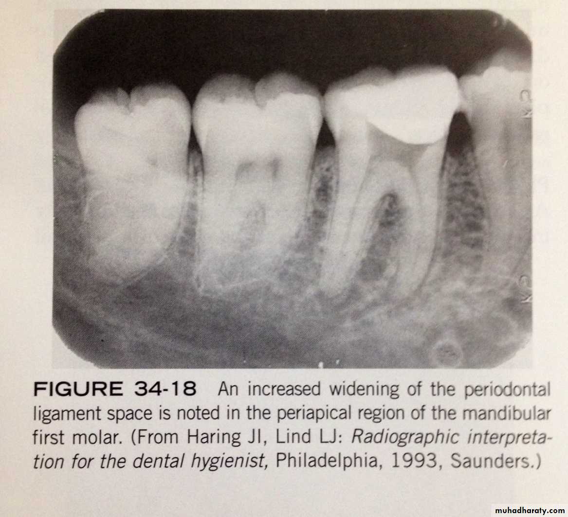
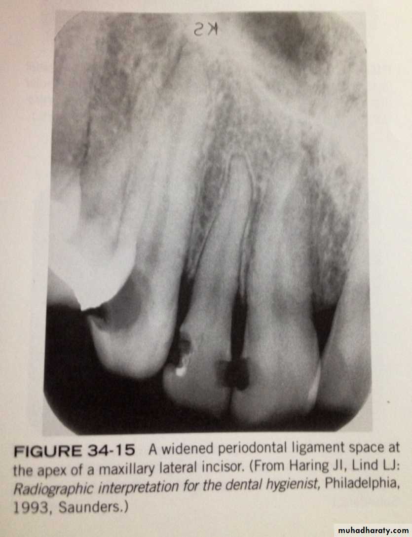
on a dental radiograph, a periapical granuloma is initially seen as widened periodontal ligament space at the root apex. with time, the widened periodontal ligament space enlarges and appears as a round or ovoid radiolucency.PERIAPICAL GRANULOMA
PERIAPICAL CYST
is a lesion that develops over a prolonged period;cystic degeneration takes place with in a periapical granuloma and results in a periapical cyst.
The periapical cyst are the most common of all tooth-related cyst and comprise 50-70% of all cyst in oral region. they are typically asymptomatic.
PERIAPICAL CYST
on dental radiograph the typical periapical cyst appear as a round or ovoid radiolucency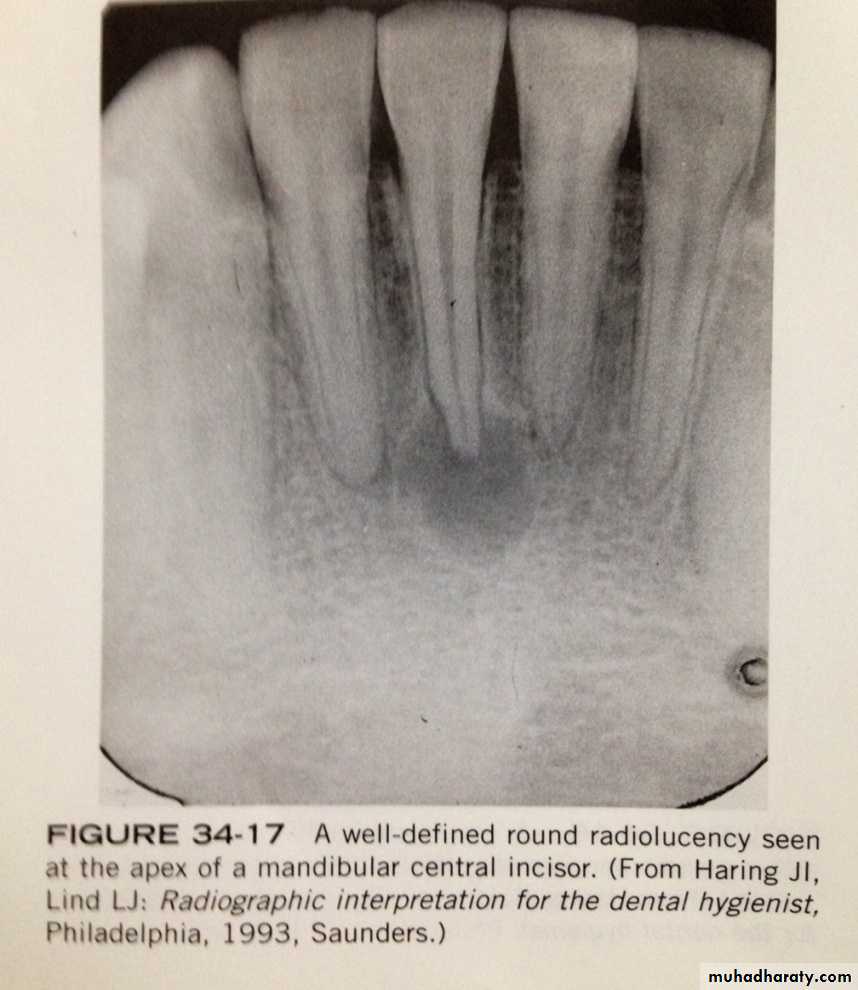
PERIAPICAL ABSCESS
is a localized collection of pus in the periapical region of a tooth that results from pulpal death.periapical abscesses may be acute or chronic.
An acute periapical abscess has features of an acute pus-producing process.
PERIAPICAL ABSCESS
An acute periapical abscess is painfull; the pain may be intense, throbbing, and constant. the tooth is nonvital and is sensitive to pressure, percussions, and heat.chronic periapical abscesses are usually asymptomatic because the pus drains through bone or the periodontal ligament space.
PERIAPICAL ABSCESS
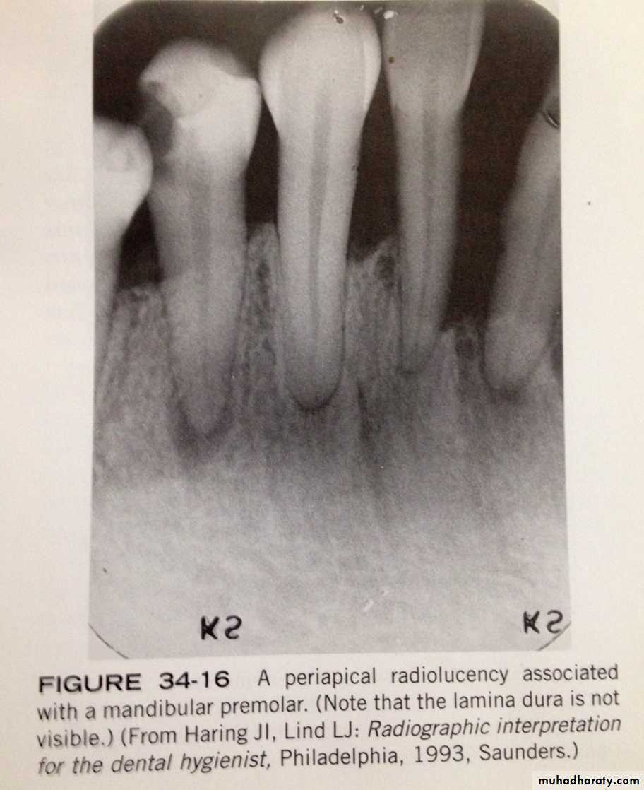
with an acute periapical abscess, no radiographic change maybe evident. early radiographic changes include an increased widenning pf the periodontal ligament space. A chronic peripacal abscess appears as a round or ovoid apical radioloucency with poorly defined margins.PERIAPICAL ABSCESS