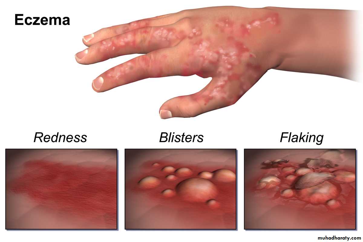
Eczema (Dermatitis )
Dr. Manar GhanemLEC. 4
17 / 11 / 2016
Objective
Define eczemaDetrmine its prevalence
List stages, types and clinical presentations of eczema
Determine etiological factors and clinical features of endogenous dermatitis.
Management plan for endogenous dermatitis
Eczema
Is the most common inflammatory skin diseaseThe characteristic components of eczematous inflammation
Erythema
Scale
Vesicle
Stages Of Eczematous Inflammation
There are three stages of eczema:acute,
subacute,And chronic
Stage
SymptomsPrimary and secondary
Treatment
acute
Intense itch
Vesicles, blisters, intense redness
Cold, wet compresses;
oral or topical
steroids;
antihistamines;
+_ antibiotics
subacute
Slight to moderate
itch, pain, ,
burning
Redness, scaling, fissuring,
Topical steroids with or without occlusion,
lubrication,
antihistamines,
+_ antibiotics,
chronic
Moderate to
intense itch
Thickened skin, skin lines
accentuated (lichenified
skin), excoriations, fissuring
Topical steroids (with occlusion for best results),
antihistamines,
antibiotics,
lubrication
Acute eczematous inflammation. Numerousvesicles on an erythematous base..
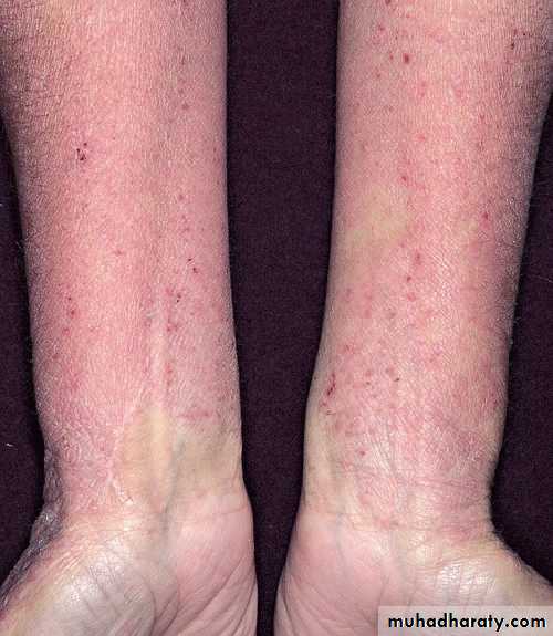
Subacute And Chronic Eczematous Inflammation.The Skin Is Dry, Red, Scaling, And ThickenedSubacute eczema. Erythema and scalingare present, the surface is dry, and the borders are indistinct.
Chronic eczematous inflammation , Accentuated skin lines differentiatethis process from psoriasis.
Classification
The classification of eczematous dermatosis is based on aetiologyExogenous
Endogenous
Why ??
Classification of dermatitis• Atopic dermatitis
• Seborrheic dermtitis
• Discoid eczema
• Asteatotic eczema
• Gravitational dermatitis
• Pompholyx
• Lichen simplex
1. Contact dermatitis
Irritant
Allergic
2. Infective dermatitis
3. Photodermatitis
4. Radiodermatitis
Endogenous
Exogenous
Atopic Dermatitis
The term atopy was introduced years ago to designate agroup of patients who had a personal or family history of
one or more of the following diseases: hay fever, asthma,
very dry skin, and eczema.
Atopic dermatitis (AD) is a chronic, pruritic inflammatory
skin disease that occurs most frequently in children,but also affects many adults.
AD is often associated with elevated serum immunoglobulin
E (IgE) levels and a personal or family history of type I allergies, allergic rhinitis, and asthma.Prevalence
Approximately 15% to 30% of children and2% to 10% of adults are affected.
About 45% of cases of atopic dermatitis begin within the first 6 months of life, 60% begin during the first year, and 85% begin before 5 years of age.
Up to 70% of children have a remission before adolescence.
Atopic dermatitis can start in adults.Pathogenesis AND IMMUNOLOGY
Increase serum IgE evelIncrease histamine level in blood and skin
Increase blood esinophiles
Decrease cell mediated immunity , Patients may develop severe diffuse cutaneous infection with the herpes simplex virus (eczema herpeticum) whether or not their dermatitis is active.
Decrease neutrophil and monocyte chemotaxis
Increase susceptibility to viral and fungal infection, staphylococcal colony may be high and bacterial infection may supervene .
The disease characteristics vary with age.:
Infants :Facial and patchy or generalized body eczema, with extensor prediliction .
Adolescents and adults :
Have eczema in flexural areas and on thehands.
The pattern of inheritance is polygenic.
Phases of atopic dermatitis infantile phase: 3months – 2 years
Face , forehead, scalp , extensor side of theCheek first place
Diaper freq spared
limbs
Itchy dry skin +/- bacterial infection
Usually progress to childhood stage but may resolve in the age of 1- 1.5 yrs
Atopic dermatitis—infant phase. Red, scaling plaques confined to the cheeks are one of the first signs of atopic dermatitis in an infant.
Atopic dermatitis—infant phase. Generalizedinfantile atopic dermatitis sparing the diaper area,
Childhood Phase 2 to 12 Years
inflammation in flexural areas (i.e., the antecubital fossae, neck, wrists, and anklesperspiration stimulates burning and intense itching and initiates the itch-scratch cycle.
Tight clothing that traps heat about the neck or extremities further aggravates the problem.The eruption begins with papules that rapidly coalesce into plaques, which become lichenified when scratched.
Constant scratching may lead to destruction of melanocytes, resulting in areas of hypopigmentation
if they have been vigorously scratched, they may be bright red and scaling with erosions.
The border may be sharp and well-defined, as it is in psoriasis, or poorly defined with papules extraneous to the lichenified areasHypopigmentation in the antecubital fossae caused by destruction of melanocytes by chronic scratching.
Atopic dermatitis. Classic appearance of confluent papules forming plaques
Atopic dermatitis—childhood phase. Diffuse inflammation on the face of a child. The eczema initially spared the perioral areaAdult Phase 12 Years to Adult
As in the childhood phase, localized inflammation with lichenificationMostly in flexural areas.
Adults may have no history of dermatitis in earlier years, but this is unusual.severe generalized atopic dermatitis. thedermatitis has generalized to involve the entire body.
Criteria for diagnosis
Must be presentPruritus
Eczema (acute, subacute, chronic)
Typical morphology and age-specific patterns*
Chronic or relapsing history
*Patterns include:
Facial, neck, and extensor involvement in infants
Current or previous flexural lesions in children and adult stage
sparing of the groin and axillary regions
Seen in most cases, adding support to the diagnosis:
Early age of onset
Personal and/or family history of atopyImmunoglobulin E reactivity
Xerosis
Essential Features
Important Features
Help to suggest the diagnosis of atopic dermatitis but are nonspecific
Atypical vascular responses (white dermographism )
Facial pallor
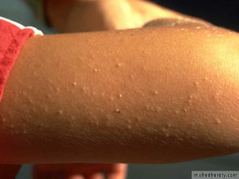
Keratosis pilaris
Pityriasis alba
Hyperlinear palms
Ichthyosis
Cataract
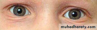
Infraorbital fold (dennie-morgan )
Persistent dry or itchy skin in adult life
Widespread dermatitis in childhoodAssociated allergic rhinitis, bronchial asthma
Family history of atopic dermatitis
Early age at onset
Female gender
Associated Features
Unfavorable
Prognostic Factors
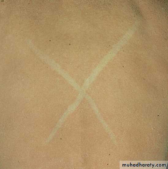
White dermographism
Keratosis pilaris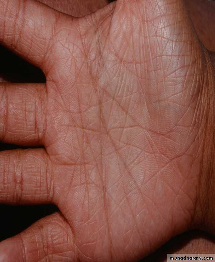
Hyperlinear palm
Pityriasis alba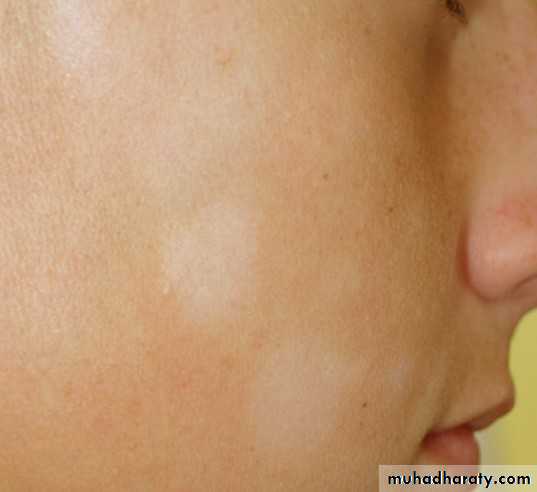
Icthyosis
Infraorbital fold• Bactereal infections ex.S.Aureus infection.
• Severe viral infections Herpes simplex infection (eczema herpeticum). Widespread infection with the herpes simplex
• Poor growth.
• Local& systemic side effect of steroids
• Negative psychological effects
Complications:
Eczema herpeticumDifferential diagnosis
ScabiesSeborrheic dermatitis
Contact dermatitis (irritant or allergic)
Ichthyosis
Cutaneous T-cell lymphoma
Psoriasis

Atopic dermatitis
ScabiesDiagnosis
Diagnosis based on the clinical features ( essential ,important , associated )
Treatment
We have to deal with the psychological impact on the patient and his family , by discussing , reassuranceAvoid triggering factor
Treat the existing lesionsPrevention
Avoid wool.Use 100% cotton.
Use soaps only in axilla, groin, feet.
Avoid perfumes or makeup that burns or itches.
Do not scratch.
Apply soothing lubricants.
Maintain cool, stable temperatures.
Do not overdress.
Avoid sweating.
Humidify the house in winter.
Avoid cigarettes.
Minimize animal dander—no cats, dogs, rodents, or birds.
Control emotional stress
Diet control is a controversial treatment method
Treatment
Topical steroid with different potency .. strong on dry chronic lesionSystemic steroid.. For generalized sever cases
For itching .. sedative antihistamine
For infection ..Antibiotic
Continuously using Vaseline to the skin
Others tacrolimus , tar, phototherapy
Other type of endogenous eczema Seborrhic dermatitis
A chronic superficial inflammation ,Common affect 3-5% of population
On hairy region (pilosebaceous unit)
Affect infant up to third months and after puberty ( two phases only ).
Etiology : Androgen increase sebum release
• Also over active normal flora( pityriosporum ovale ) which increase activity in seborrhic area
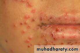
Clinical feature
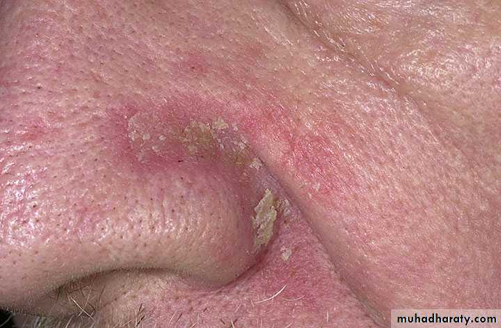
Erythematous patch or plaque covered by greasy yellowish scale, indistinct margin, hair loss uncommon .Most common on scalp, eyebrow , face , mustache , nasolabial folds
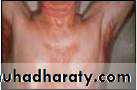
Also central chest( presternal area ) , axillae and groinItchy lesion
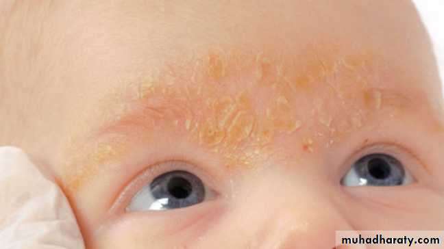
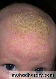
In infant it cover the scalp known as cradle cap
Napkin area is often affected which differ from ?But it spread beyond the area covered by napkin ( not as in contact dermatitis )
Acute onset or wide spread exacerbation of seborrhic dermatitis is commonly seen in HIV infection.
SEBORRHIC DERMATITIS ( INFANT STAGE )
SD ADULT STAGE
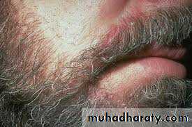
SD MUSTACHE
NASOLABIAL FOLD SD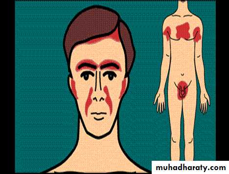
Differential Diagnosis
• Psoriasis vulgaris.• Dermatophytosis (tinea capitis, tinea faciale, tinea corporis),
• Candidiasis (intertriginous).
• Tinea amiantacea.
• Contact dermatitis
• Diagnosis: clinical
treatment
• A-general measures:
• Regular bathing.
• Avoid irritant & oily applications.
• B-topical therapy:
• 1-For scalp shampoos containing 2 % ketoconazole,
• Ketoconazole or any antifungal cream for the face and body.
• 2-corticosteroids for more severe cases;
• Hydrocortisone or low-potency corticosteroid solution, lotion, or gel( for scalp)
• 1 % or 2.5% hydrocortisone cream for other sites.
• *the main treatment of seborrhoeic eczema of scalp in infancy is emollients.
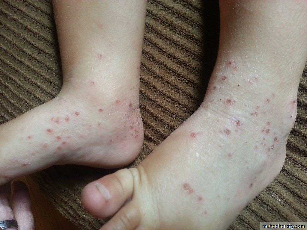
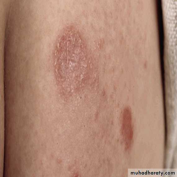
Nummular ( Discoid eczema )
• A chronic, intensely pruritic, coin-shaped erythematous scaly plaques,• During winter months;
• Often seen in atopic individuals.
• Age: two peaks in incidence: young adults and old age.
• Plaques may be :
• Exudative and crust( wet type).
• Or dry scaly (dry type).
• Distribution: lower legs, trunk, hands and fingers or generalized.
• Pathophysiology: unknown.
• Differential diagnosis :
Psoriasis
Tinea corporis
Herald patch of pityriasis rosea .
Diagnosis: clinical, biopsy shows eczema
Treatment :Avoid irritant as soap , wool
Emollient as vasaline
medium potency topical steroid
If superadded infection so topical and even systemic antibiotic if wide spread
If itching antihistamine
Pityriasis alba
Thought to be photo allergyAppear on exposed parts
On children and adolescents
Self limiting
Reassure the family with mild topical steroid, and sun avoidance

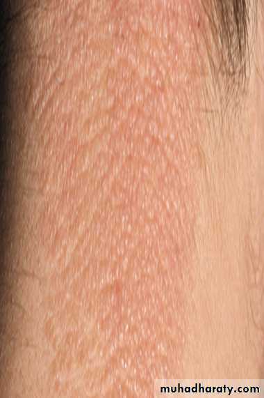
Asteatotic Dermatitis
• Eczematous lesion that occurs in the winter and in old persons• On the legs, arms, and hands but also may be on the trunk.
• Itchy, eyhematous scaling dry, “cracked,” fissured skin
• Very often the eruption results from too frequent bathing ,frequent washing with soap , especially in winter when the humidity is low, also in patient taking diuretics
Treatment
Avoid overbathing with soapAvoid soap and irritant wool
Emollient vasaline twice dailySteroid may be used
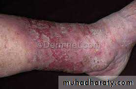
Gravitational eczema
Previously misnamed as stasis or varicosity dermatitis ( thought to be due to stasis , but there is NO stasis .Due to Increased hydrostatic ( venous ) pressure and capillary damage with GOOD blood supply , especially seen on the leg
Due to faulty valve and increase capillary pressure with widening of the epithelial pores
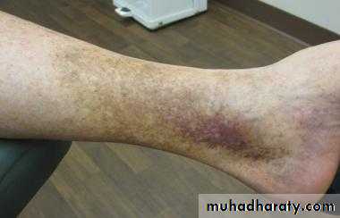
lead to:
extravasated fluids lead to edema
and RBCs lead to hemosiderosis , brownish black discoloration
Fibrinogen which will converted into fibrin lead to vasoconstriction that cause bad nutrition , so any truma may lead to persistant leg ulcer
Most common site is proximal to the medial malleolus
As an itchy ill-defined, erythematous patches with fine scaling, sometimes with excoriations, on lower legs, especially around varicosities.
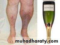
• Complications:
• 1 contact dermatitis from medication• 2 infection.
• 3 ulcer
• 4 inverted champagne bottle look to the leg may result from prolong disease ulceration and fibrosis.
• Treatment:
Leg elevation,
Weight reduction
Topical steroid
Treatment of secondary bacterial infection ( avoid topical neomycin becouse it cause sensitization ) .
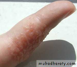
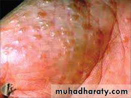
pomphylox
Lesions: very itchy deep seated tiny vesicles along the sides of the fingers , palms, sides toes, and soles.• may coalesce and become bullae. which weep and become painful dry, hyperkeratotic and fissured.
Age: Majority under 40 years (range 12 to 40 years).
Sex: Equal ratio.
Associations:
It is more common in atopics
Treatment: with potent topical steroid under cover , Antihistamine
Lichen Simplex Chonicus (Neurodermatitis)
• Definition: A pruritic eczematous condition resulting from habitual continued rubbing and scratching at a localized area of the skin, associated with a period of anxiety.• Age: over 20 years.
• Sex: more frequent in women.
• Lesion: characteristicly itchy lichenified plaque, well defined, unilateral flesh coloured, pink or hyper pigmented.
• Pruritus,often in paroxysms, and it becomes a pleasure to scratch.
• Often the rubbing becomes, reflexive and a subconscious habit.
• Lightly stroking the involved skin with a cotton swab generates a strong desire to scratch the skin
• The constant scratching leads to a vicious cycle of:
• scratch → release histamine → itch → scratching
• Distribution:, especially: back of neck (female), just below elbow, back of hand, genitalia, buttock, lower leg.
• Diagnosis: clinical, biopsy - rarely required, shows eczema.
• Differential diagnosis: hypertrophic lichen planus.
• Treatment:
• Relive anxiety
• stop scratching ??
• super potent topical steroids under occlusion