The Third Week of Development
The Trilaminar Germ DiscThird Week of Development:
Trilaminar Germ Disc
dr.haythem ali alsayigh
Department Of Human Anatomy And Histolog
1
PROBLEMS TO SOLVE1
1. A 22-year-old woman consumes large quantities of alcohol at a party and loses consciousness; 3 weeks later, she misses her second consecutive period. A pregnancy test is positive. Should she be concerned about the effects of her bingedrinking episode on her baby?2
PROBLEMS TO SOLVE2
2. An ultrasound scan detects a large mass near the sacrum of a 28-week female fetus. What might the origin of such a mass be, and what type of tissue might it contain?3
PROBLEMS TO SOLVE3
3. On ultrasound examination, it was determined that a fetus had well-developed facial and thoracic regions, but caudal structures were abnormal. Kidneys were absent, lumbar and sacral vertebrae were missing, and the hindlimbs were fused. What process may have been disturbed to cause such defects?
4
PROBLEM 4
4. A child has polysplenia and abnormal positioning of the heart. How might these two abnormalities be linked developmentally, and when would they have originated? Should you be concerned that other defects might be present? What genes might have caused this event, and when during embryogenesis would it have been initiated?5
GASTRULATION This process is the formation of the mesoderm. and endoderm
event occurring duringthe third week of gestation is gastrulation the process that establishes all three germ layers (ectoderm, mesoderm, and endoderm) in the embryo.
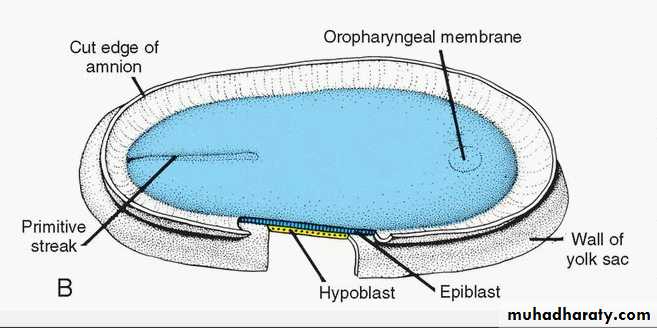
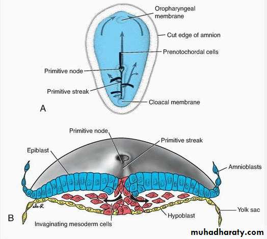
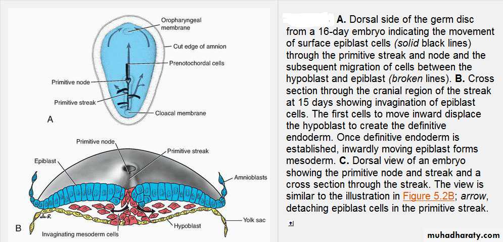
Begins with the formation of the primitive streak on the caudal region of the dorsal surface of the epiblast.
6
GASTRULATION
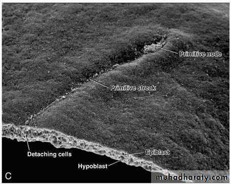
in a 15- to 16-day embryo, it is clearly visible as a narrow groove with slightly bulging regions on either sideThe cephalic end of the streak, the primitive node, consists of a slightly elevated area surrounding the small primitive pit
7
8
9
GASTRULATION
The epiblastic cells migrate toward the streak and on arrival these epiblast cells become flask shaped and invaginated inside the streak. This inward movement is known as invagination.10
GASTRULATION
Cell migration and specification are controlled by (fibroblast growth factor 8) (FGF8), which is synthesized by streak cells themselves.This growth factor controls cell movement by down regulating E-cadherin, a protein that normally binds epiblast cells together.
FGF8 then controls cell specification into the mesoderm by regulating Brachyury (T) expression
11
GASTRULATION
Once the cells have invaginated, some displace the hypoblast, creating the embryonic Endoderm, and others come to lie between the epiblast and newly created endoderm to form Mesoderm. Cells remaining in the epiblast then form EctodermThus, the epiblast, through the process of gastrulation, is the source of all of the germ layers
12
GASTRULATION
As more and more cells move between the epiblast and hypoblast layers, they begin to spread laterally and cranially
Gradually, they migrate beyond the margin of the disc and establish contact with the extraembryonic mesoderm covering the yolk sac and amnion
13
GASTRULATION
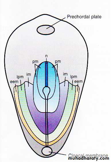
In the cephalic direction, they pass on each side of the prechordal plate.The prechordal plate itself forms between the tip of the notochord and the oropharyngeal membrane and is (derived from some of the first cells that migrate through the node in the midline and move in a cephalic direction).
Later, the prechordal plate will be important for induction of the forebrain ().
The oropharyngeal membrane at the cranial end of the disc consists of a small region of tightly adherent ectoderm and endoderm cells that represents the future opening of the oral cavity.
14
15
GASTRULATION
The invaginating mesodermal cells spread laterally and anteriorly to be in contact with the extraembryonic mesoderm, anteriorly these cells surround the prechordal plate which is the region lying just behind the buccopharyngeal membrane.16
FORMATION OF THE NOTOCHORD
Prenotochordal cells invaginating in the primitive node move forward cranially in the midline until they reach the prechordal plate17
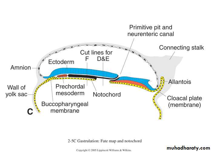
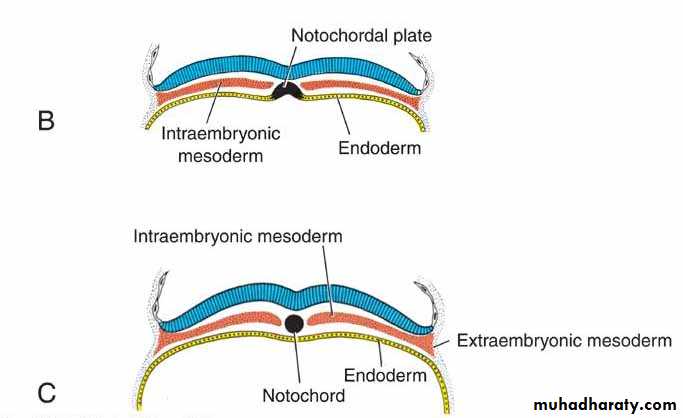
These prenotochordal cells become intercalated in the hypoblast so that for a short time, the midline of the embryo consists of two cell layers that form the notochordal plate ().
As the hypoblast is replaced by endoderm cells moving in at the streak, cells of the notochordal plate proliferate and detach from the endoderm.
They then form a solid cord of cells, the definitive notochord (, which underlies the neural tube and serves as the basis for the axial skeleton
18
Because elongation of the notochord is a dynamic process, the cranial end forms first, and caudal regions are added as the primitive streak assumes a more caudal position.
The notochord and prenotochordal cells extend cranially to the prechordal plate (an area just caudal to the oropharyngeal membrane) and caudally to the primitive pit. At the point where the pit forms an indentation in the epiblast, the neurenteric canal temporarily connects the amniotic and yolk sac cavities
19
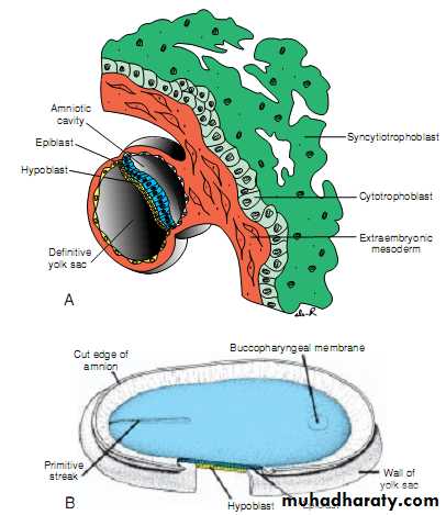
the formation of the cloacal membrane
this membrane is formed of a tightly adherent ectoderm and endoderm at the caudal end of the germ disc with no intervening mesoderm(ruptured at the 7th week). Its structure is similar to that of the buccopharyngeal membrane (ruptured at the 4th week)20
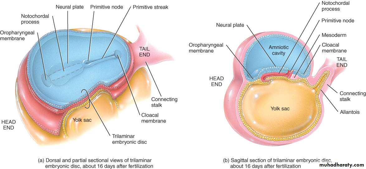
formation of the allantoenteric diverticulum
the allantois; which is a small diverticulum from the posterior wall of yolk sac extending into the connecting stalk during day 16.21
Although in some lower vertebrates the allantois serves as a reservoir for excretion products of the renal system,
in humans, it remains rudimentary but may be involved in abnormalities of bladder development
22
Fate map during gastrulation
The primitive streak is organized rostrocaudally for the formation of the mesoderm; therefore, invaginating epiblast cells develop into:1.notochord at the cranial end of the node at the primitive pit.
2.paraxial mesodermal at the lateral margins of the node and the cranial part of the streak.
3.the intermediate mesoderm at the middle of the streak.
4.lateral plate of mesoderm at the caudal part of the streak.
5.EEM extraembryonic mesoderm at the most caudal part
23
24
Dorsal view of the germ disc showing the primitive streak and a fate map for epiblast cells.
Specific regions of the epiblast migrate through different parts of the node and streak to form mesoderm.
Thus, cells migrating at the cranial most part of the node will form the notochord (n);
25
those migrating more posteriorly through the node and cranial most aspect of the streak will form paraxial mesoderm (pm; somitomeres and somites);
those migrating through the next portion of the streak will form intermediate mesoderm (im; urogenital system);
those migrating through the more caudal part of the streak will form lateral plate mesoderm (lpm; body wall); and
those migrating through the most caudal part will contribute to extraembryonic mesoderm (eem; chorion).
26
Growth of the germ disc
The flat germ disc is initially rounded, then after, it elongate with widening of the cephalic part. Therefore, the shape of the germ disc appears to be broader in the cephalic region than the narrow caudal region. This cephalic growth is produced by the cephalic migration of the cells invaginating at the primitive streak. Therefore, cephalic germ layers start their differentiation while the caudal germ layers are still developing. After the forth week of development, the invagination decrease and the primitive streak regresses and disappears.27
Growth Of The Embryonic Disc
The embryonic disc, initially flat and almost round, gradually becomes elongated, with a broad cephalic and a narrow caudal endExpansion of the embryonic disc occurs mainly in the cephalic region; the region of the primitive streak remains more or less the same size.
Growth and elongation of the cephalic part of the disc are caused by a continuous migration of cells from the primitive streak region in a cephalic direction.
Invagination of surface cells in the primitive streak and their subsequent migration forward and laterally continues until the end of the fourth week.
At that stage, the primitive streak shows regressive changes, rapidly shrinks, and soon disappears.
28
Growth Of The Embryonic Disc
That the primitive streak at the caudal end of the disc continues to supply new cells until the end of the fourth week has an important bearing on development of the embryo.In the cephalic part, germ layers begin their specific differentiation by the middle of the third week, whereas in the caudal part, differentiation begins by the end of the fourth week. Thus gastrulation, or formation of the germ layers, continues in caudal segments while cranial structures are differentiating, causing the embryo to develop cephalocaudally.
29
30
Establishment of the Body Axes
Anteroposterior
Dorsoventral
Left-Right,
The anteroposterior axis:
is signaled by cells at the anterior (cranial) margin of the embryonic disc.(AVE), expresses genes essential for head formation, including the transcription factors OTX2, LIM1, and HESX1 and the secreted factor cerberus and lefty. These genes establish the cranial end of the embryo before gastrulation.
The primitive streak itself is initiated and maintained by expression of Nodal a member of the transforming growth factor-β (TGF-β) family (TGFB) expression will induce and maintain primitive streak. Once the streak is formed, a number of genes regulate formation of dorsal and ventral mesoderm and head and tail structures
Establishment of the Body Axes
2. Dorsoventral axes:bone morphogenetic protein-4 (BMP-4) TGF-β, is secreted throughout the embryonic disc. In the presence of this protein and fibroblast growth factor (FGF), mesoderm will be ventralized to contribute to
kidneys (intermediate mesoderm),
blood and body wall mesoderm(lateral plate mesoderm).
In fact, all mesoderm would be ventralized if the activity of BMP-4 were not blocked by other genes expressed in the node. For this reason, the node is the organizer.
Thus, chordin (activated by the transcription factor Goosecoid ), noggin, and follistatin antagonize the activity of BMP-4. As a result, cranial mesoderm is dorsalized into notochord, somites, and somitomeres
Establishment of the Body Axes
Later these three genes are expressed in the notochord and are important in neural induction in the cranial region.HNF-3β(hepatocyte nuclear factor) maintains the node and later induces regional specificity in the forebrain and midbrain areas. Without HNF-3β, embryos fail to gastrulate properly and lack forebrain and midbrain structures.
Goosecoid activates inhibitors of BMP-4 and contributes to regulation of head development. Overexpression or underexpression of this gene results in severe malformations of the head region, including duplications
Establishment of the Body Axes
Dorsalization in the caudal region is egulated by; Brachyury (T) gene, this gene also block ventralization to prduce dorsalization. Decreased activity of this gene leads to caudal dysgenesis (sirenomelia) of variable gedrees as dsgenesis of the lower limbs, vertebral column, kidnies,anus, or genitalia. This dysgenesis occur in diabetic mothers
Establishment of the Body Axes
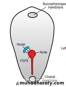
3. Left–right sidedness:When the primitive streak appears, (FGF8) is secreted by cells in the node and primitive streak and induces expression of Nodal but only on the left side of the embryo.
Establishment of the Body Axes
Later, as the neural plate is induced, FGF8 maintains Nodal expression in the lateral plate mesoderm, as well as Lefty-2, and both of these genes upregulate PITX2 which is responsible for establishing left sidedness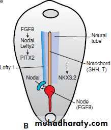
snail
Establishment of the Body Axes
Lefty-1 is expressed on the left side of thefloor plate of the neural tube and may act as a barrier to prevent left-sided signals from crossing over.
Sonic hedgehog (SHH) may also function in this role as well as serving as a repressor for left-sided gene expression on the right
Establishment of the Body Axes
The Brachyury (T) gene, encoding a transcription factor secreted by the notochord, is also essential forexpression of Nodal, Lefty-1, and Lefty-2
expression of the transcription factor Snail is restricted to the right lateral plate mesoderm
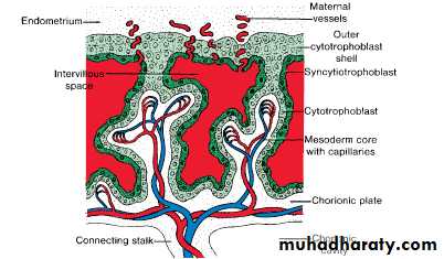
Further Development of theTrophoblast
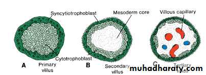
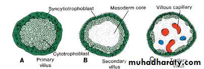
By the beginning of the third week, the trophoblast is characterized by primary villi that consist of a cytotrophoblastic core covered by a syncytial layer40
Further Development of theTrophoblast
During further development, mesodermalcells penetrate the core of primary villi and grow toward the decidua. Forming the
secondary villus
41
syncytiotrophoblast
cytotrophoblast
Further Development of theTrophoblast
By the end of the third week, mesodermal cells in the core of the villus begin to differentiate into blood cells and small blood vessels, forming the villous capillary system .The villus is now known as a tertiary villus ordefinitive placental villus
42
syncytiotrophobast
cytotrophoblast
Further Development of theTrophoblast
Capillaries in tertiary villi make contact with capillaries developing in mesoderm of the chorionic plate and in the connecting stalk which in turn, establish contact with the intraembryonic circulatory system, connecting the placenta and the embryo43
Further Development of theTrophoblast
Meanwhile, cytotrophoblastic cells in the villi penetrate progressively into the overlying syncytium until they reach the maternal endometrium. Here they establish contact with similar extensions of neighboring villous stems, forming a thin outer cytotrophoblast shell.This shell gradually surrounds the trophoblast entirely and attaches the chorionic sac firmly to the maternal endometrial tissue
44
Further Development of theTrophoblast
45
Further Development of theTrophoblast
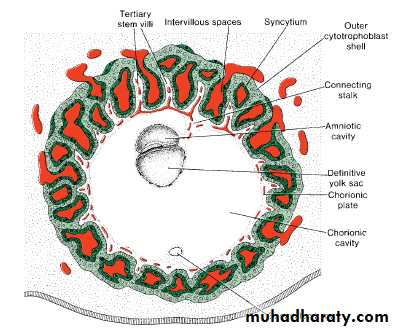
Villi that extend from the chorionic plate to the decidua basalis are called stem or anchoring villi.Those that branch from the sides of stem villi are free (terminal) villi, through which exchange of nutrients and other factors will occur.
The chorionic cavity, meanwhile, becomes larger, and by the 19th or 20th day, the embryo is attached to its trophoblastic shell by a narrow connecting stalk which later develops into the umbilical cord,which forms the connection between placenta and embryo.
46
Thank you
The end47