True bacteria – Rods - Gram positive rods
Non-spore forming bacteria
Corynebacterium, Listeria, Propionibacterium
Corynebacterium
The genus Corynebacterium includes C. diphtheriae, the cause of the diphtheria,
and Diphtheriodes, as a large genus of the normal flora (the skin, the conjunctival sac,
the mouth, the vagina).
Corynebacterium diphtheriae
Diphtheria, caused by C. diphtheriae is an acute respiratory or
cutaneous disease and may be a life-threatening illness. The
vaccination protocols and widespread immunization beginning in
early childhood has made the disease rare in developed countries
while is a serious disease in those countries where the population
has not been immunized.
Epidemiology
C. diphtheriae is found in the throat and nasopharynx of
carriers and in patients with diphtheria. The disease is a local
infection, so the organism is primarily spread by:
1- respiratory droplets by carriers.
2-direct contact with an infected individual
3-contaminated waste.
Pathogenesis
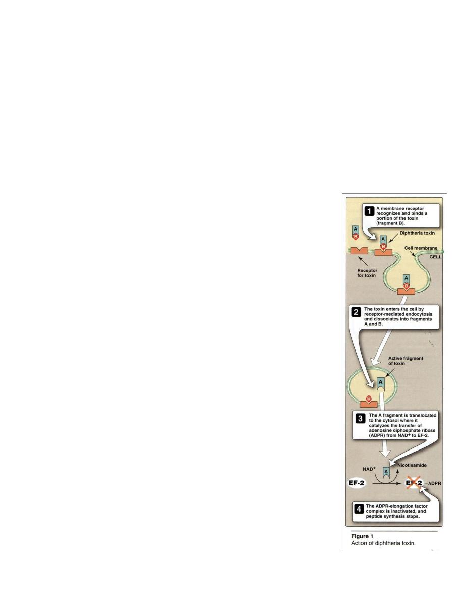
Diphtheria is caused by the effects of a exotoxin that inhibits
eukaryotic protein synthesis. The toxin is composed of two

fragments A and B(Figure 1). Fragment B binds to cell membranes (by the receptor in
cell membrane) and then mediates the delivery of fragment A to it’s target. Inside the
cell, fragment A separates from fragment B, and catalyzes the transfer of adenosine
diphosphate ribose (ADPR) from nicotine adenine dinucleotid (NAD
+
) to polypeptide
chain elongation factor EF-2 ,then,the complex(ADPR+EP-2)is inactivated and peptide
synthesis stops.
Clinical Significance
Infection has two forms respiratory and cutaneous or in an asymptomatic carrier
1-Respiratory diphtheria
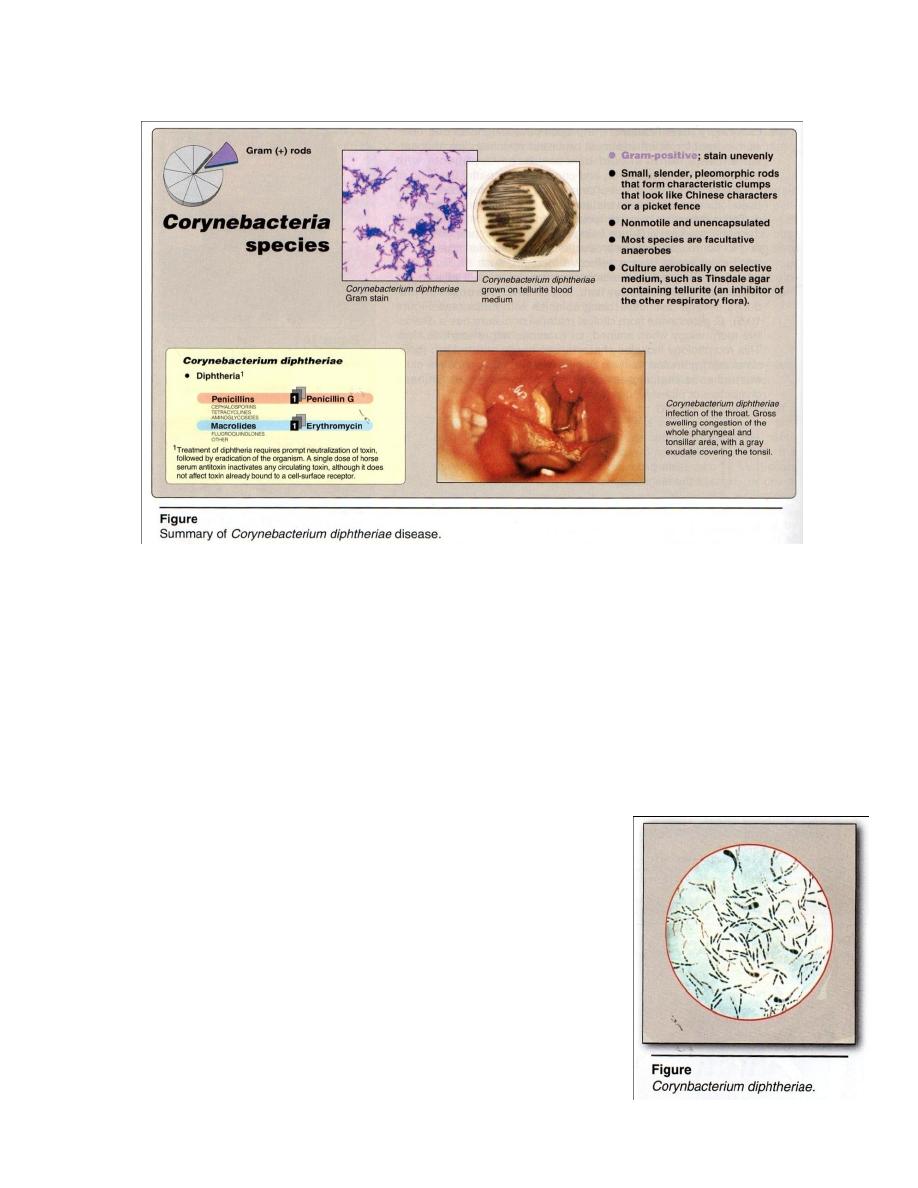
Diphtheria consist of (1) local infection of the throat, produces a thick, grayish,
adherent exudate (pseudomembrane) that is composed of cell debris from the mucosa
and inflammatory products (Figure 2), coats the throat and may extend into the nasal
passages or down ward in the respiratory tract, leading to suffocation.
(2) generalized symptoms occur as the disease progresses (marked swelling of
the lymph nodes in the neck), involve the heart and peripheral nerves, lead to congestive
heart failure and permanent heart damage and neuritis of cranial nerves and paralysis of
muscle groups (such as those control movement of the palate or the eye) are seen late in
the disease.
2- Cutaneous diphtheria
A puncture wound or cut in the skin can result in introduction of C. diphtheriae
into the subcutaneous tissue, leading to a chronic, non healing ulcer with a gray
membrane.
Immunity
Diphtheria toxin is antigenic and stimulates the production of Abs that neutralize
the toxin’s activity. Formalin treatment of the toxin produces a toxoid that remains the
antigenicity but not the toxicity of the molecule, which used for immunization against
the disease.

Diagnostic laboratory tests
1- Clinical observation → Diphtheria should be considered in patients with =
pharyngitis, low –grade fever,cervical adenopathy (swelling of the neck),erythema of
the pharynx and adherent gray pseudomembranes.
2- Microscopic examination: G+ rods ,small, slender, pleomorphic,that tend to stain
unevenly,form clumps that look like Chinese characters , nonmotile, noncapsulated
and do not form spores. Albert’s stain is important to
diagnostic C. because its contain volutin granules (C.
contains
accumulation
of
phosphate
granules,
metachromatic granules) that stain deeply give rod a beaded
appearance beside the bacilli looks like bipolar under the
microscope(see figure 3) and arranged in pairs or groups
and from various angles with each other like L, V, X, Y, Z
thus called Chinese letter arrangement.

3-Macroscopic examination: Culture = Most spp. are facultative anaerobic and those
found associated with humans grow aerobically on standard laboratory media such as
blood agar. C. diphtheriae can be isolated easily from a selective medium such as
Tinsdale’s agar (Figure 2) which contains potassium tellurite, an inhibitor of other
respiratory flora, and on which C. produces black colonies with halos (to reduction of
tellurite salt). On rich media (blood, serum or egg) colonies are small, granular, moist,
glistening irregular edges.
Biochemical reaction = Catalas +, oxidase - , gelatin- , Urase - , phosphatase -,
ferment glucose and maltose with acid production only.
Fermentation of starch, glycogen and dextrin, is useful to differentiate between
the 3 types of C. diphtheriae (on selective media containing tellurite, 3 types of C.
diphtheriae : Gravis, Mitis, Intermedius, all of them capable of producing same toxin
and clinical disease). Gravis ferments starch, glycogen and dextrin, while the two other
types don’t ferment starch and glycogen,ferment only dextrin.
Serologic test : by precipitin reaction to demonstrate toxin production.
Treatment
Requires (1) neutralization of toxin: by A single dose of horse serum antitoxin
inactivates any circulating toxin, not bound toxin to a cell-surface receptor.
(2) eradication of the organism: by several antibiotics, such as erythromycin
or penicillin (Figure 2), which slows the spread of infection and, by killing the organism,
prevents further toxin production.
Prevention
By vaccine contains diphtheria toxoid as a part of the DTaP vaccine (Diphtheria
+ Tetanus + a Pertussis vaccine)which neutralize unbound toxin preventing the disease
from progressing. The initial series of injections should be started in children (2 years ).
Booster injections of diphtheria toxoid (with tetanus toxoid) should be given at 10 year
intervals throughout life. The control of an epidemic outbreak of diphtheria involves
rigorous immunization and a search for healthy carriers among patient contacts.

Diphtheroids
Several other Corynebacterium spp., that morphologically resemble to C.
diphtheriae , are common commensals of the nose, throat, nasopharynx, skin,
urinary tract and conjunctiva, and are generally unable to produce exotoxin, but a
few cause disease in immunosuppressed individuals.
Listeria
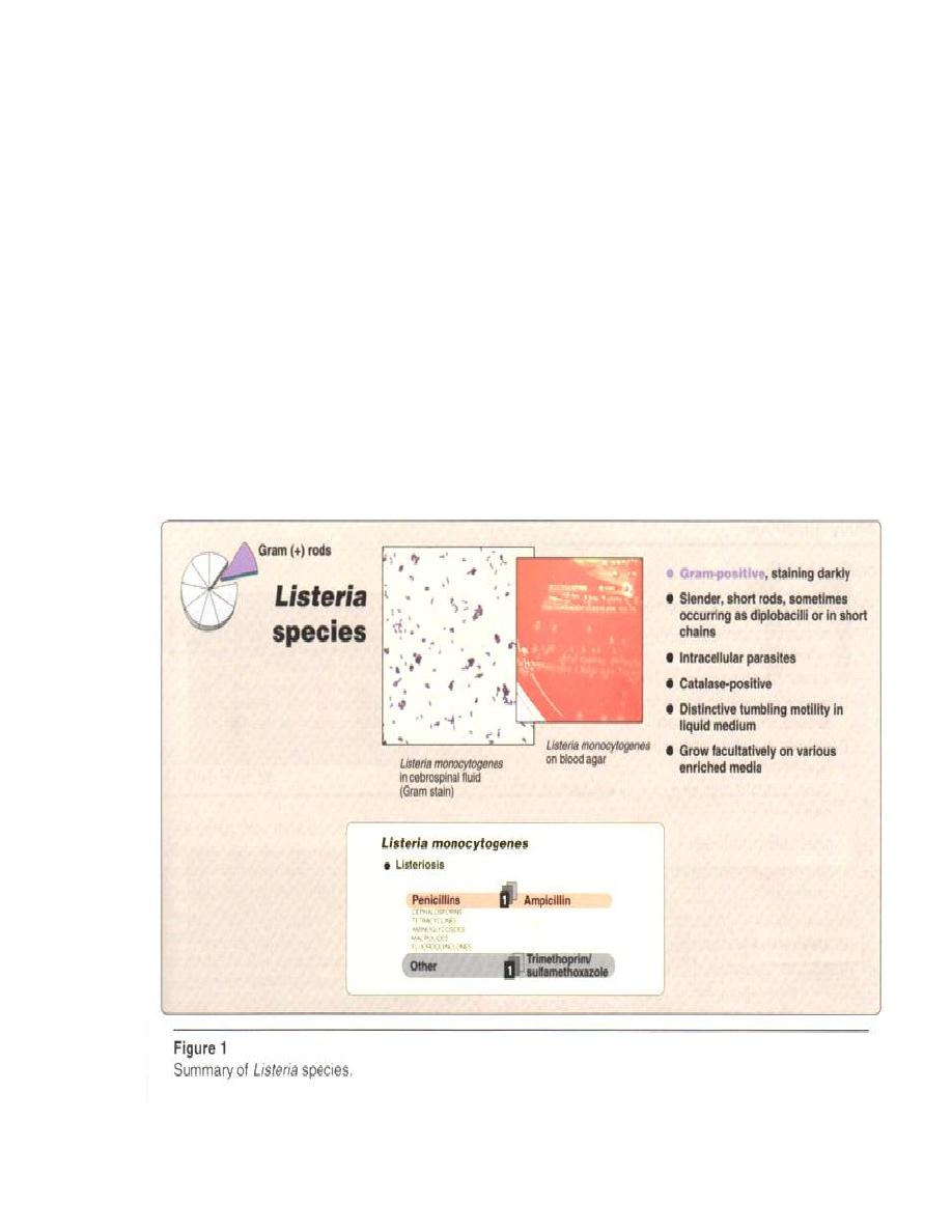
Listeria species are intracellular parasites that may be seen within host cells
in tissue samples (Figure 1) . Listeria monocytogenes is the only species that infects
humans, other species are widespread among animals.

Epidemiology
Listeria infections are usually food borne, 2 - 3 %
of processed dairy products (ice cream and cheese), 20 -
30 % of ground meats and poultry (growth at 4°C in
food).
1 – 15 % of healthy humans are asymptomatic
intestinal carriers of the organism. Listeria infections are
most common in pregnant women + fetuses or newborns
+ immunocompromised individuals, such as the elderly
or patients receiving corticosteroids.
Antigenic classification
Three serotypes are present isolates from humans ,
Ia , Ib , Irb .
Irb causes an epidemic associated with cheese
made from anpasteurized milk,cause meningitis between
birth 3 week of life with high mortality rate .
Pathogenesis
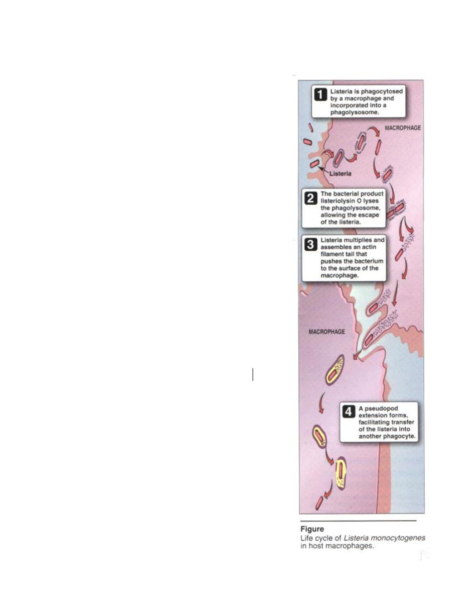
It’s a facultative intracellular parasite, attaches to
and enters a variety of mammalian cells, by
phagocytosis(macrophage)and
incorporated
into
a
phagolysosome (see figure) .
It escapes from the phagolysosome by elaborating
a toxin called Listeriolysin O as virulent factors
.
L. monocytogenes grows in the cytosol, and
assembles an actin filament tail that pushes the
bacterium to the surface of the macrophage .
A pseudopod extension forms , facilitating
transfer of the Listeria into a neighboring phagocyte.
Listeria produced phospholipases mediate the passage
of the organism directly to a neighboring cell, allowing
avoidance of cells of the immune system.

Clinical significance
Septicemia and meningitis are the most commonly reported forms of L.
monocytogenes infection (listeriosis).
Pregnant women, usually in the third trimester, may have a milder illness as well
as in asymptomatic vaginal colonizaition, the organism can be transmitted to a newborn
(cause of newborn meningitis) or to the fetus and initiate abortion. Immunocompromised
individuals are susceptible to serious generalized infections, as meningocephalitis and
bacteremia .
Diagnostic laboratory tests
Specimens = blood, cerebrospinal fluid, and other clinical specimens by standard
bacteriologic procedures.
Microscopic examination = G+ rods, sometimes they occur as diplobacilli or in short
chains, do not form spores and they are intracellular parasites that may be seen within
host cells in tissue samples. Its motile with tumbling motility by light microscopy in
liquid medium after growth at 25°C , distinguish it from Corynebacterium
(nonmotile) which may be confused morphologically with Listeria.
Macroscopic examination = It's facultative anaerobes on a variety of enriched media.
On blood agar, L. monocytogenes produces a small colony with β- hemolysis( figure 1).
Biochemical reaction = catalase + distinguish it from Streptococci (catalase - )
which may be confused morphologically with Listeria.
Treatment
Ampicillin and trimethoprim /sulfamethoxazole are used (Figure 1). Ampicillin
and gentamicin also used after therapy.
Prevention
By proper food preparation and handling.
Propionibacterium
Propionibacterium acnes is classified as a Corynebacterium anaerobic
diphtherods, form part of the normal flora of the skin.They cause acne by produce
lipase which split off free fatty acids from skin ,then produce tissue inflammation
which contribute to acnes . Sometimes associated with endocarditis or infections of
plastic implants. It's produce propione acid from carbohydrate fermentation . Its G+ rods
, non spore forming , anaerobic microaerophilic rods of diphtheroid like morphology.

