Anatomy of the Larynx
Dr. Abdullah AlkhalilFJMC, H.S
MRCS-ENT(UK), DOHNS(LONDON)
1
General principles of development
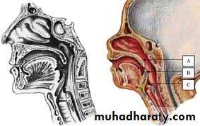
The development of the larynx can be divided into prenatal and postnatal stages.At birth, the larynx is located high in the neck between the C1 and C4 vertebrae, allowing concurrent breathing or vocalization and deglutition.
By age 2 years, the larynx descends inferiorly; by age 6 years, it reaches the adult position between C4 and C7 vertebrae. This new position provides a greater range of phonation (because of the wider supraglottic pharynx) at the expense of losing this separation of function, i.e., deglutition and breathing.
2

3
General principles of development
The larynx develops from the endodermal lining and the adjacent mesenchyme of the foregut between the fourth and sixth branchial arches.At 20 days' gestation, the foregut is first identifiable with a ventral laryngotracheal groove. It continues to deepen until its lateral edges fuse.
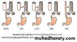
Trachea becomes separated from the esophagus by the tracheoesophageal septum with a persistent slit like opening into the pharynx
This fusion occurs in the caudal-to-cranial direction, and incomplete fusion results in development of persistent communication between the larynx or trachea and the esophagus
4

5
6
General principles of development
The larynx grows rapidly during the first 3 years of life, while the arytenoids remain approximately the same size.Beginning at age 18-24 months, the larynx descends in the neck to achieve its final position at vertebrae C4-C7 by age 6 years.
The larynx elongates as the hyoid, thyroid, and cricoid cartilages separate from each other.
The cricoid cartilage continues to develop during the first decade of life, gradually changing from a funnel shape to a wider adult lumen; therefore, it is no longer the narrowest portion of the upper airway.
7
8
Functions of the larynx
Sound production (phonation). NOTE: phonation = speech.Regulates amount of air entering the lungs.
Acts as a valve to prevent food in the pharynx from entering the trachea.
Provides a sphincter as part of the Valsalva maneuver (“bearing down”, lifting heavy objects).9
Functions of the larynx
Sound is generated in the larynx, and that is where pitch and volume are manipulated. The strength of expiration from the lungs also contributes to loudness.Fine manipulation of the larynx is used to generate a source sound with a particular fundamental frequency, or pitch. This source sound is altered as it travels through the vocal tract, configured differently based on the position of the tongue, lips, mouth, and pharynx.
10
Functions of the larynx
The vocal folds can be held close together (by adducting the arytenoid cartilages) so that they vibrate. The muscles attached to the arytenoid cartilages control the degree of opening. Vocal fold length and tension can be controlled by rocking the thyroid cartilage forward and backward on the cricoid cartilage (either directly by contracting the cricothyroids or indirectly by changing the vertical position of the larynx), by manipulating the tension of the muscles within the vocal folds, and by moving the arytenoids forward or backward. This causes the pitch produced during phonation to rise or fall. In most males the vocal folds are longer and with a greater mass, producing a deeper pitch.11
12
Functions of the larynx
During swallowing, the backward motion of the tongue forces the epiglottis over the glottis' opening to prevent swallowed material from entering the larynx which leads to the lungs; the larynx is also pulled upwards to assist this process. Stimulation of the larynx by ingested matter produces a strong cough reflex to protect the lungs.13
Laryngeal components
Cartilages.Extrinsic ligaments.
A cavity (airway) lined by a mucosa.
Fibro-elastic membranes.
Intrinsic skeletal muscles.
Nerves, arteries, veins, and lymphatics.
14
15
16
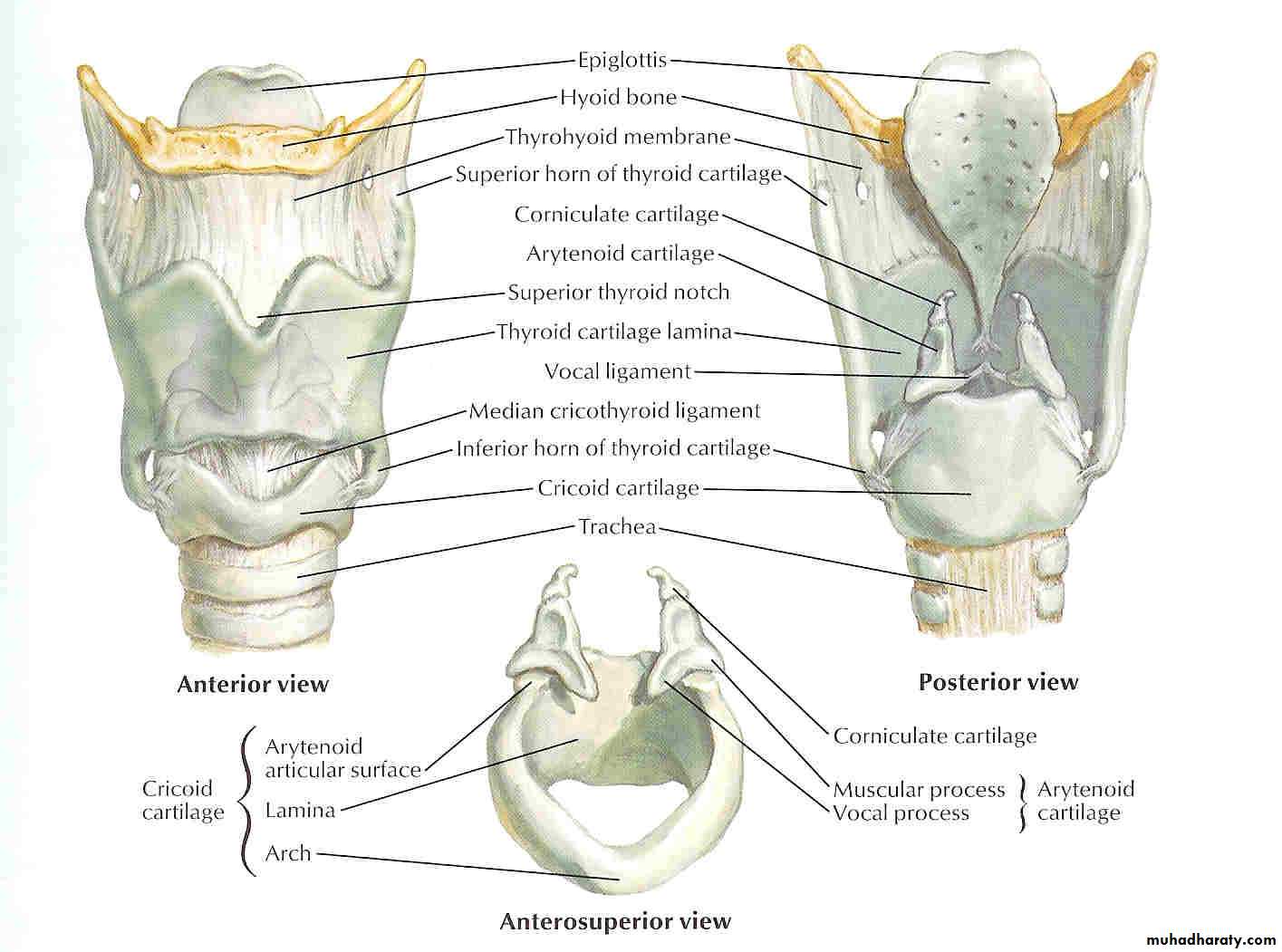
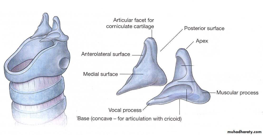
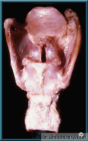
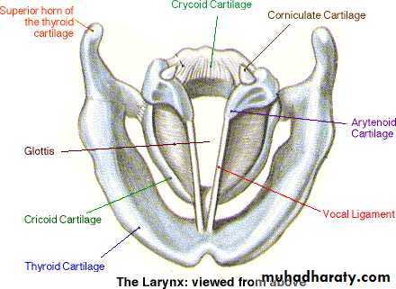
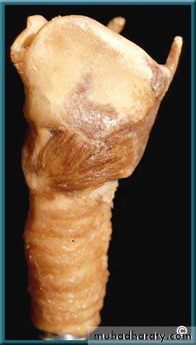
The “skeleton” of the larynx
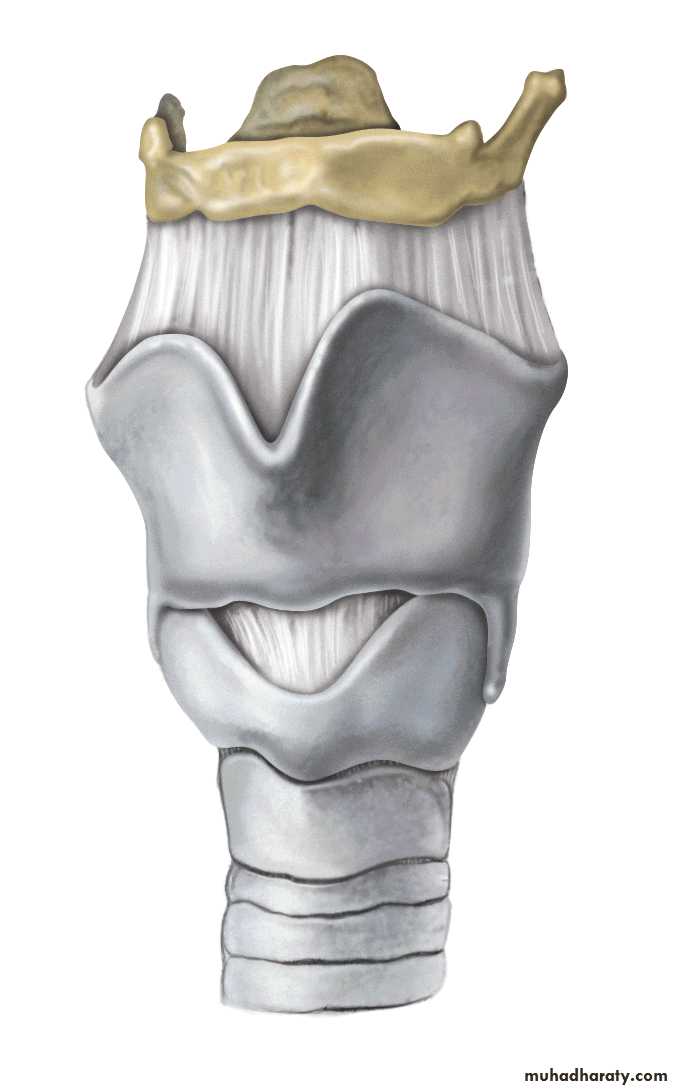
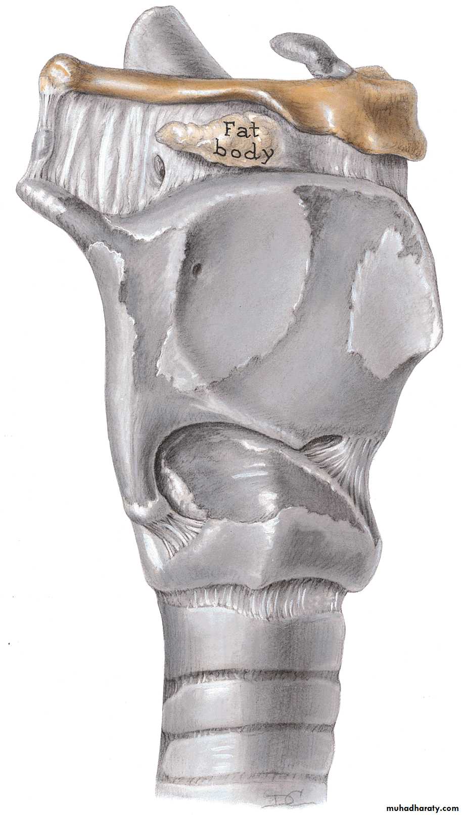
5 Major cartilages:
Thyroid (1)Cricoid (1)
Epiglottis (1)
Arytenoid (2)
Covered with mucous membranes and skeletal muscles.
Cricothyroid joint (synovial)
17
The “skeleton” of the larynx
All major cartilages are composed of hyaline cartilage EXCEPT the epiglottis = Elastic cartilage
Arytenoid cartilages are paired
They are shaped like pyramids perched atop the cricoid cartilage
Crico-arytenoid joints (synovial)
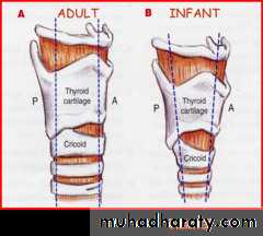
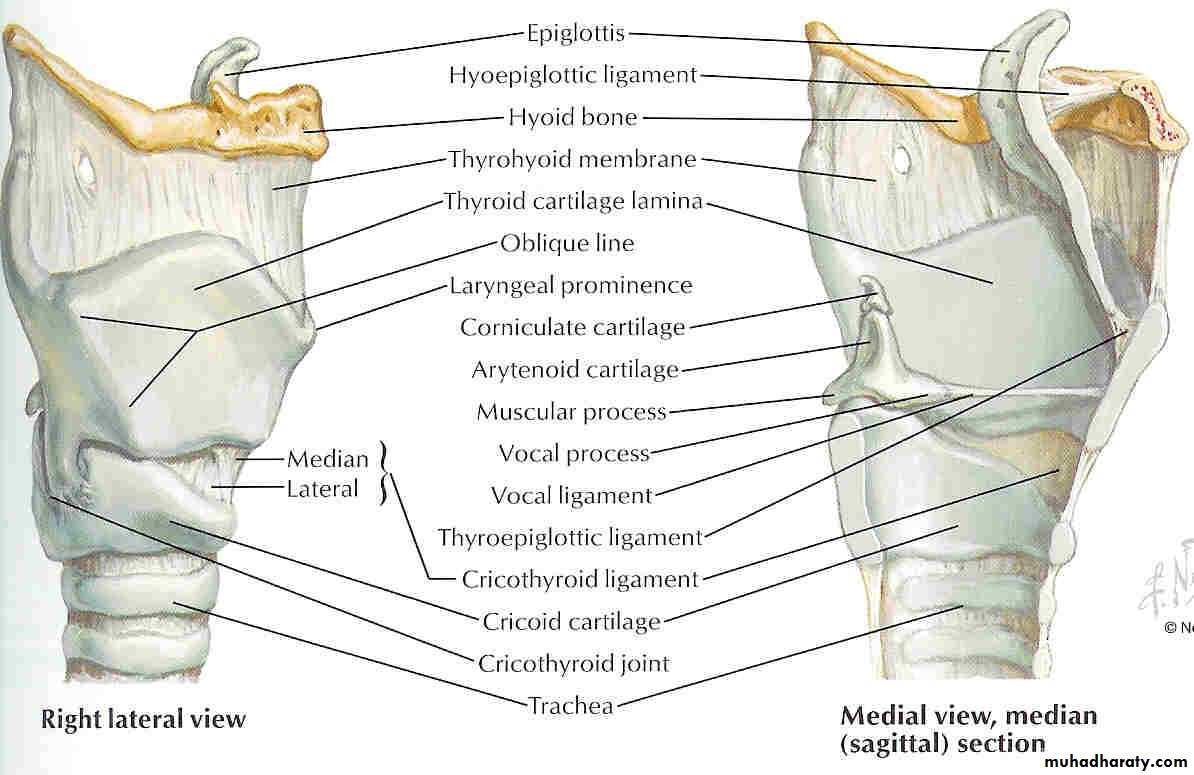
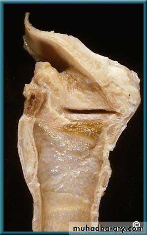
18Thyroid Cartilage
Shield shaped, open posteriorly, angulated anteriorlyAngulation more acute in males
Its function is to shield larynx from injury and provide an attachment to vocal cords
19
Adam’s Apple
20
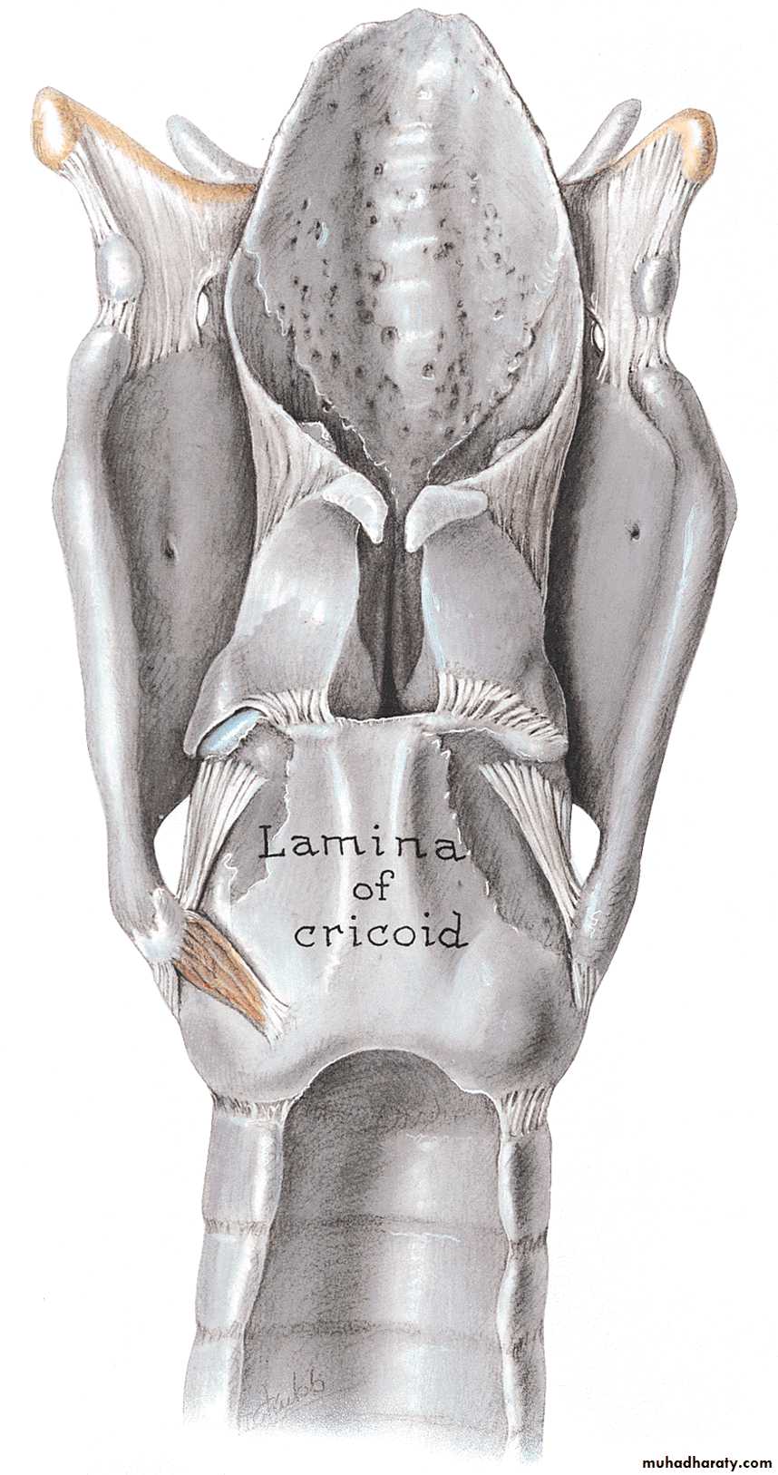
Cricoid Cartilage
Signet ring shapedStronger than thyroid cartilage.
Lamina – 2 to 3 cm from above downwards, considerably broader than anterior arch.

21
Important from structural & functional point of view
Base for entire larynxSupport to arytenoid
Attachment to intrinsic muscles
Only part of cartilagenous framework that forms continuous 360 degree ring
Once injured or strictured , difficult to resect while preserving laryngeal function
22
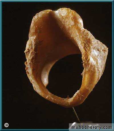
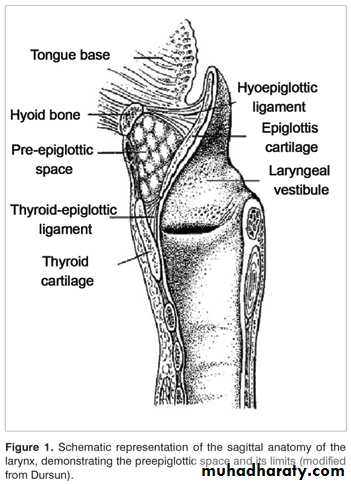
Epiglottis
Thin leaf shaped fibro-cartilage, situated in midlineUpper free end broad & rounded, projects up behind base of tongue
Narrow base called pitiole
This attachment forms lower limit of pre-epiglottis space
23
Half of epiglottis projects above hyoid
This part has a laryngeal and lingual surfaces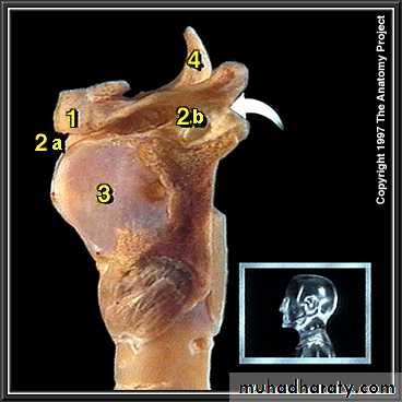
24
Infrahyoid portion has no free anterior surface
Forms posterior wall of PESEpiglottic cartilage contains many pits filled with mucous glands
Little barrier between infrahyoid portion and PES
25
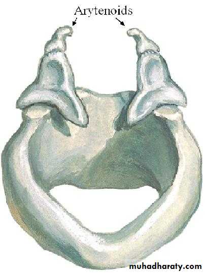
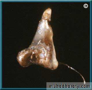
Arytenoids
Paired cartilages, pyramidal in shapeBase articulated with cricoid
PCA & LCA muscles attach on muscular process
Anterior angle elongated into vocal process which receives insertion of vocal ligament
26
The arytenoid cartilages have vocal and muscular processes
Anterior Posterior
27Minor laryngeal cartilages
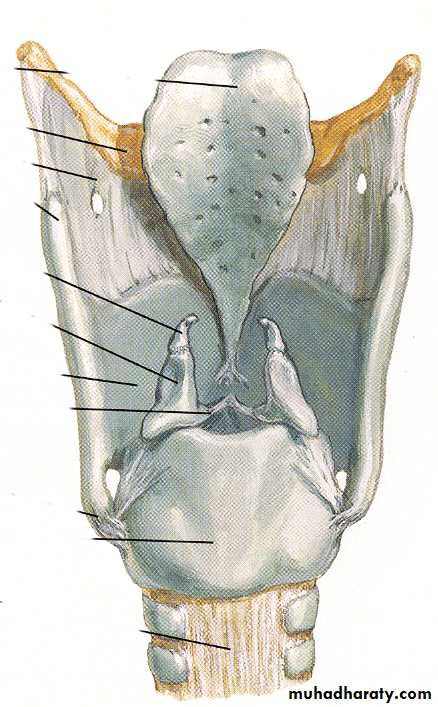
Cuneiform cartilages
Corniculate cartilagesProbably support the ary-epiglottic folds – they produce swellings called cuneiform and corniculate tubercles.
28
Laryngeal Subsites
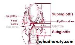
29
Supraglottis
Consists of ventricles, false cords, laryngeal surface of epiglottis, aryepiglottic folds and the mucosal expanse.Posterior tapering shape reduces area of mucosa in posterior region
So majority of SG tumors are epiglottic
30
Glottis
Consists of true cords, anterior commissure and posterior commissureNarrow triangular space between the true cords is called rima glottis
Anterior 2/3 is membranous
Posterior third consists of vocal processes of arytenoids
Posterior 1/3 of cords and covering mucosa are called posterior commissure
31
Sub-glottis
Begins about 5mm below free margins of VCConsists of a mobile upper and fixed lower part
32
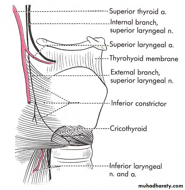
Extrinsic ligaments
Thyrohyoid ligament
Attaches the larynx to the hyoid – this explains why the larynx is elevated during swallowing.
The thyrohyoid ligaments has apertures for the internal branch of the superior laryngeal nerve and superior laryngeal vessels.
Median cricothyroid ligament (a.k.a. – cricothyroid ligament.
33Extrinsic ligaments
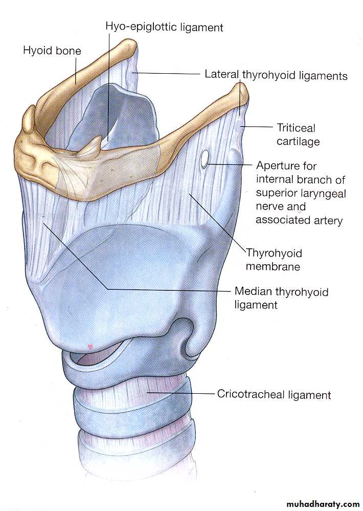
Hyo-epiglottic ligament
Cricotracheal ligament34
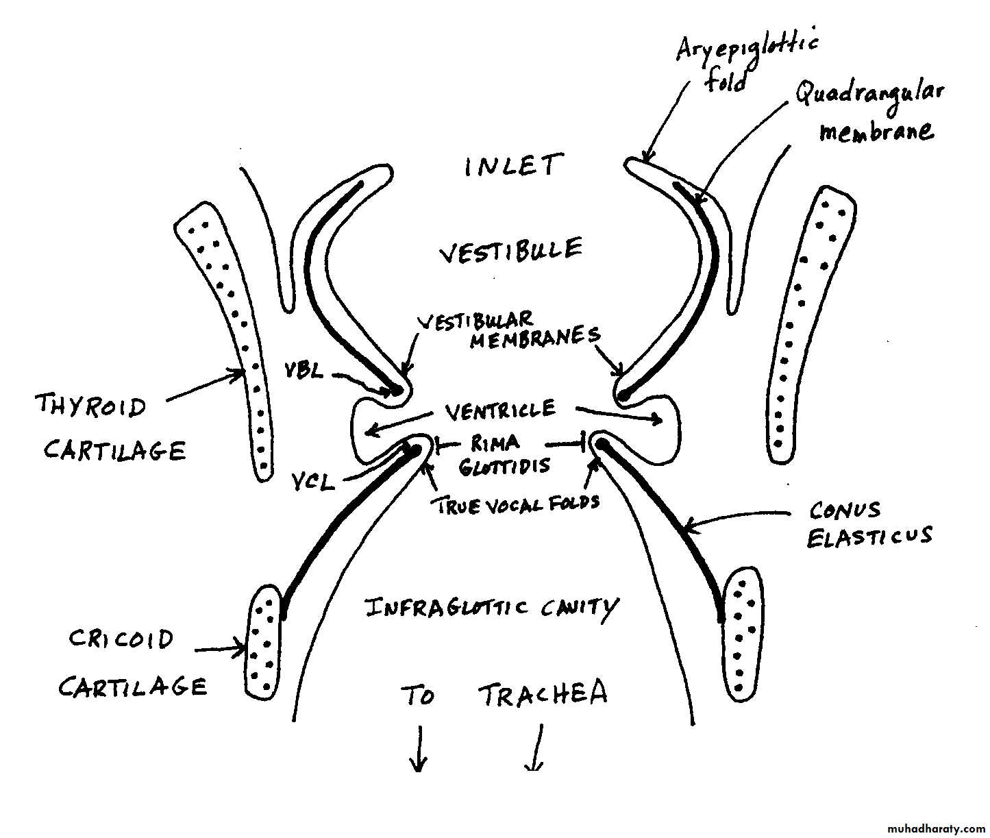
Laryngeal inlet (Aditus)
The inlet is surrounded by the:
Ary-epiglottic folds.Epiglottis.
Note the cuneiform and corniculate tubercles – features of the ary-epiglottic folds.
35
Cavity of the larynx
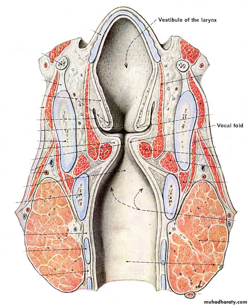
Coronal section

Sagittal section
VestibuleSaccule
VentricleInfraglottic cavity
Trachea
36The walls of the laryngeal cavity contains two vocal folds
Vestibular folds ( false vocal folds).
Vocal folds ( true vocal folds, vocal cords).37
The circumscribed region of the laryngeal cavity that contains the (true) vocal folds is called the glottis.
The spaces between the left and right vestibular and vocal folds are called the rima vestibuli and rima glottidis, respectively.
Rima vestibuli
Rima glottidis38
39
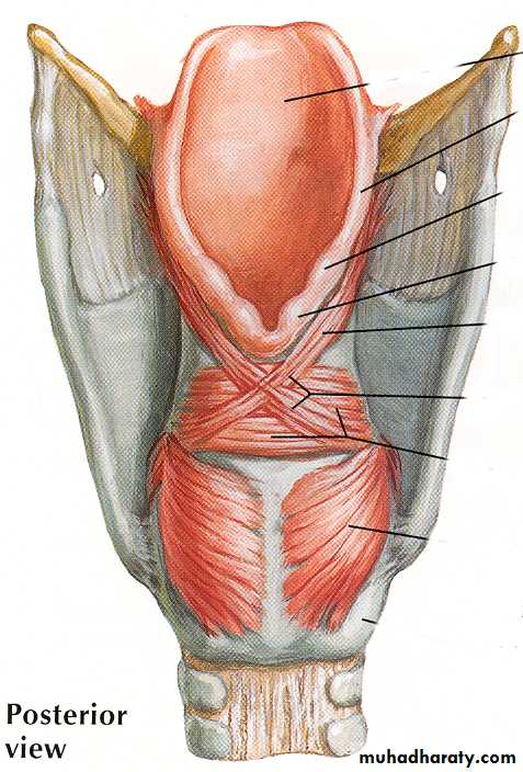
Intrinsic muscles of the larynx
Perform 4 basic actions: Abduct the vocal cords.
Adduct the vocal cords.
Tense/relax the vocal cords.
Close the laryngeal inlet.
Skeletal muscles innervated by the vagi
Recurrent laryngeal nerve
Superior laryngeal nerve“Muscles of phonation” do these tasks
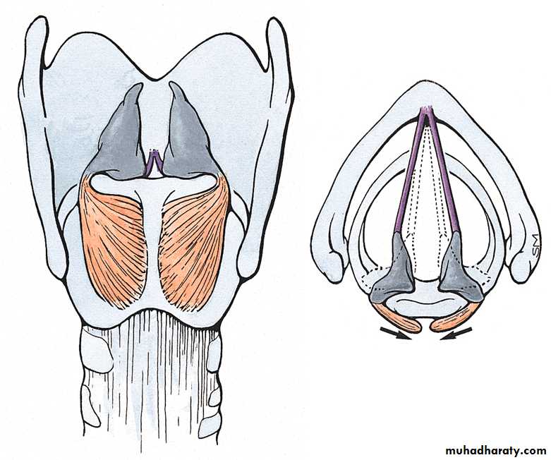
40Muscles that abduct the vocal cords
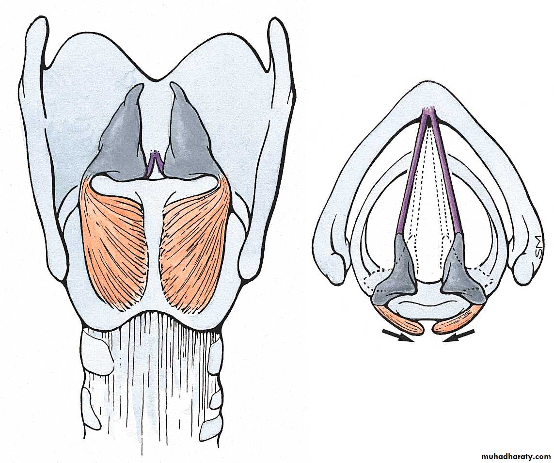
Posterior crico-arytenoid muscles
41
Abductor of Larynx
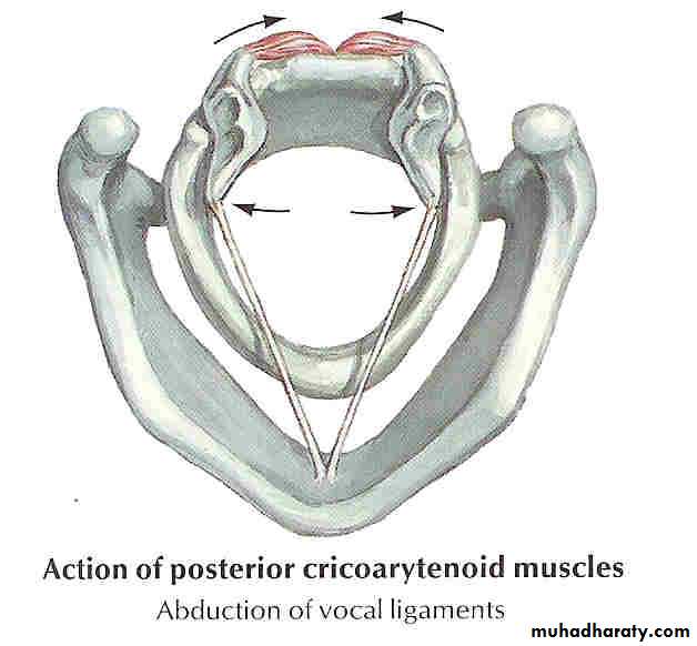
42
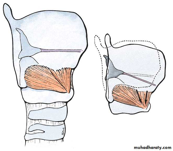
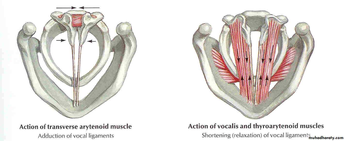
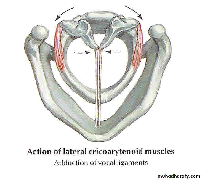
Muscles that adduct the vocal cords
Lateral crico-arytenoid muscles & transverse arytenoid muscles.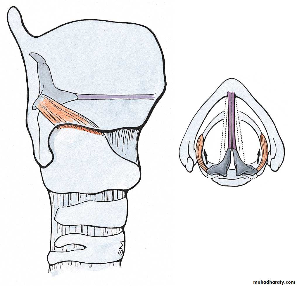
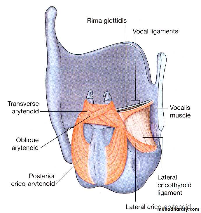
43
Adductors of the Vocal Folds
44
Muscles that tense the vocal cords
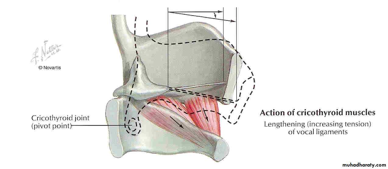
Cricothyroid muscles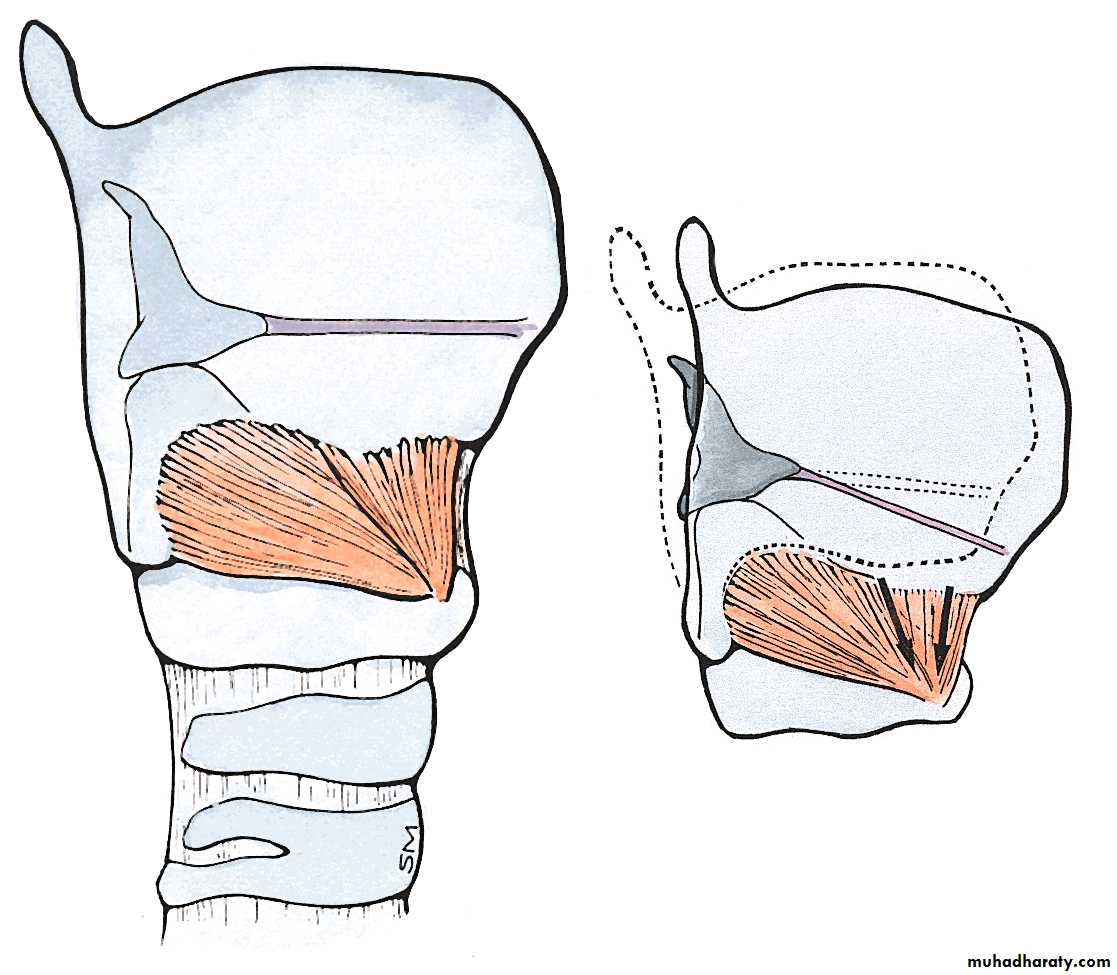
Thyroid cartilage tilts forward on cricoid cartilage – vocal cords are stretched and become taut.
45
Cricothyroid Muscle
46
Muscles that relax the vocal cords
Thyro-arytenoid muscles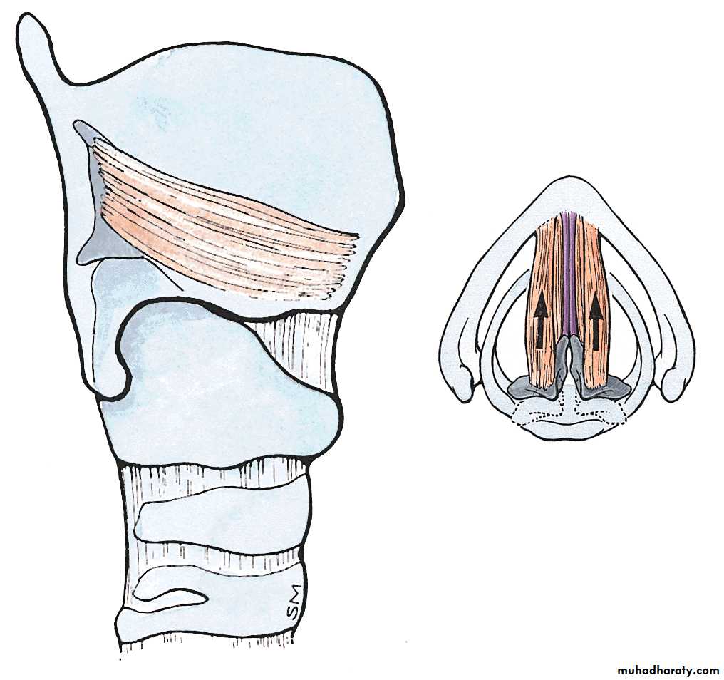
These muscles draw the thyroid cartilage closer to the arytenoid cartilages – thus the vocal cords are no longer taut and relax.
47
The vocalis muscles perform fine, regional adjustments of tension
These muscle fibers attach to the vocal processes of the arytenoid cartilage and thyroid angle They insert at various places along the vocal ligaments.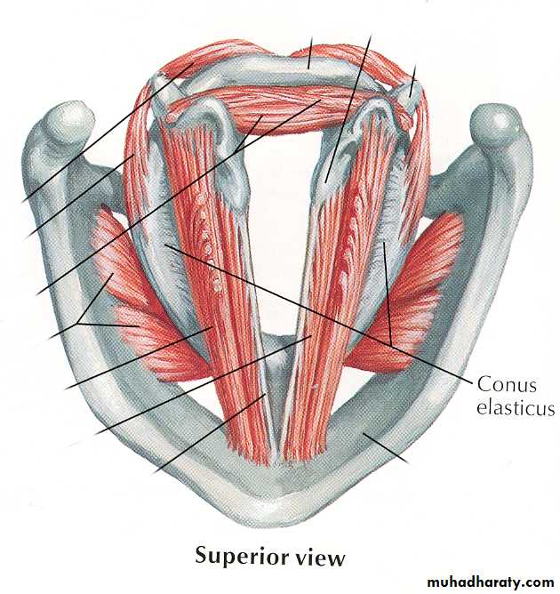
Subparts of the vocal ligaments can be tensed and relaxed.
48Muscles that close the laryngeal inlet
Transverse arytenoid & ary-epiglottic musclesThese muscles draw the ary-epiglottic folds, the epiglottis, and arytenoid cartilages together – narrowing the inlet
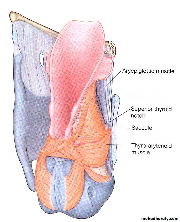
49
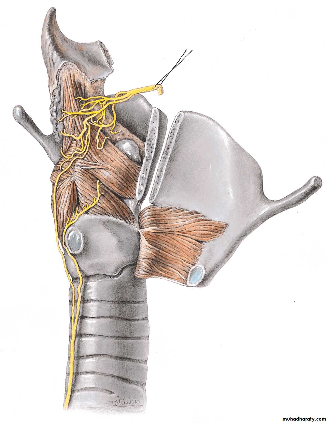
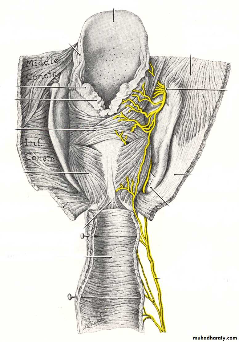
Nerve Supply: Derived from the Vagus
Superior Laryngeal Nerve -It leaves the vagus nerve high in the neckInternal -It provides sensation of the glottis and supraglottis, which includes the pharynx, underside of the epiglottis and the larynx above the cords. Remember: SIS-superior internal sensory.
External -It supplies motor function to the cricothyroid muscle which tenses the vocal cords and could cause laryngopasm.
50
Recurrent Laryngeal Nerve -It provides sensation to the subglottic area which includes the larynx below the vocal cords and upper esophagus. It provides motor function to the intrinsic muscles of the larynx.
It branches from the vagus in the mediastinum and turns back up into the neck. On the right, it travels inferior to the subclavian and loops up, and on the left it travel inferior to the aorta and loops up.
51

52
Motor supply to intrinsic muscles of larynx
Recurrent laryngeal nerve supplies all laryngeal muscles EXCEPT the cricothyroid Supplied by external branch of superior laryngeal nerve.
53
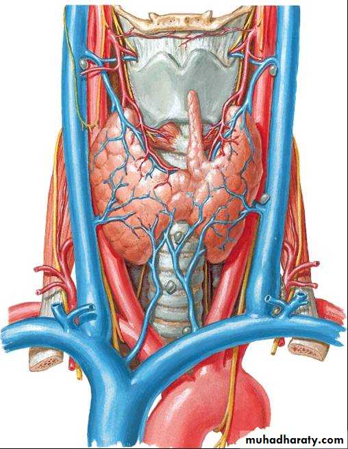
ARTERIAL SUPPLY
Sup. Laryngeal A. from Sup. Thyroid arteryInf. Laryngeal A. from Inf. Thyroid artery
54
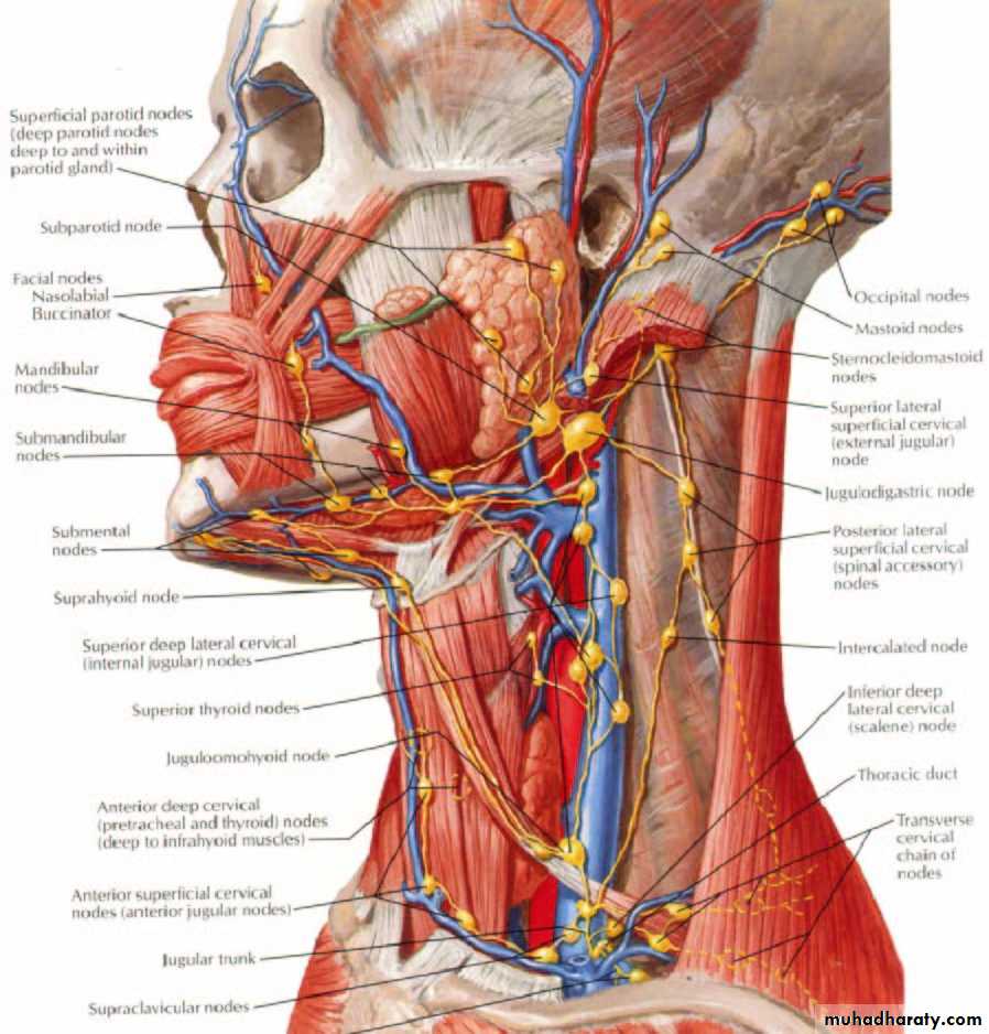
Lymphatic drainage of larynx
Lymph vessels follow blood vessels – ultimately draining to deep cervical nodes.Above the vocal folds To superior group of deep cervical nodes.
Below the vocal folds To paratracheal and inferior group of deep cervical nodes.
55
END
56