Laryngeal Carcinoma
Dr. Abdullah AlkhalilMRCS-ENT(UK), DOHNS(London)
FJMC, H.S
1
Overview
11,000 new cases of laryngeal cancer per year.Accounts for 25% of head and neck cancer and 1% of all cancers
One-third of these patients eventually die of their disease
Most prevalent in the 6th and 7th decades of life
2
Overview
4:1 male predilectionDownward shift from 15:1 post WWII
Due to increasing public acceptance of female smoking
More prevalent among lower socioeconomic class, in which it is diagnosed at more advanced stages
3
Subtypes
Glottic Cancer: 59%
Supraglottic Cancer: 40%
Subglottic Cancer: 1%Most subglottic masses are extension from glottic carcinomas
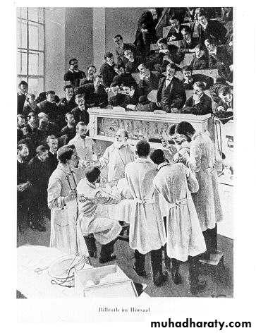
4History
The first laryngectomy for cancer of the larynx was performed in 1883 by BillrothPatient was successfully fed by mouth and fitted with an artificial larynx
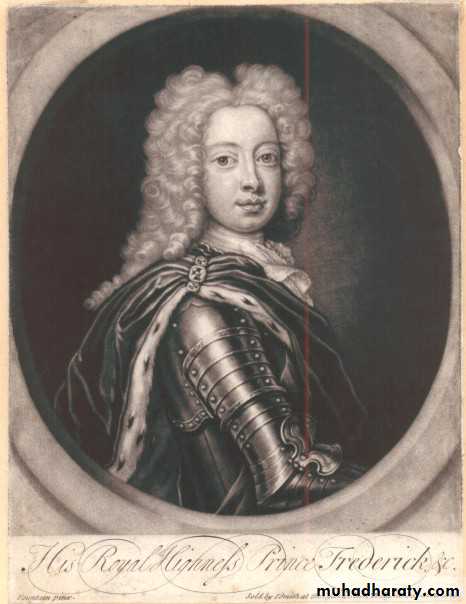
In 1886 the Crown Prince Frederick of Germany developed hoarseness as he was due to ascend the throne.
5
Crown Prince Frederick of Germany
6
History
Was evaluated by Sir Makenzie of London, the inventor of the direct laryngoscope
Frederick’s lesion was biopsied and thought to be cancer
He refused laryngectomy and later died in 1888
7
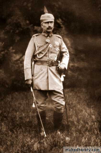
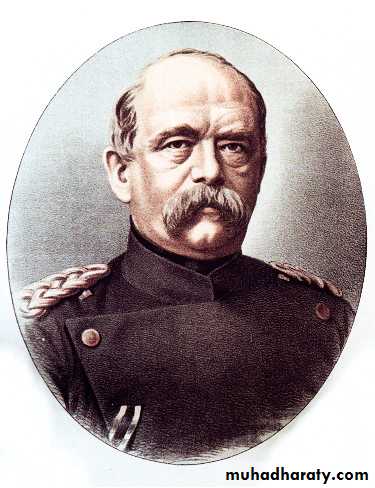
History
Frederick was succeeded by Kaiser Wilhelm II, who along with Otto von Bismark militarized the German Empire and led them into WW ICould an Otolaryngologist have prevented WW I?
8
Risk Factors


9
Risk Factors
Prolonged use of tobacco and excessive EtOH use primary risk factorsThe two substances together have a synergistic effect on laryngeal tissues
90% of patients with laryngeal cancer have a history of both
10
Risk Factors
Human Papilloma Virus 16 &18Chronic Gastric Reflux
Occupational exposures
Prior history of head and neck irradiation
11
Histological Types
85-95% of laryngeal tumors are squamous cell carcinomaHistologic type linked to tobacco and alcohol abuse
Characterized by epithelial nests surrounded by inflammatory stroma
Keratin Pearls are pathognomonic
12
Histological Types
Verrucous Carcinoma
Fibrosarcoma
Chondrosarcoma
Minor salivary carcinoma
Adenocarcinoma
Oat cell carcinoma
Giant cell and Spindle cell carcinoma
13
Anatomy
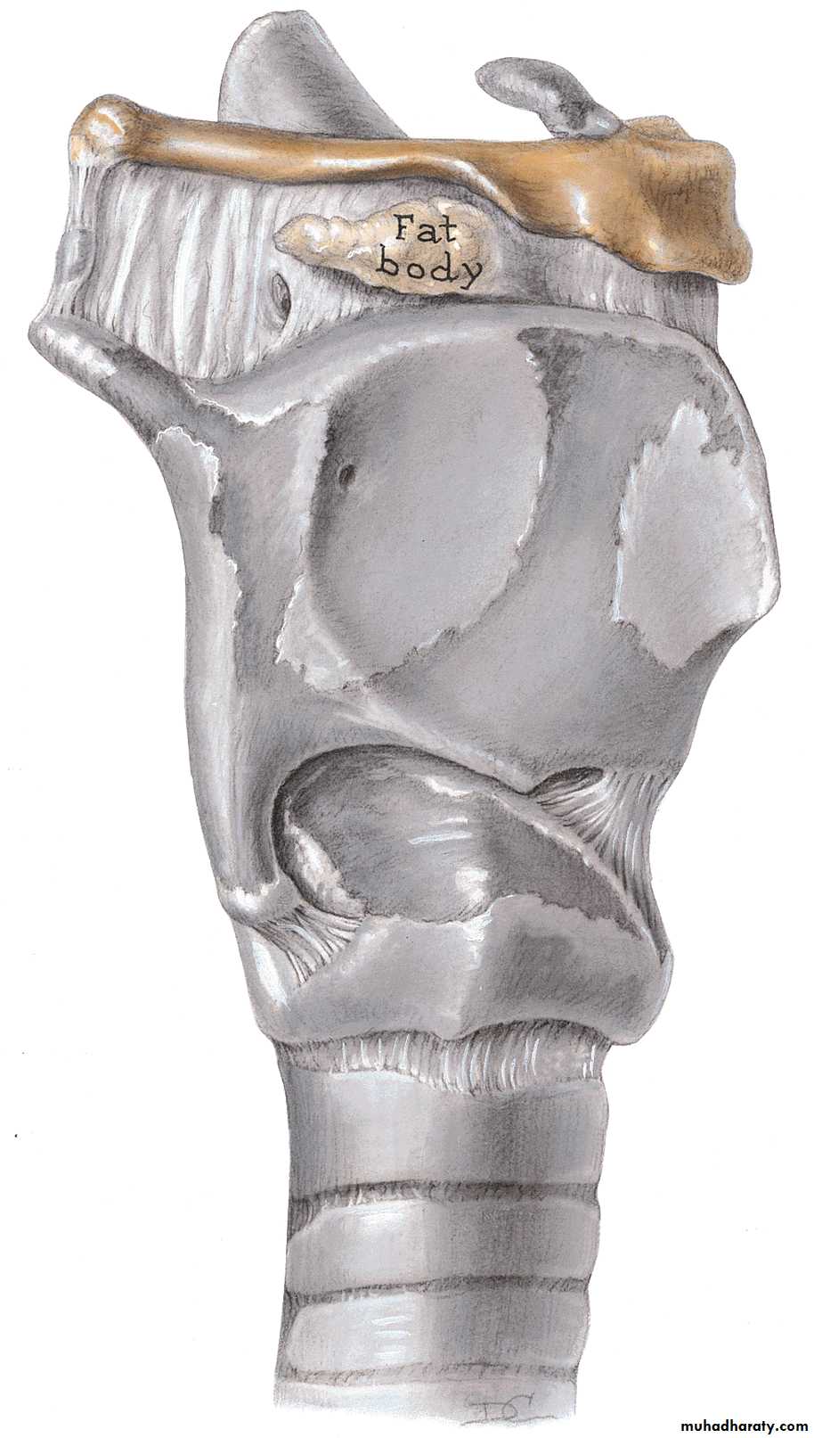
14
Anatomy
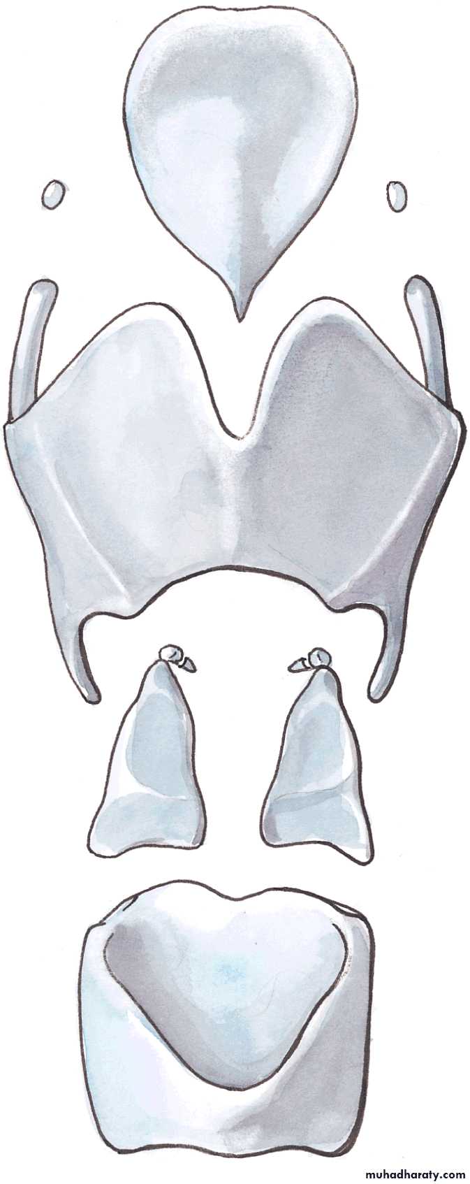
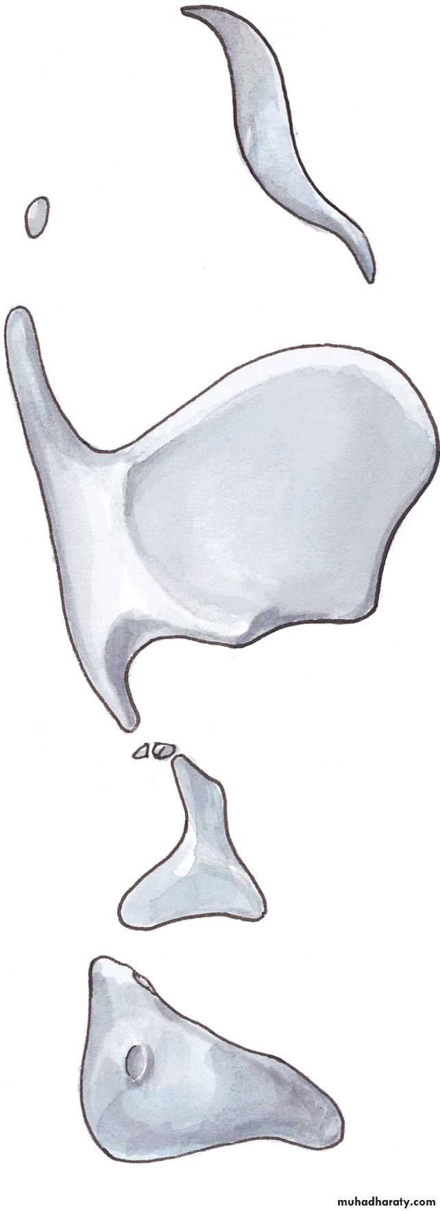
15
Anatomy
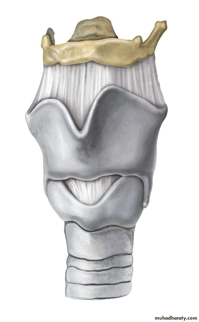
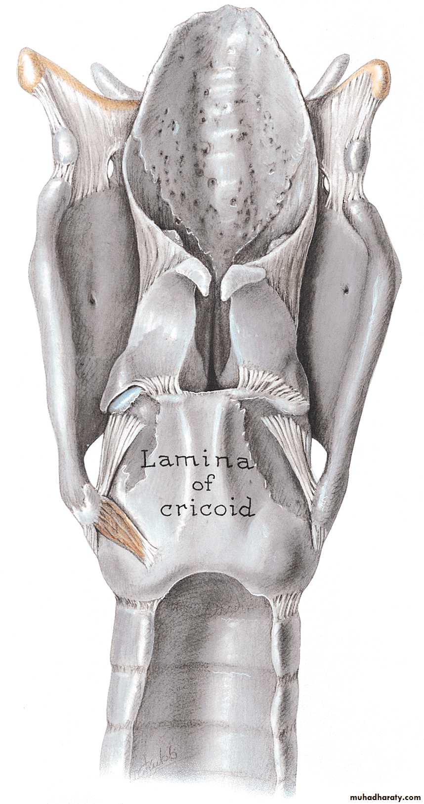
16
Anatomy
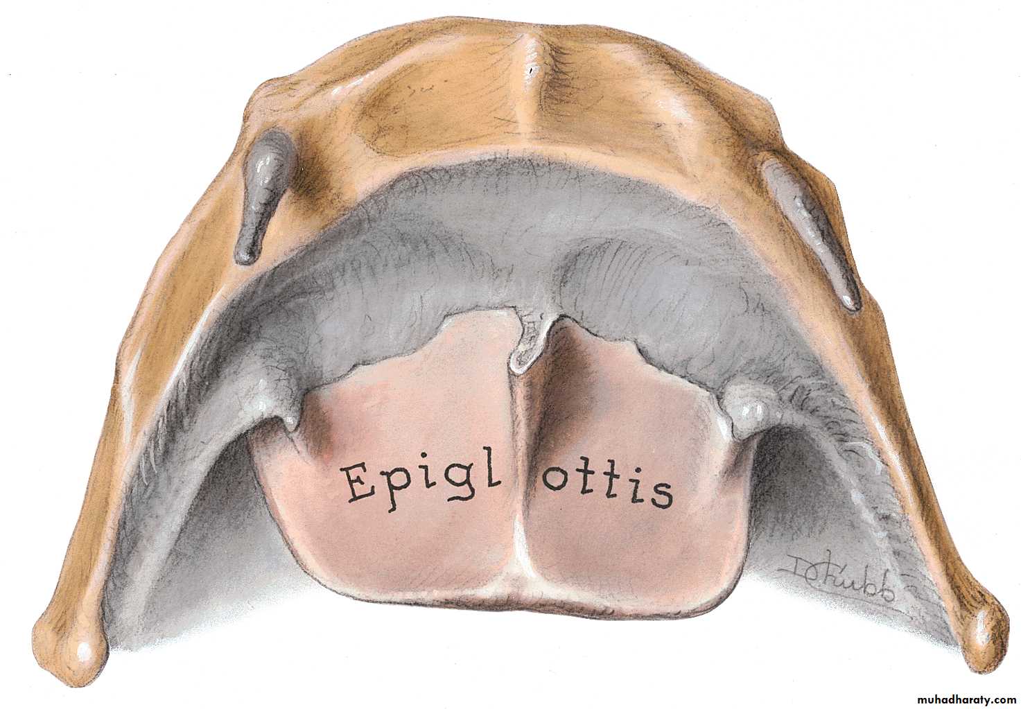
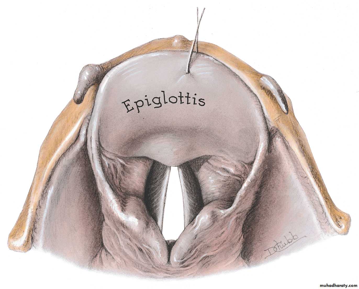
17
Anatomy
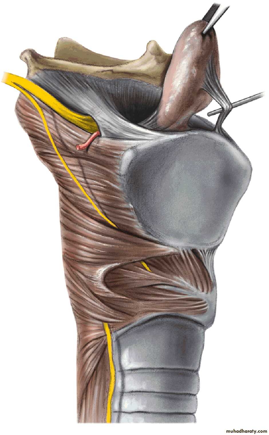
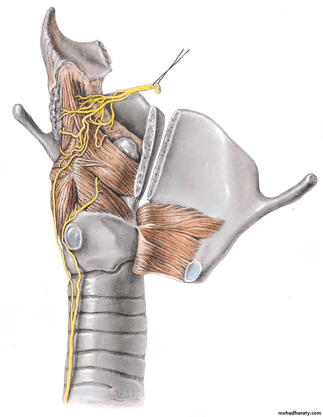
18
Anatomy
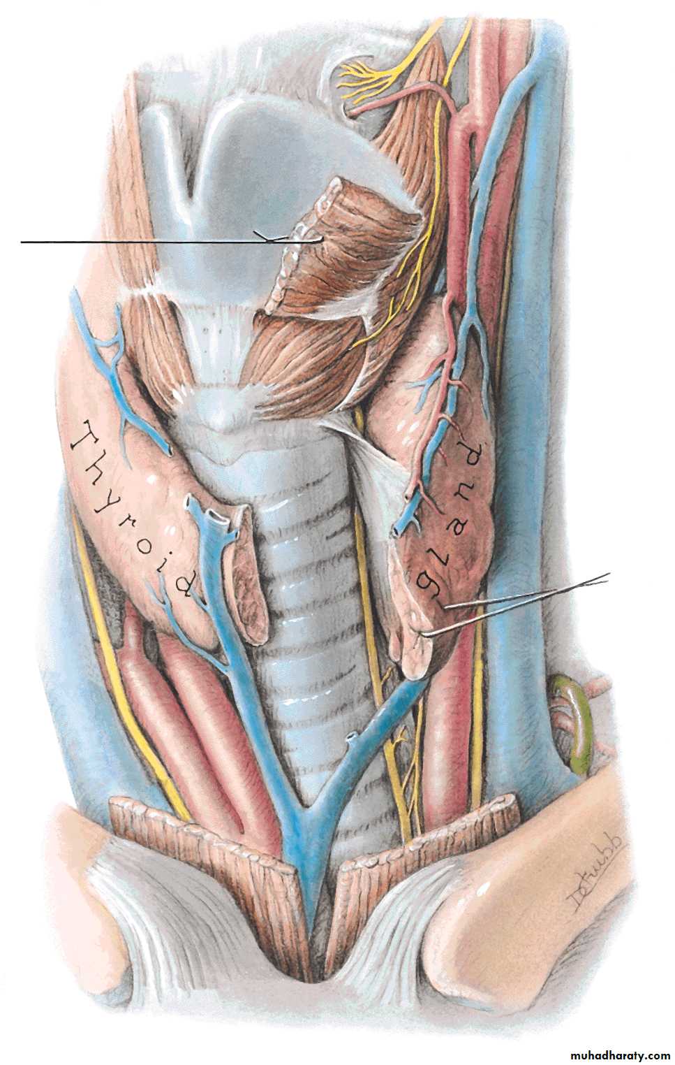
19
Anatomy
20
Anatomy
21
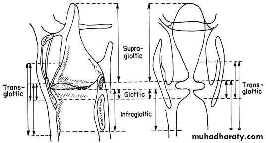
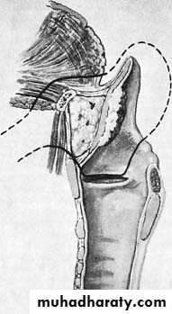
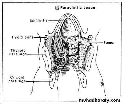
Natural History
Supraglottic tumors more aggressive:Direct extension into pre-epiglottic space
Lymph node metastasis
Direct extension into lateral hypopharnyx, glossoepiglottic fold, and tongue base
22
Natural History
Glottic tumors grow slower and tend to metastasize late owing to a paucity of lymphatic drainageThey tend to metastasize after they have invaded adjacent structures with better drainage
Extend superiorly into ventricular walls or inferiorly into subglottic space
Can cause vocal cord fixation
23
Natural History
True subglottic tumors are uncommonGlottic spread to the subglottic space is a sign of poor prognosis
Increases chance of bilateral disease and mediastinal extension
Invasion of the subglottic space associated with high incidence of stomal reoccurrence following total laryngectomy (TL)
24
Presentation
HoarsenessMost common symptom
Small irregularities in the vocal fold result in voice changes
Changes of voice in patients with chronic hoarseness from tobacco and alcohol can be difficult to appreciate
25
Presentation
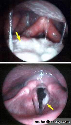
Patients presenting with hoarseness should undergo an indirect mirror exam and/or flexible laryngoscope evaluationMalignant lesions can appear as friable, fungating, ulcerative masses or be as subtle as changes in mucosal color
Videostrobe laryngoscopy may be needed to follow up these subtler lesions
26
Presentation
Good neck exam looking for cervical lymphadenopathy and broadening of the laryngeal prominence is requiredThe base of the tongue should be palpated for masses as well
Restricted laryngeal crepitus may be a sign of post cricoid or retropharyngeal invasion
27
Presentation
Other symptoms include:Dysphagia
Hemoptysis
Throat pain
Ear pain
Airway compromise
Aspiration
Neck mass
28
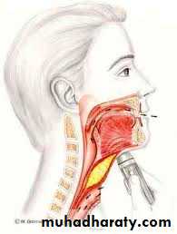
Work up
Biopsy is required for diagnosis
Performed in OR with patient under anesthesia
Other benign possibilities for laryngeal lesions include: Vocal cord nodules or polyps, papillomatosis, granulomas, granular cell neoplasms, sarcoidosis, Wegner’s granulomatosis
29
Work up
Other potential modalities:Direct laryngoscopy
Bronchoscopy
Esophagoscopy
Chest X-ray
CT or MRI
Liver function tests with or without US
PET ?
30
31
Staging- Primary Tumor (T)
• TX• Minimum requirements to assess primary tumor cannot be met
• T0
• No evidence of primary tumor
• Tis
• Carcinoma in situ
32
Staging- Supraglottis
• T1• Tumor limited to one subsite of supraglottis with normal vocal cord mobility
• T2
• Tumor involves mucosa of more than one adjacent subsite of supraglottis or glottis, or region outside the supraglottis (e.g. mucosa of base of the tongue, vallecula, medial wall of piriform sinus) without fixation
• T3
• Tumor limited to larynx with vocal cord fixation and or invades any of the following: postcricoid area, preepiglottic tissue, paraglottic space, and/or minor thyroid cartilage erosion (e.g. inner cortex)
• T4a
• Tumor invades through the thyroid cartilage and/or invades tissue beyond the larynx (e.g. trachea, soft tissues of neck including deep extrinsic muscles of the tongue, strap muscles, thyroid, or esophagus)
• T4b
• Tumor invades prevertebral space, encases carotid artery, or invades mediastinal structures
33
Staging- Glottis
• T1
• Tumor limited to the vocal cord (s) (may involve anterior or posterior commissure) with normal mobilty
• T1a
• Tumor limited to one vocal cord
• T1b
• Tumor involves both vocal cords
• T2
• Tumor extends to supraglottis and/or subglottis, and/or with impaired vocal cord mobility
• T3
• Tumor limited to the larynx with vocal cord fixation and/or invades paraglottic space, and/or minor thyroid cartilage erosion (e.g. inner cortex)
• T4a
• Tumor invades through the thyroid cartilage, and/or invades tissues beyond the larynx (e.g. trachea, soft tissues of the neck including deep extrinsic muscles of the tongue, strap muscles, thyroid, or esophagus
• T4b
• Tumor invades prevertebral space, encases carotid artery, or invades mediastinal structures
34
Staging- Subglottis
• T1• Tumor limited to the subglottis
• T2
• Tumor extends to vocal cord (s) with normal or impaired mobility
• T3
• Tumor limited the larynx with vocal cord fixation
• T4a
• Tumor invades cricoid or thyroid cartilage and/or invades tissues beyond larynx (e.g. trachea, soft tissues of the neck including deep extrinsic muscles of the tongue, strap muscles, thyroid, or esophagus)
• T4b
• Tumor invades prevertebral space, encases carotid artery, or invades mediastinal structures
35
Staging- Nodes
• N0
• No cervical lymph nodes positive
• N1
• Single ipsilateral lymph node ≤ 3cm
• N2a
• Single ipsilateral node > 3cm and ≤6cm
• N2b
• Multiple ipsilateral lymph nodes, each ≤ 6cm
• N2c
• Bilateral or contralateral lymph nodes, each ≤6cm
• N3
• Single or multiple lymph nodes > 6cm
36
Staging- Metastasis
• M0• No distant metastases
• M1
• Distant metastases present
37
Stage Groupings
• 0
• Tis
• N0
• M0
• I
• T1
• N0
• M0
• II
• T2
• N0
• M0
• III
• T3
• N0
• M0
• T1-3
• N1
• M0
• IVA
• T4a
• N0-2
• M0
• T1-4a
• N2
• M0
• IVB
• T4b
• Any N
• M0
• Any T
• N3
• M0
• IVC
• Any T
• Any N
• M1
38
Treatment
Premalignant lesions or Carcinoma in situ can be treated by surgical stripping of the entire lesion
CO2 laser can be used to accomplish this but makes accurate review of margins difficult
39
Treatment
Early stage (T1 and T2) can be treated with radiotherapy or surgery alone, both offer the 85-95% cure rate.Surgery has a shorter treatment period, saves radiation for recurrence, but may have worse voice outcomes
Radiotherapy is given for 6-7 weeks, avoids surgical risks but has own complications
40
Treatment
XRT complications include:Mucositis
Odynophagia
Laryngeal edema
Xerostomia
Stricture and fibrosis
Radionecrosis
Hypothyroidism
41
Treatment
Advanced stage lesions often receive surgery with adjuvant radiationMost T3 and T4 lesions require a total laryngectomy
Some small T3 and lesser sized tumors can be treated with partial larygectomy
42
Treatment
Adjuvant radiation is started within 6 weeks of surgery and with once daily protocols lasts 6-7 weeksIndications for post-op radiation include: T4 primary, bone/cartilage invasion, extension into neck soft tissue, perineural invasion, vascular invasion, multiple positive nodes, nodal extracapsular extension, margins<5mm, positive margins, CIS margins, subglottic extension of primary tumor.
43
Treatment
Chemotherapy can be used in addition to irradiation in advanced stage cancersTwo agents used are Cisplatinum and 5-flourouracil
Cisplatin thought to sensitize cancer cells to XRT enhancing its effectiveness when used concurrently.
44
Treatment
Induction chemotherapy with definitive radiation therapy for advanced stage cancer is another optionStudies have shown similar survival rates as compared to total laryngectomy with adjuvant radiation but with voice preservation.
Role in treatment still under investigation
45
Treatment
Modified or radical neck dissections are indicated in the presence of nodal disease
Neck dissections may be performed in patients with supra or subglottic T2 tumors even in the absence of nodal disease
N0 necks can have a selective dissection sparing the SCM, IJ, and XI
N1 necks usually have a modified dissection of levels II-IV
46
Surgical Options
47
Hemilaryngectomy
No more than 1cm subglottic extension anteriorly or 5mm posteriorlyMobile affected cord
Minimal anterior contralateral cord involvement
No cartilage invasion
No neck soft tissue invasion
48
Supraglottic laryngectomy
T1,2, or 3 if only by preepiglottic space invasionMobile cords
No anterior commissure involvement
FEV1 >50%
No tongue base disease past circumvallate papillae
Apex of pyriform sinus not invloved
49
Supracricoid Laryngectomy
Resection of true vocal cords, supraglottis, thyroid cartilageLeave arytenoids and cricoid ring intact
Half of patients remain dependent on tracheostomy
50
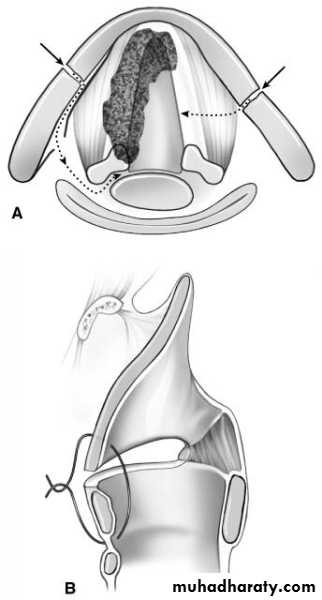
Total Larygectomy
Indications:
T3 or T4 unfit for partial
Extensive involvement of thyroid and cricoid cartilages
Invasion of neck soft tissues
Tongue base involvement beyond circumvallate papillae
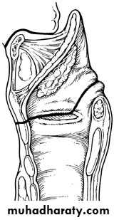
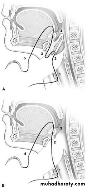
51
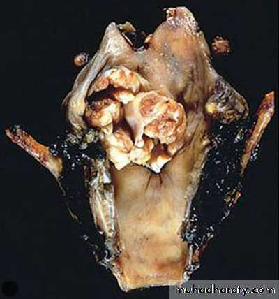
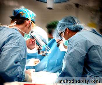
Total Laryngectomy
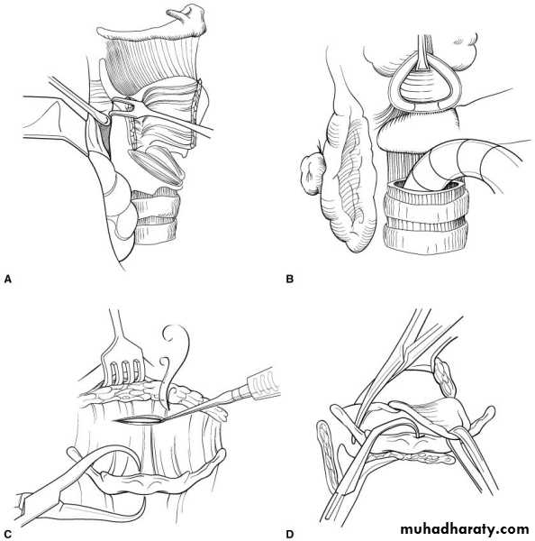
52
Total Laryngectomy
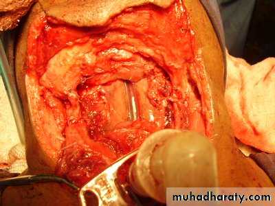
53
Total Laryngectomy
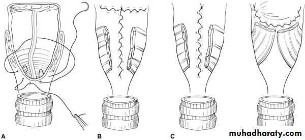
54
Total Laryngectomy
55
Complication of Surgery
Vocal problemSwallowing problem
Loss of taste and smell
Fistula development
Cranial nerve injury
Vascular Injuries and Events
Dropped Shoulder
Hypothyroidism
56
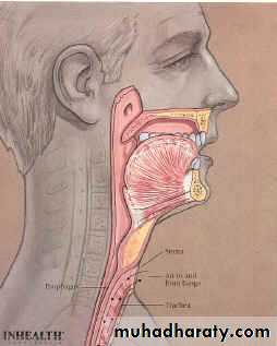
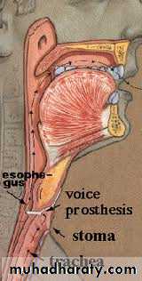
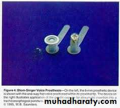
Voice Rehabilitation
Tracheostomal prosthesisElectrolarynx
Pure esophageal speech
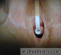
57Tracheoesophageal voice prosthesis
58
Electrolarynx

59
Prognosis
After initial treatment patients are followed at 4-6 week intervals. After first year decreases to every 2 months. Third and fourth year every three months, with annual visits after that
• 5 year survival
• Stage I
• >95%
• Stage II
• 85-90%
• Stage III
• 70-80%
• Stage IV
• 50-60%
60
Prognosis
Patients considered cured after being disease free for five yearsMost laryngeal cancers reoccur in the first two years
Despite advances in detection and treatment options the five year survival has not improved much over the last thirty years
61
62