Head /Neck AnatomyII year medical students
Dr.Adel Sahib Al-Mayaly
otolaryngologist/ head & neck surgeon
Expert comment
A clear understanding of the anatomy is the key to being successful surgeon.Attention please

The neckTopography
It is the region between:-1- The lower margin of the mandible above
2- the suprasternal notch and the upper border of the clavicle below.
Posteriorly: Cl-7 vertebrae.
Bone of Neck
cervical part of vertebral column, is the skeletal support of neckis convex forward
supports skull.
Extend from base of skull to the
superior thoracic inlet.
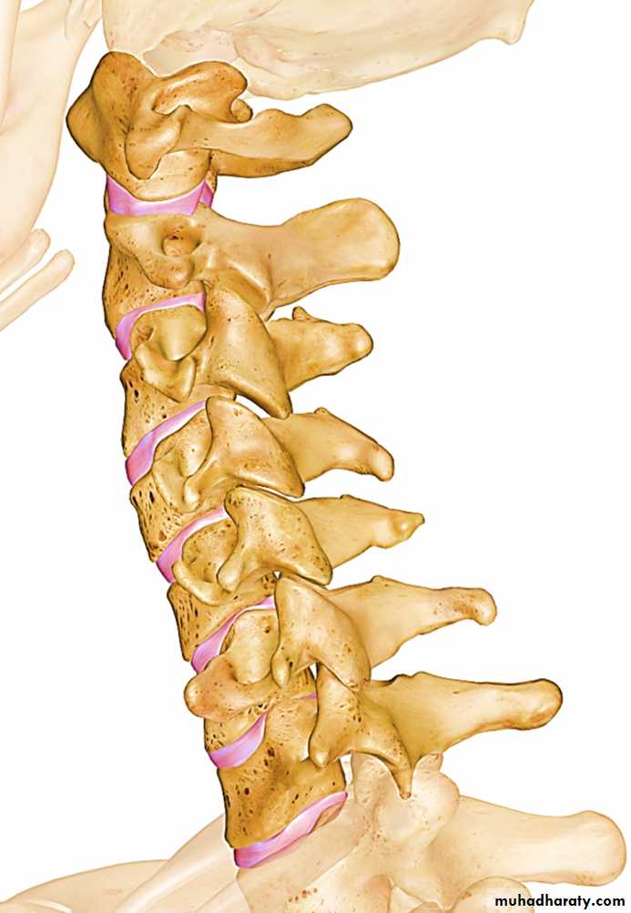
Cervical vertebrae
Behind the vertebrae is a mass of extensor muscles (main support of cervical spine) supplied by posterior primary ramiIn front is a smaller group of flexor muscles supplied by anterior primary rami.
In midline are parts of respiratory system, (larynx & trachea),
And parts of alimentary system,
pharynx & esophagus.
Carotid sheaths on the sides
COMPARTMENTS of NECK
Anterior Compartment of the Neck
Visceral, muscular, and neurovascular components located anterior to the prevertebral fascia.
Viscera
1. Pharynx
2. Cervical esophagus
3. Thyroid gland
4. Trachea
5. Larynx
Muscles
1. Platysma
2. SCM
3. Trapezius
4. Suprahyoid group
5. Infrahyoid group
Viscera
1. Pharynx
2. Cervical esophagus
3. Thyroid gland
4. Trachea
5. Larynx
Compartments
Vascular
1. Carotid arterial system2. Jugular venous system
3. Regional lymphatic system
Nerves
1. Recurrent laryngeal
2. Vagus (CN X)
3. Hypoglossal (CN XII)
4. Spinal accessory
5. Cervical sympathetic trunk
6. Facial (CN VII), cervical and ramus division
7. Cervical sensory plexus
Posterior Compartment of the Neck
Osseous, muscular, and neural components located posterior to the prevertebral fascia.Muscles
1. Scalene group
2. Levator scapulae
3. Splenius of the head
4. Semispinal of the head
5. Rhomboids
Nerves
1. Brachial plexus
2. Cervical plexus
3. Phrenic nerve
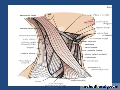
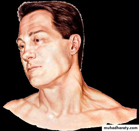
Landmarks of the neck
Sternocleidomastoid
Suprasternal fossa
Supraclavicular fossa.
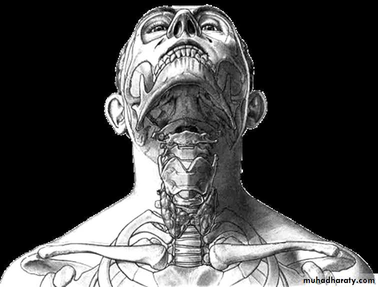
Landmarks of the neck
Hyoid boneThyroid cartilage
Cricoid cartilage
Cervical part of trachea
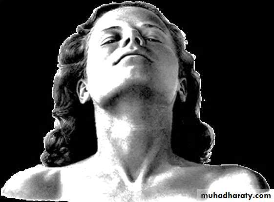
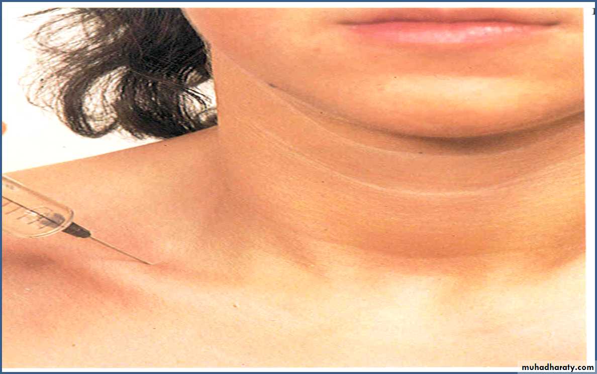
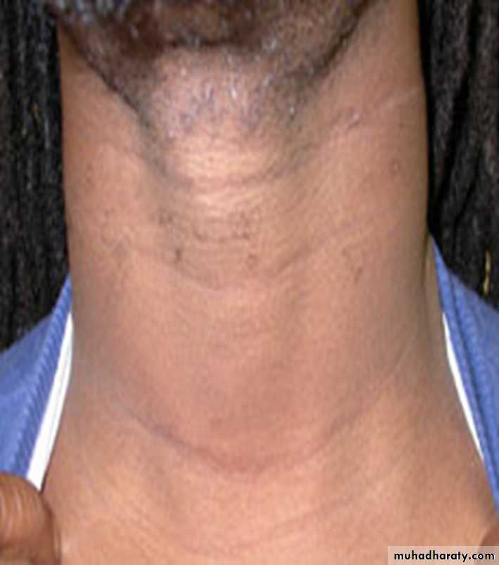
Skin of Neck
It runs horizontally around neck.is flexible and thin.
The upper segments have a greater density of hair in adult males.
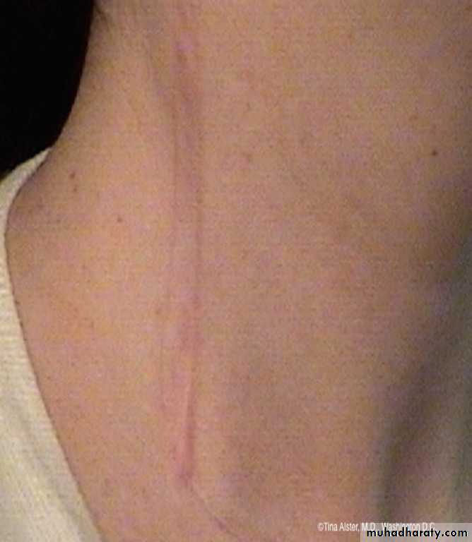
Skin creases
lines of cleavage of skin are constant
They correspond to the collagen fibres of dermis
Incision along creases; thin scar
Incision across the flexion skin creases; excessive scaring.

If an incision :
along a cleavage line narrow scar,crosses lines wide or heaped-up scar.
Fascia of Neck
1-Superficial Fascia2-deep Fascia
Superficial Fascia
Thin layer of loose connective tissue.
It invests the platysma muscle.
It replaces the subcutaneous layer elsewhere.
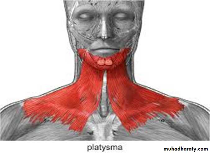
Platysma muscle:
• Is embedded in the subcutaneous tissue.• Varies in thickness.
• Extends from the superior thoracic region over the clavicle and the anterolateral surface of the neck
• to the lateral surface of the mandible.
Contents
1- Cutaneous nerves.2- External jugular vein.
3- Superficial cervical lymph nodes.
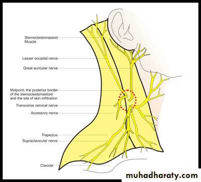
cutaneous nerves5 nerves
1- The skin overlying the back of the neck, the back of the scalp; as high as the vertex;(posterior rami of cervical nerves 2 to 5).
2-The skin of the front and sides of the neck;
(by anterior rami of cervical nerves 2 to 4)
The 1st cervical nerve has no cutaneous branch.
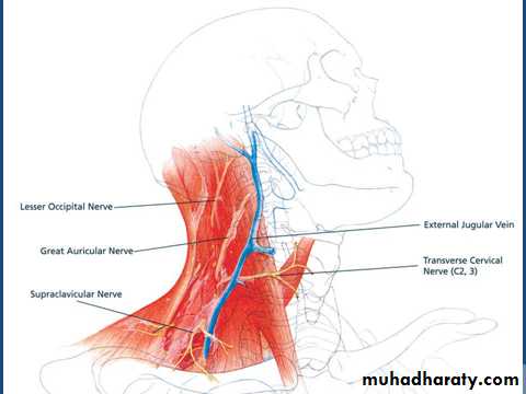
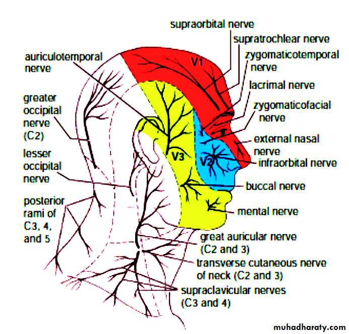
Cutaneous nerves
The greater occipital nerve (C2):-Back of neck & back of occipital region up to the vertex.
The lesser occipital nerve (C2).
Lateral part of occipital region & med. Surface of auricle.
Great auricular nerve:- (C2 - C3) Angle of mandible, parotid region & both surfaces of auricle.
Cutaneous Nerves;
4- The transverse cutaneous nerve (C2 ,C3)the skin on the anterior and lateral surfaces of the neck,
5- The supraclavicular nerves (C3 and 4):-
Medial , intermediate & lateral supraclavicular
nerves
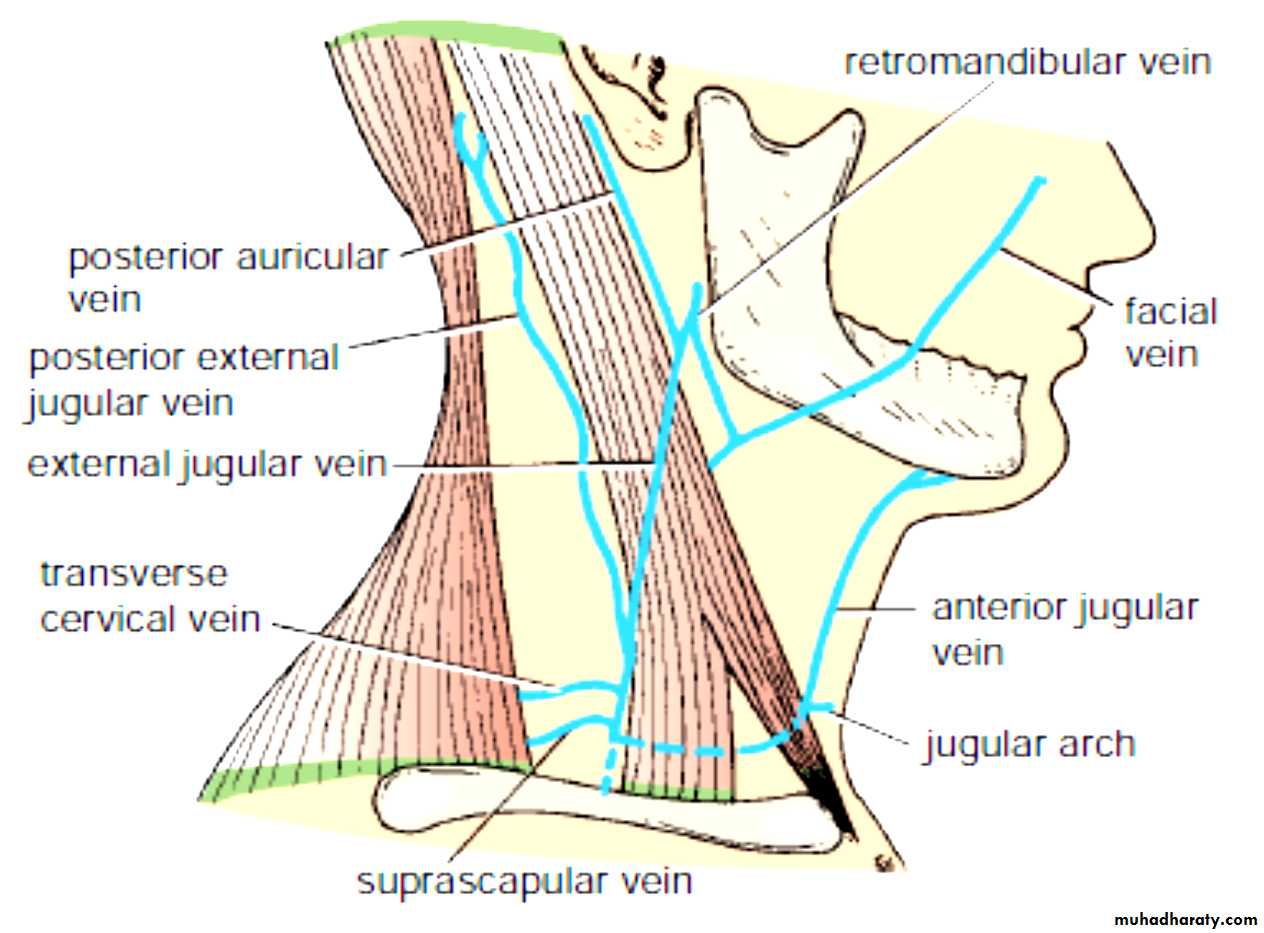
Superficial Veins
External Jugular Vein:-Formation:-
Begins just behind the angle of the mandible by the union of:-
1- posterior auricular vein.
2- posterior division of the retromandibular vein.
Course:- It descends obliquely across the SCM muscle
Termination:- It ends in the posterior triangle by join subclavian vein.
External jugular vein
Tributaries of EJV:-1- Posterior EJV:- from the back of the scalp
2-Transverse cervical vein from the skin and the fascia over posterior ∆
3-Suprascapular vein from the back of the scapula
4-Anterior jugular vein
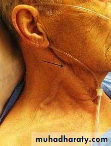
C L I N I C A L N O T E S
Anterior Jugular Vein
It begins just below the chin, by the union of several small veinIt runs down the neck close to the midline.
above the suprasternal notch; the veins of the two sides are united by a transverse trunk
called the jugular arch.
The vein then turns sharply laterally to drain to EJV.
Cross section of the neck at the level of the 6th cervical vertebra
Investing layerAttachments –
Superiorly to the external occipital protuberance, the superior nuchal lines, the mastoid tip and the zygomatic arch.
Anteriorly this is attached to the mandible, hyoid & down to the suprasternal area.
Inferiorly attached to the acromion & spine of scapula, the clavicle and the sternum
Posteriorly to ligamentum nuchae & spinous processes of cervical spines.
Superficial layer
It splits to enclose the trapezius, sternocleidomastoidn and the parotid & submandibular glands.
Middle or visceral layer
Encircles the trachea, thyroid g. and theEsophagus & slings to the hyoid bone.
Movement of the hyoid during swallowing
elevates the thyroid g. so that thyroid lumps
characteristically move up during deglutition.
Deep cervical fascia
The condensation of connective tissue surrounding the deeper structures of the neck.It is made up of 3 layers:-
1- superficial layer (investing layer).
2- middle layer (visceral layer).
3- deep layer (prevertebral layer).
Deep or prevertebral layer
It is a tough membrane that covers the prevertebral muscles.It forms a basis upon which the viscera of the neck slides during swallowing
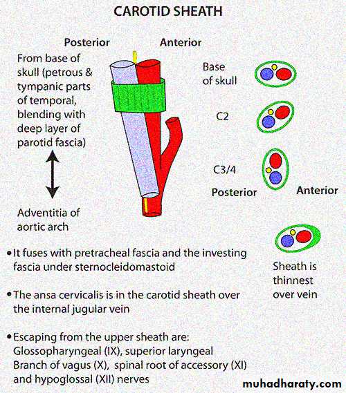
Carotid sheaths
They are tube of connective tissue surrounding the
1- carotid A. ( common carotid below the bifurcation, external & internal carotid above).
2- internal jugular vein)
3- vagus nerve.
It extends from base of skull (around the opening of carotid canal) to arch of aorta.
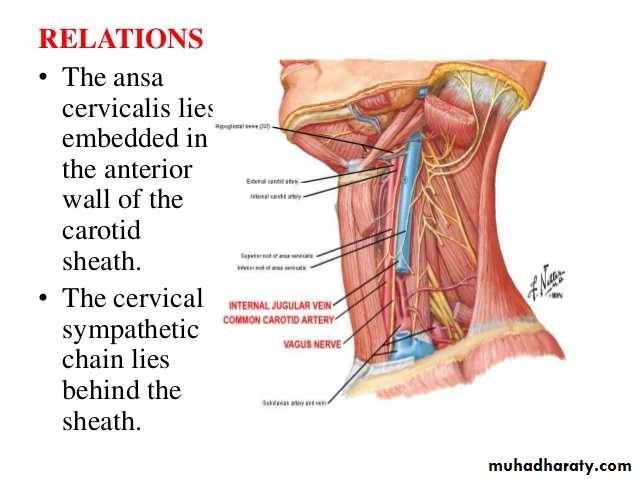
Embedded within the wall of carotid sheath is the ansa cervicalis nerve while immediately behind is the sympathetic trunk.
Triangles of neck