Spinal Cord Lec:3
• Assis.Professor Dr. Farah Nabil Abbas• MBChB, MSc, PhD
Muscle Sensory Receptors
Proper control of muscle function requires:Excitation by anterior motor neurons
Continuous sensory feedback information from each muscle – muscle status - that is:
• What is the length of the muscle?
• What is its tension?
• How rapidly length or tension changing?
Muscle spindles & Golgi tendon organs.
Length-Monitoring System & Stretch Reflex
Changes in muscle length monitored by stretch receptors embedded in them.2 receptor types respond to:
How much the muscle stretched.
Magnitude of stretch and speed with which it occurs.
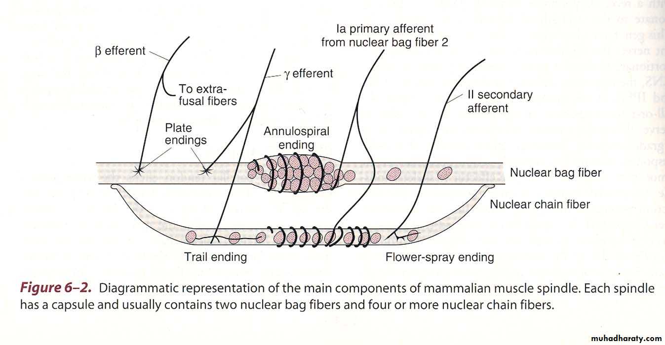
Receptors consist of nerve fiber endings wrapped around modified muscle fibers.
Length-Monitoring System & Stretch Reflex
Several muscle fibers enclosed in a connective tissue capsule “muscle spindle”.
Each spindle is 3-10 mm long.
Build around 3-12 very small modified intrafusal fibers attached to glycocalyx of surrounding large "extrafusal fiber“.
Intrafusal Skeletal Muscle Fibers
Small skeletal muscle fiber• Central region - no or few actin and myosin filaments does not contract when ends do (Function as sensory receptors).
• End portions that do contract - excited by small gamma motor neurons.
2 types
Intrafusal Skeletal Muscle Fibers
• Nuclear-bag fibers1-3/spindle
Central portion - Large number of nuclei congregated in an expanded bag.
Innervated by primary (Ia) nerve endings.
Intrafusal Skeletal Muscle Fibers
(2) Nuclear-chain fibers3-9/spindle.
1/2 in diameter & 1/2 as long as first type.
Nuclei aligned in a chain.
Innervation: 1ry & 2ndary nerve endings (II).
Ends connected to nuclear-bag fibers.
Intrafusal Skeletal Muscle Fibers
Sensory fibers which originate in central portion and stimulated by stretching of this mid portion of spindle are of 2 types:• Primary Endings (Annulospiral) Fibers
• Secondary (Flowerspray) Fibers
Primary Endings (Annulospiral) Fibers
Large fibers encircles the receptor center.Termination of type Ia afferent fibers.
17 µm Transmit at CV = 70-120 m/sec.
Stretching muscle Rapid discharge
Sustained stretch Less rapid discharge.
Respond to changes in length and change in rate of stretch.
Excited by both chains of intrafusal fibers.
Secondary (Flowerspray) Fibers
Termination of 1 or 2 small type II fibers.Diameter 8 µm.
Innervate receptor region on one side of primary ending.
Discharge at increasing rate through out period when the muscle is stretched.
Respond to change in length alone.
Excited by nuclear chain intrafusal fibers
Stretching m. spindle stimulation of 1ry and 2ndary nerve endings.
Muscle spindles are parallel to extrafusal fibers.
Muscle stretching by external force pulls on the intrafusal fibers stretching them + activating their receptor endings reflex contraction of muscle extrafusal fibers.
More or faster muscle stretching greater rate of receptor firing.
In contrast:Contraction of the extrafusal fibers muscle shortening remove tension on the spindle slow firing rate of stretch receptor.
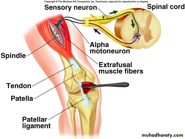
Muscle spindle afferent neurons Pathways
On entering the NS, they divide:1) 1st path:
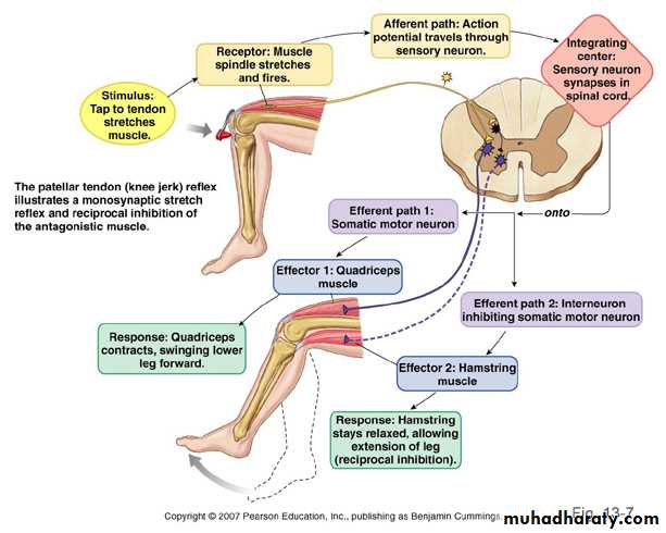
Directly stimulates motor neuron going back to the stretched muscle completing a reflex arc known as the "stretch reflex".
Stretch reflex is most familiar in the form of the knee jerk.
Muscle spindle afferent neurons Pathways
2) 2nd Path:Afferent nerve fibers end on interneuron.
When activated inhibit motor neuron controlling antagonistic muscles (knee jerk – inhibition of knee flexor muscles).
Reciprocal Innervation: Activation of one muscle with simultaneous inhibition of its antagonistic muscle.
Muscle spindle afferent neurons Pathways
3) 3rd Path:Afferent nerve fibers activate motor neurons of synergistic muscles - muscles whose contraction assists the intended motion (knee jerk-other extensor muscles).
Note: The activated muscles are on same side of body as receptors ipsilateral response.
Muscle spindle afferent neurons Pathways
4) 4th Path:
Afferent nerve fibers continue to the brain stem.
Synapse with interneuron forms next link in the pathway that conveys information about muscle length to brain areas dealing with motor control.
Stretch Reflex
• Muscle SpindleSense Organ
• Ia fiber primary (annulospiral) and II fibers secondary (flower spray) ending
Afferent Neuron
CNS (spinal cord)
Synapse
Alpha Motor Neuron
Efferent Neuron
Muscle (extrafusal muscle)
• Effector
Clinical Examples of Stretch Reflex
Knee jerk or patellar reflexAnkle jerk
Biceps reflex
Triceps reflex
Knee Jerk
• Tapping pattelar tendon stretched.
• Thigh muscles stretched + activation of stretched receptors within these muscles.
• Action potentials are generated in afferent nerve fibers.
• Transmitted to motor neurons that control these muscles stimulating motor units
Knee Jerk
• Shortened thigh muscles.• Patient's lower leg is extended.
During normal movement:
• Stretch receptors rarely all activated at same time
• Stretch receptors not activated so strongly.
• Movement occurs in response to integration of many types of local descending controls.
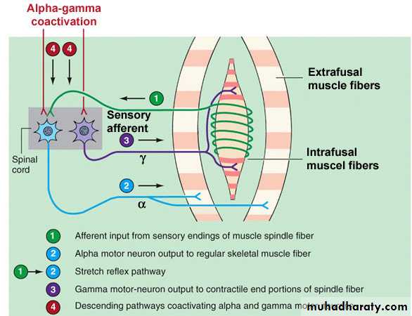
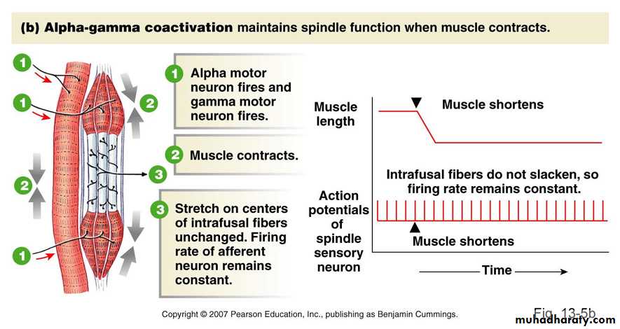
Alpha-Gamma Coactivation
Shortening muscle intrafusal fibers stretching .At this time, spindle stretch receptors go completely slack stop firing APs.
But:
In this situation: No indication of any further changes in muscle length.
Alpha-Gamma Coactivation
Physiologically:To prevent this loss of information
Stimulation of two ends of each intrafusal muscle fiber contract during shortening of the extrafusal muscle fibers.
Thus: maintaining tension in the central region of the intrafusal fibers, where stretch receptors are located.
Alpha-Gamma Coactivation
Intrafusal muscle fibersNot large enough or strong enough to shorten a whole muscle and move joints
Job: maintain tension on spindle stretch receptors.
Alpha and gamma motor neurons
Activated by interneurons in their vicinity and by neurons of descending pathways.
Coactivated - excited at almost same time during many voluntary and involuntary movements.
Tension-Monitoring System and Golgi Tendon Reflex
Inputs to motor neurons of a muscles various degrees of tension depending on:Muscle length
Load on the muscle
Degree of muscle fatigue
Necessary feedback to inform the motor control systems of tension achieved.
Vision & Golgi tendon organs which monitors how much tension is being exerted by contracting MUs.