The peritoneum,omentum, mesenteryand retroperitonealspace
Dr. Rabah alsamrraeTHE PERITONEUM
The peritoneal membrane is conveniently divided into two parts – the visceral peritoneum surrounding the viscera and the parietal peritoneum lining the other surfaces of the cavity. The peritoneum has a number of functionsFunctions of the peritoneum
■ Pain perception (parietal peritoneum)
■ Visceral lubrication
■ Fluid and particulate absorption
■ Inflammatory and immune responses
■ Fibrinolytic activity
The parietal portion is richly supplied with nerves and, when irritated, causes severe pain accurately localised to the affected area.The visceral peritoneum, in contrast, is poorly supplied with nerves and its irritation causes vague pain that is usually located to the midline.
The peritoneal cavity is the largest cavity in the body, the surface area of its lining membrane (2 m2 in an adult) being nearly equal to that of the skin. The peritoneal membrane is composed of flattened polyhedral cells (mesothelium), one layer thick, resting upon a thin layer of fibroelastic tissue.
Beneath the peritoneum, supported by a small amount of areolar tissue, lies a network of lymphatic vessels and rich plexuses of capillary blood vessels from which all absorption and exudation must occur. In health, only a few millilitres of peritoneal fluid is found in the peritoneal cavity. The fluid is pale yellow, somewhat viscid and contains lymphocytes and other leucocytes; it lubricates the viscera, allowing easy movement and peristalsis.
In the peritoneal space, mobile gas-filled structures float upwards, as does free air (‘gas’). In the erect position, when free fluid is present in the peritoneal cavity, pressure is reduced in the upper abdomen compared with the lower abdomen. When air is introduced, it rises, allowing all of the abdominal contents to sink. When a visceral perforation occurs, the free fluid that spills into the peritoneal cavity runs downwards, largely directed by the normal peritoneal attachments. For example, spillage from a perforated duodenal ulcer may run down the right paracolic gutter
ACUTE PERITONITIS Most cases of peritonitis are caused by an invasion of the peritoneal cavity by bacteria.Bacterial peritonitis is usually polymicrobial, both aerobic and anaerobic organisms being present. The exception is primary peritonitis (‘spontaneous’ peritonitis), in which a pure infection with streptococcal, pneumococcal or Haemophilus bacteria occurs.
Bacteriology
Bacteria from the gastrointestinal tract
The number of bacteria within the lumen of the gastrointestinal tract is normally low until the distal small bowel is reached, whereas high concentrations are found in the colon. However, disease (e.g. obstruction, achlorhydria, diverticula) may increase proximal colonisation. The biliary and pancreatic tracts are normally free from bacteria, although they may be infected in disease,e.g. gallstones. Peritoneal infection is usually caused by two or more bacterial strains. Gram-negative bacteria contain endotoxins (lipopolysaccharides) in their cell walls that have multiple toxic effects on the host, primarily by causing the release of tumour necrosis factor (TNF) from host leucocytes. Systemic absorption of endotoxin may produce endotoxic shock with hypotension and impaired tissue perfusion. Other bacteria such as Clostridium welchii produce harmful exotoxins
Bacteroides are commonly found in peritonitis. These Gram negative, non-sporing organisms, although predominant in the lower intestine, often escape detection because they are strictly anaerobic and slow to grow on culture media unless there is an adequate carbon dioxide tension in the anaerobic apparatus . In many laboratories, the culture is discarded if there is no growth in 48 hours. These organisms are resistant to penicillin and streptomycin but sensitive to metronidazole, clindamycin ,lincomycin and cephalosporin compounds. Since the widespread use of metronidazole (Flagyl), Bacteroides infections have greatly diminished.
Route of infection
Infecting organisms may reach the peritoneal cavity via a number of routes■ Gastrointestinal perforation, e.g. perforated ulcer, diverticular perforation
■ Exogenous contamination, e.g. drains, open surgery, trauma
■ Transmural bacterial translocation (no perforation), e.g.inflammatory bowel disease, appendicitis, ischaemic bowel
■ Female genital tract infection, e.g. pelvic inflammatory disease
■ Haematogenous spread (rare), e.g. septicaemia
Even in patients with non-bacterial peritonitis (e.g. acute pancreatitis, intraperitoneal rupture of the bladder or haemoperitoneum),the peritoneum often becomes infected by transmural spread of organisms from the bowel, and it is not long (often a matter of hours) before a bacterial peritonitis develops. Most duodenal perforations are initially sterile for up to several hours , and many gastric perforations are also sterile at first; intestinal perforations are usually infected from the beginning. The proportion of anaerobic to aerobic organisms increases with the passage of time. Mortality reflects:
• the degree and duration of peritoneal contamination;
• the age of the patient;
• the general health of the patient;
• the nature of the underlying cause
Localised peritonitis
Anatomical, pathological and surgical factors may favour the localisation of peritonitisAnatomical
The greater sac of the peritoneum is divided into (1) the subphrenic spaces, (2) the pelvis and (3) the peritoneal cavity proper. The last is divided into a supracolic and an infracolic compartment by the transverse colon and transverse mesocolon, which deters the spread of infection from one to the other. When the supracolic compartment overflows, as in a peptic ulcer perforates, it does so over the colon into the infracolic compartment or by way of the right paracolic gutter to the right iliac fossa and hence to the pelvis.
Pathological
The clinical course is determined in part by the manner in which adhesions form around the affected organ. Flakes of fibrin appear and cause loops of intestine to become adherent to one another and to the parietes. Peristalsis is retarded in affected bowel and this helps to prevent distribution of the infection. The greater omentum, by enveloping and becoming adherent to inflamed structures, forms barrier to the spread of infection.
Surgical
Drains placed during operation to assist localisation (and exit) of intra-abdominal collections
Diffuse peritonitis
A number of factors may favour the development of diffuse peritonitis:
• Speed of peritoneal contamination is a prime factor. If an inflamed appendix or other hollow viscus perforates before localisation has taken place, there is a gush of contents into the peritoneal cavity, which may spread over a large area almost instantaneously. Perforation proximal to an obstruction or from sudden anastomotic separation is associated with severe generalised peritonitis and a high mortality rate.
• Stimulation of peristalsis by the ingestion of food or even water hinders localisation. Violent peristalsis occasioned by the administration of a purgative or an enema may cause the widespread distribution of an infection that would otherwise have remained localised.
• The virulence of the infecting organism may be so great as to render the localisation of infection difficult or impossible.
• Young children have a small omentum, which is less effective in localising infection.
• Disruption of localised collections may occur with injudicious handling, e.g. appendix mass or pericolic abscess.
• Deficient natural resistance (‘immune deficiency’) may result from use of drugs (e.g. steroids), disease [e.g. acquired immune deficiency syndrome (AIDS)] or old age.
Clinical features
Localised peritonitisLocalised peritonitis is bound up intimately with the causative condition, and the initial symptoms and signs are those of that condition. When the peritoneum becomes inflamed, the temperature , and especially the pulse rate, rise. Abdominal pain increases and usually there is associated vomiting. The most
important sign is guarding and rigidity of the abdominal wall over the area of the abdomen that is involved, with a positive ‘release’ sign (rebound tenderness). If inflammation arises under the diaphragm, shoulder tip (‘phrenic’) pain may be felt. In cases of pelvic peritonitis arising from an inflamed appendix in the pelvic
position or from salpingitis, the abdominal signs are often slight; there may be deep tenderness of one or both lower quadrants alone, but a rectal or vaginal examination reveals marked tenderness of the pelvic peritoneum. With appropriate treatment, localised peritonitis usually resolves; in about 20% of cases, an abscess follows. Infrequently, localised peritonitis becomes diffuse.
Conversely, in favourable circumstances, diffuse peritonitis can become localised, most frequently in the pelvis or at multiple sites within the abdominal cavity
Diffuse (generalised) peritonitis
Diffuse (generalised) peritonitis may present in differing ways dependent on the duration of infection.Early
Abdominal pain is severe and made worse by moving or breathing. It is first experienced at the site of the original lesion and spreads outwards from this point. Vomiting may occur. The patient usually lies still. Tenderness and rigidity on palpation are found typically when the peritonitis affects the anterior abdominal
wall. Abdominal tenderness and rigidity are diminished or absent if the anterior wall is unaffected, as in pelvic peritonitis or, rarely, peritonitis in the lesser sac. Patients with pelvic peritonitis may complain of urinary symptoms; they are tender on rectal or vaginal examination. Infrequent bowel sounds may still be heard for a few hours but they cease with the onset of paralytic ileus. The pulse rises progressively but, if the peritoneum is deluged with irritant fluid, there is a sudden rise. The temperature
changes are variable and can be subnormal.
Late
If resolution or localisation of generalised peritonitis does not occur, the abdomen remains silent and increasingly distends. Circulatory failure ensues, with cold, clammy extremities, sunken eyes, dry tongue, thready (irregular) pulse and drawn and anxious face (Hippocratic facies). The patient finally lapses into unconsciousness. With early diagnosis and adequate treatment, this condition is rarely seen in modern surgical practice
Diagnostic aids
Investigations may elucidate a doubtful diagnosis, but the importance of a careful history and repeated examination must not be forgotten.
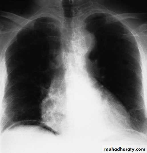
• A radiograph of the abdomen may confirm the presence of dilated gas-filled loops of bowel (consistent with a paralytic ileus) or show free gas, although the latter is best shown on an erect chest radiograph. If the patient is too ill for an ‘erect’ film to demonstrate free air under the diaphragm, a lateral decubitus film is just as useful, showing gas beneath the abdominal wall.
• Serum amylase estimation may establish the diagnosis of acute pancreatitis provided that it is remembered that moderately raised values are frequently found following other abdominal catastrophes and operations, e.g. perforated duodenal ulcer.
• Ultrasound and computerised tomography (CT) scanning are increasingly used to identify the cause of peritonitis.
• Peritoneal diagnostic aspiration may be helpful but is usually unnecessary. Bile-stained fluid indicates a perforated peptic ulcer or gall bladder; the presence of pus indicates bacterial peritonitis. Blood is aspirated in a high proportion of patients with intraperitoneal bleeding
Gas under the diaphragm in a patient with free perforationand peritonitis
General care of the patient
Correction of circulating volume and electrolyte imbalance Patients are hypovolaemic with electrolyte disturbances.The plasma volume must be restored and electrolyte concentrations corrected. Central venous catheterisation and pressure monitoring may be helpful.If the patient’s recovery is delayed for more than 7–10 days,intravenous nutrition is required.Gastrointestinal decompression
A nasogastric tube is passed into the stomach and aspirated. Intermittent aspiration is maintained until the paralytic ileus has resolved. If the abdomen is soft and not tender, and bowel sounds return, oral feeding may be introduced.
Antibiotic therapy
As the infection is usually a mixed one, initial treatment with parenteral broad-spectrum antibiotics active against aerobic and anaerobic bacteria should be given.
Correction of fluid loss
A fluid balance chart must be started so that daily output by gastric aspiration and urine is known. Additional losses from the lungs, skin and in faeces are estimated, so that the intake requirements can be calculated and seen to have been administered.
Analgesia
The patient should be nursed in the sitting-up position and must be relieved of pain before and after operation. If appropriate expertise is available, epidural infusion may provide expertise. Freedom from pain allows early mobilisation and adequate physiotherapy in the postoperative period, which help to prevent basal pulmonary collapse, deep vein thrombosis and pulmonary embolism.Vital system support
Specific treatment of the cause
If the cause of peritonitis is amenable to surgery, operation must be carried out as soon as the patient is fit for anaesthesia. This is usually within a few hours. In peritonitis caused by pancreatitis or salpingitis, or in cases of primary peritonitis
of streptococcal or pneumococcal origin, non-operative treatment is preferred provided the diagnosis can be made with confidence.
Peritoneal lavage
In operations for general peritonitis it is essential that, after the cause has been dealt with, the whole peritoneal cavity is explored with the sucker and, if necessary, mopped dry until all seropurulent exudate is removed. The use of a large volume of saline (1–2 litres) containing dissolved antibiotic (e.g. tetracycline) has been shown to be effective
Complications
Acute intestinal obstruction due to peritoneal adhesions
This usually gives central colicky abdominal pain with evidence of small bowel gas and fluid levels sometimes confined to the proximal intestine on radiography. Bowel sounds are increased. It is more common with localised peritonitis. It is essential to distinguish this from paralytic ileus.
Paralytic ileus
There is usually little pain, and gas-filled loops with fluid levels are seen distributed throughout the small and large intestines on abdominal imaging. In paralytic ileus, bowel sounds are reduced or absent.
Abdominal and pelvic abscesses
Abscess formation following local or diffuse peritonitis usually occupies one of the situations . The symptoms and signs of pus may be vague and consist of nothing more than lassitude, anorexia and malaise; pyrexia (often low-grade), tachycardia, leucocytosis, raised C-reactive protein and localised tenderness are also common Later, a palpable mass may develop that should be monitored by marking out its limits on the abdominal wall and meticulous daily examination. More commonly is monitored by repeat ultrasound or CT scanning. In most cases, with the aid of antibiotic treatment, the abscess or mass gradually reduces in size until, finally, it is undetectable. In others, the abscess fails to resolve or becomes larger, then it must be drained. If facilities are available ,ultrasound- or CT-guided drainage may avoid further operation.
Open drainage of an intraperitoneal collection another option
Pelvic abscess
The pelvis is the commonest site of an intraperitoneal abscess because the vermiform appendix is often pelvic in position and the fallopian tubes are frequent sites of infection. A pelvic abscess can also occur as a sequel to any case of diffuse peritonitis and is common after anastomotic leakage following colorectal surgery.The most characteristic symptoms are diarrhoea and the passage of mucus in the stools. Rectal examination reveals a bulging of the anterior rectal wall, which, when the abscess is ripe, becomes softly cystic. Left to nature, a proportion of these abscesses burst into the rectum, after which the patient nearly always recovers
rapidly. If this does not occur, the abscess should be drained deliberately. In women, vaginal drainage through the posterior fornix is often chosen. In other cases, when the abscess is definitely pointing into the rectum, rectal drainage is
employed. If any uncertainty exists, the presence of pus should be confirmed by ultrasound or CT scanning with needle aspiration if indicated. Laparotomy is almost never necessary. Rectal drainage of a pelvic abscess is far preferable to suprapubic drainage, which risks exposing the general peritoneal cavity to infection. Drainage
tubes can also be inserted percutaneously or via the vagina or rectum under ultrasound or CT guidance
Intraperitoneal abscess
AnatomyThe complicated arrangement of the peritoneum results in the formation of four intraperitoneal spaces in which pus may collect
Left subphrenic space
Left subhepatic space/lesser sac
The commonest cause of infection here is complicated acute pancreatitis.
Right subphrenic space
Right subhepatic space
This lies beneath the right lobe of the liver in Rutherford Morison’s pouch. It is the deepest space of the four and the commonest site of a subphrenic abscess, which usually arises from appendicitis, cholecystitis, a perforated duodenal ulcer or following upper abdominal surgery.
Clinical features
The symptoms and signs of subphrenic infection are frequently non-specific and it is well to remember the aphorism, ‘pus somewhere, pus nowhere else, pus under the diaphragm’.
Symptoms
Sweating, wasting and anorexia are present. There is sometimes epigastric fullness and pain, or pain in the shoulder on the affected side, because of irritation of sensory fibres in the phrenic nerve, referred along the descending branches of the cervical plexus. Persistent hiccoughs may be a presenting symptom.
Signs
A swinging pyrexia is usually present. If the abscess is anterior, abdominal examination will reveal some tenderness, rigidity or even a palpable swelling. Sometimes the liver is displaced downwards but more often it is fixed by adhesions. Examination of the chest is important and, in the majority of cases, collapse of the lung or evidence of basal effusion or even an empyema is
found.
Investigations
A number of the following investigations may be helpful:• Blood tests usually show a leucocytosis and raised C-reactive protein.
• A plain radiograph sometimes demonstrates the presence of gas or a pleural effusion. On screening, the diaphragm is often seen to be elevated (so called ‘tented’ diaphragm) and its movements impaired.
• Ultrasound or CT scanning is the investigation of choice and permits early detection of subphrenic collections
Treatment
If suppuration seems probable. If skilled help is available it is usually possible to insert a percutaneous drainage tube under ultrasound or CT control. The same tube can be used to instil antibiotic solutions or irrigate the abscess cavity. If an operative approach is necessary and a swelling can be detected in the subcostal region or in the loin, an incision is made over the site of maximum tenderness or over any area where oedema or redness is discovered.
SPECIAL FORMS OF PERITONITIS
PostoperativeThe patient is ill with raised pulse and peripheral circulatory failure. Following an anastomotic dehiscence, the general condition of a patient is usually more serious than if the patient had suffered leakage from a perforated peptic ulcer with no preceding operation. Local symptoms and signs are less definite. Abdominal pain may not be prominent and is often difficult to assess because
of normal wound pain and postoperative analgesia. The patient’s deterioration may be attributed wrongly to cardiopulmonary collapse, which is usually concomitant. Peritonitis follows abdominal operations more frequently than is realised. The principles of treatment do not differ from those of peritonitis of other origin. Antibiotic therapy alone is inadequate; no antibiotic can stay the onslaught of bacterial peritonitis caused by leakage from a suture line, which must be dealt with by operation.
Bile peritonitis
Unless there is reason to suspect that the biliary tract was damaged during operation, it is improbable that bile as a cause of peritonitis will be thought of until the abdomen has been opened. Unless the bile has extravasated slowly and the collection becomes shut off from the general peritoneal cavity, there are signs of diffuse peritonitis. After a few hours a tinge of jaundice is not unusual. Laparotomy (or laparoscopy) should be undertaken with evacuation of the bile and peritoneal lavage. The source of bile leakage should be identified. Aleaking gall bladder is excised or a cystic duct ligated. An injury to the bile duct may simply be drained or alternatively intubated; later, reconstructive operation is often required. Infected bile is more lethal than sterile bile. A ‘blown’ duodenal stump should be drained as it is too oedematous to repair, but sometimes it can be covered by a jejunal patch. The patient is often jaundiced from absorption of peritoneal bile, but the surgeon must ensure that the abdomen is not closed until any obstruction to a major bile duct has been either excluded or relieved. Bile leaks after cholecystectomy or liver trauma may be dealt with by percutaneous (ultrasound-guided) drainage and endoscopic biliary stenting to reduce bile duct pressure. The drain is removedwhen dry and the stent at 4–6 weeks.
TUBERCULOUS PERITONITIS
Acute tuberculous peritonitis
Tuberculous peritonitis sometimes has an onset that so closely resembles acute peritonitis that the abdomen is opened. Straw coloured fluid escapes and tubercles are seen scattered over the peritoneum and greater omentum. Early tubercles are greyish and translucent. They soon undergo caseation and appear white or yellow and are then less difficult to distinguish from carcinoma Occasionally, they appear like patchy fat necrosis. On opening the abdomen and finding tuberculous peritonitis, the fluid is evacuated, some being retained for bacteriological studies. A portion of the diseased omentum is removed for histological confirmation of the diagnosis and the wound closed without drainage.
Chronic tuberculous peritonitis
The condition presents with abdominal pain (90% of cases), fever
(60%), loss of weight (60%), ascites (60%), night sweats (37%)
and abdominal mass (26%)
Origin of the infection
Infection originates from:• tuberculous mesenteric lymph nodes;
• tuberculosis of the ileocaecal region;
• a tuberculous pyosalpinx;
• blood-borne infection from pulmonary tuberculosis, usually
the ‘miliary’ but occasionally the ‘cavitating’ form.
Varieties of tuberculous peritonitis
four varieties of tuberculous peritonitis: ascitic,encysted, fibrous and purulent.
Ascitic form
The peritoneum is studded with tubercles and the peritoneal cavity becomes filled with pale, straw-coloured fluid. The patient is usually brought for advice because of distension of the abdomen. Pain is often absent; which may be associated with constipation or diarrhoea. On inspection, dilated veins may be seen coursing beneath the skin of the abdominal wall. In boys, congenital hydroceles sometimes appear, resulting from the patent processi vaginales becoming filled with ascitic fluid. Because of the increased intra-abdominal pressure, an umbilical hernia occur
Diagnosis
Laparoscopy is useful by allowing inspection of the peritoneal cavity. Areas of caseation can be biopsied for histology and microbiological studies. A chest radiograph should always be taken before laparoscopy or laparotomy is performed.Encysted form
The encysted (loculated) form is similar to the ascitic form except that one part of the abdominal cavity alone is involved. Thus, a localised intra-abdominal swelling is produced.
Fibrous form
The fibrous form is characterised by the production of widespread adhesions, which cause coils of intestine, especially the ileum, to become matted together and distended. These distended coils act as a ‘blind loop’ and give rise to steatorrhoea, wasting and attacks of abdominal pain. Anti-tuberculous therapy will often rapidly cure the condition without the need for surgery.
Purulent form
The purulent form is rare. When it occurs, usually it is secondary
to tuberculous salpingitis.
THE OMENTUM
Rutherford Morison called the greater omentum ‘the abdominal policeman’. The greater omentum attempts, often successfully, to limit intraperitoneal infective and other noxious processes . For instance, an acutely inflamed appendix is often found
wrapped in omentum, and this saves many patients from developing diffuse peritonitis. Some sufferers of herniae are also greatly indebted to this structure, for it often plugs the neck of a hernial sac and prevents a coil of intestine from entering and becoming strangulated. The omentum is usually involved in tuberculous peritonitis and carcinomatosis of the peritoneum.
THE MESENTERY
Mesenteric injuryA wound of the mesentery can follow abdominal contusion and is a cause of haemoperitoneum.
Seatbelt syndrome
If a car accident occurs when a seatbelt is worn, sudden deceleration can result in a torn mesentery. If there is any bruising of the abdominal wall, or even marks of clothing impressed into the skin, laparotomy may be indicated.
Diagnostic peritoneal lavage
Diagnostic peritoneal lavage may be helpful in this situation.
THE RETROPERITONEAL SPACE
Pus or blood in the retroperitoneal space tends to track to the corresponding iliac fossa. If a retroperitoneal abscess develops, it should be evacuated by the nearest route through the abdominal wall, avoiding opening the peritoneum. Should the retroperitoneal collection be found at laparotomy, it must be drained by a counterincision in the flank. Pus may develop from a renal orspinal source and is sometimes tuberculous (‘cold abscess’); tracking can develop alongside the psoas muscle and appear in the groin, where it must be distinguished from other swellings (e.g. hernia). Retroperitoneal haematoma may be caused by a fractured spine or pelvis, a leaking abdominal aneurysm, acute pancreatitis or a ruptured kidney