طفيليات / د.اكرام / طب عام ثالث
عملي 9/3/2017د.إكرام الحسو عمليParasitology Lec:1
9-3-2017
Medically important Cestodes (Tapeworms) of human beings
Echinococcus Spp.
E. granulosus
E. multilocularis
E. vogeli
E.granulosis
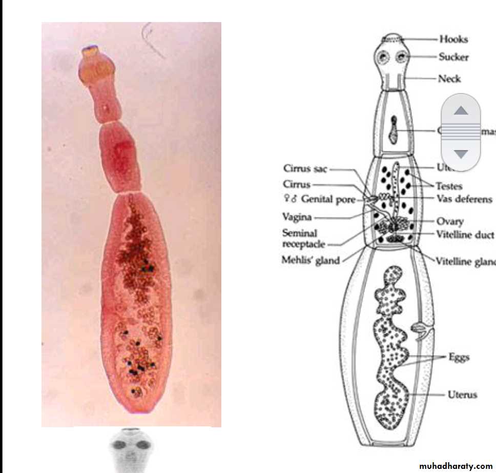
Morphology of adult worm
2-6 mm long.
The body consist of:
head (scolex) with 4 suckers and rostellum with two rows of hooks.
three segments (proglottids):Immature, mature and gravid.
The mature segment: contain fully developed male and female sexual organs.
The gravid segment: contain only uterus full with eggs.
Morphology of the egg (Taenia spp. Egg ).
Spherical, 35-45 um in diameter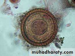
Hexacanth Embryo centrally located
Radially staited shellInfective stage to human, sheep & cattle .
The same egg of Echinococcus Spp., Taenia saginata & T. solium
Types of hydatid cysts : Unilocular, Osseous & Multilocular or alveolar.
Unilocular type:It is the larval stage of E. granulosus composed of single cavity. The majority of human H. cysts are unilocular type. It may reach 15 cm or more in diameter after 10 to 20 Years.
Morphology:
consist of three layers with central fluid contain protoscolices
The three layers :
1- Inner nucleated germinal layer .
2- non-nucleated laminated layer.
3- Outer (fibrous layer):This layer formed as a result of defense mechanism of the host against the parasite.
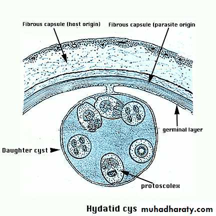
Layers of hydatid cyst
TransmissionIngestion of uncooked vegetables and fruits(eg: lettuce &parsley) & drinking water contaminated with eggs.
Handling soil or animal hair that containing eggs.
Diagnosis
Clinical manifestation: depend on the size and site of hydatid cyst. At the beginning the disease is asymptomatic. As the cyst continue to grow pressure effect usually results.
2. Imaging techniques:
X-ray picture: useful to detect the calcified cyst.
Ultrasound scan.
MRI & CT scan.
3. Serology tests : eg:
Indirect immunofluescence.
Enzyme-linked immunosorbent assay (ELISA).
Polymerase chain reaction (PCR).
Taenia saginata &Taenia solium
Taenia saginata Beef tapewormTaenia solium Pork tapeworm
Disease : Taeniasis.
Morphology of adult
1. They are flat , hermaphroditic worms , live in small intestine of man.
2. Adult worms consist of a head , neck and segmented body (proglottids) may reach thousands.
3. The head (scolex) contain suckers (T. saginata), or suckers & hooks (T. solium).
4. Mature segment contain both male & female sexual organs.
5. Gravid segment contain only uterus full with eggs which usually passed with eggs out with feces as diagnostic stages.
6. The length of adult worms is usually 4-10m for T. saginata and 2 - 7 meter for T. solium.
7 . T. saginata usually have 1,000 to 2,000 segments,
while T. solium have an average of 1,000 segments.
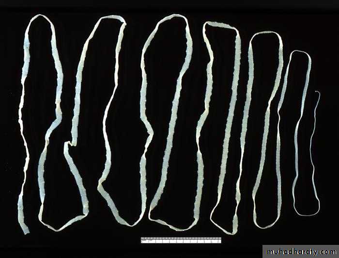
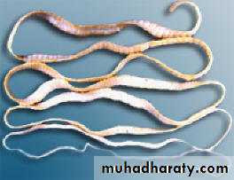
Adult worm of Taenia saginata Adult worm of Taenia solium
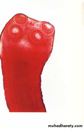
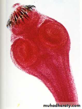
Scolex of T. saginata Scolex of T. Solium
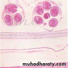
Cysticercoids larva:
It is bladder like cyst called bladder worm with invagenated or evagenated scolex.They are infective stages to human and they are two type :
1. C. Bovis: scolex without hooks present in the muscles of cattles.
2. C.cellulosae : scolex with hookspresent in the muscles of pigs


Cysticercus bovis Cysticercus cellulosae
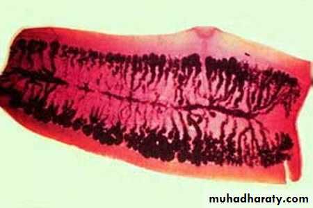
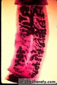
Gravid segment of T.Saginata Gravid segment of T.Solium
Gravid segment of T.Saginata:
gravid segments ;longer than wide consist of median uterus with 15-30 lateral uterine branches with unilateral genital pore .
Gravid segment of T.Solium :
7 – 13 lateral uterine branches.
Transmission
Human infection with Taenia spp occur by eating under cooked beef or pork containing cysticercoids larva.Laboratory Diagnosis
Microscopic identification of scolex and segments in feces are diagnostic for taeniasis.
The gravid segments can differtiate between T.saginata and T. solium but the eggs are morphologically identical, and indistinguishable between taenia species and from eggs of E.granulosus .