


Fracture maxilla
طب اسنان \ خامس
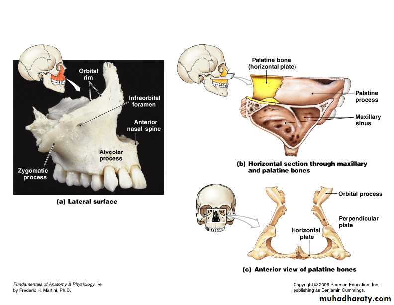
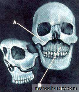
جراحة فم \ د. وفاء م(11)The Maxillary Bones
The largest facial bonesFunctions of the Maxillary Bones
Support upper teeth
Form upper jaw and hard palate
Contain maxillary sinuses (largest sinuses)
The facial skeleton can be divided into 3 parts
The upper part (forehead). The frontal bone.The lower third (mandible).
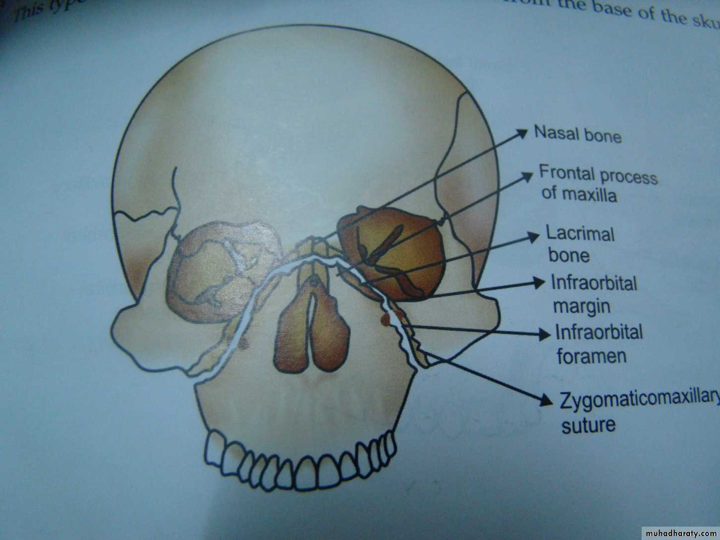
The middle 3rd ( the midface) that extend below the frontal bone .The middle 3rd consist of the following bones : maxilla, zygoma, zygomatic process of the temporal bone, nasal bones, lacrimal bones, vomer, ethmoid bone, inferior conchae, palatine bones, and the pterygoid plate of sphenoid.
Radiographic evaluation
Interpretation of occipitomental view:
McGrigor and Campbell 5 lines as followed :
1- The first line runs across zygomatico frontal sutures, supra orbital margin and the frontal sinus.
2- The 2nd line runs across the zygomatic arches, the infra orbital margin and the nasal bone.
3- The 3rd line passes along the condyle , coronoid process and the maxillary sinus.
4- the 4th line runs across the ramus and the occlusal plane.
5- the 5th line runs along the inferior mandibular border.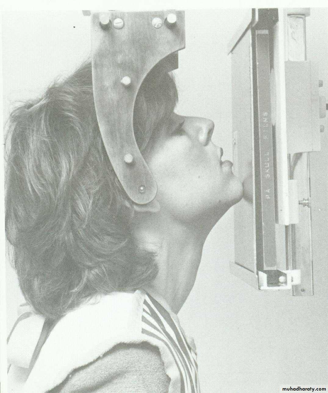
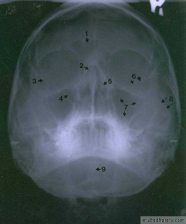
1. Frontal sinus
2. Ethmoid air cells3. Innominate line
4. Superior orbital fissure
5. Sphenoid sinus
6. Lesser wing of sphenoid
7. Maxillary sinus
8. Zygomatic arch
9. Dens (odontoid process) of axis
Rowe and Williams classification
A- fracture not involving the occlusion:
1- central region: nasal bone, frontal process of the maxilla, ethmoid bone and frontal bone.
2- lateral region : fracture involving the zygomatic bone , arch , and maxilla excluding the dento-alveolar component.
B- fracture involving the occlusion.
1- dento alveolar.2- subzygomatic : lefort I and lefort II.
3- suprazygomatic : lefort III or high level or craniofacial dysjunction.
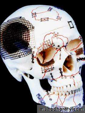
Lefort Classification
Weakest areas of midfacial complex when assaulted from a frontal direction at different levels (Rene’ Lefort, 1901)Lefort I: above the level of teeth(Guerin's fracture).
Lefort II: at level of nasal bones ( pyramidal) or subzygomatic fracture.
Lefort III: at orbital level (Craniofacial Dysjunction)
Lefort IV fracture of the frontal bone.
Differentiating Leforts
Pull forward on maxillary teethLefort I: Maxilla only moves
Lefort II: Maxilla & base of nose moves
Lefort III: Whole face moves
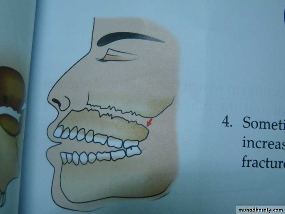
Maxillary FracturesLeFort I
Definition:
Horizontal fracture of the maxilla at the level of the nasal fossa.
Allows motion of the maxilla while the nasal bridge remains stable.
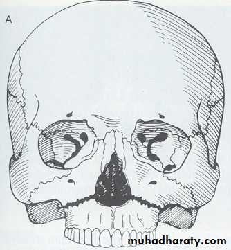
It separates the palate and tooth bearing segments , bilaterally from the midface).
It run through the lower 3rd of nasal septum , then through the lateral wall of the nose or the anterior nasal aperture , below the zygomatic buttress , across the lower third of pterygoid plate.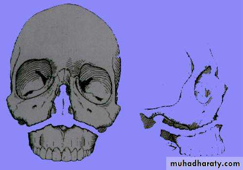
Lefort I FractureTransverse Maxillary
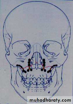
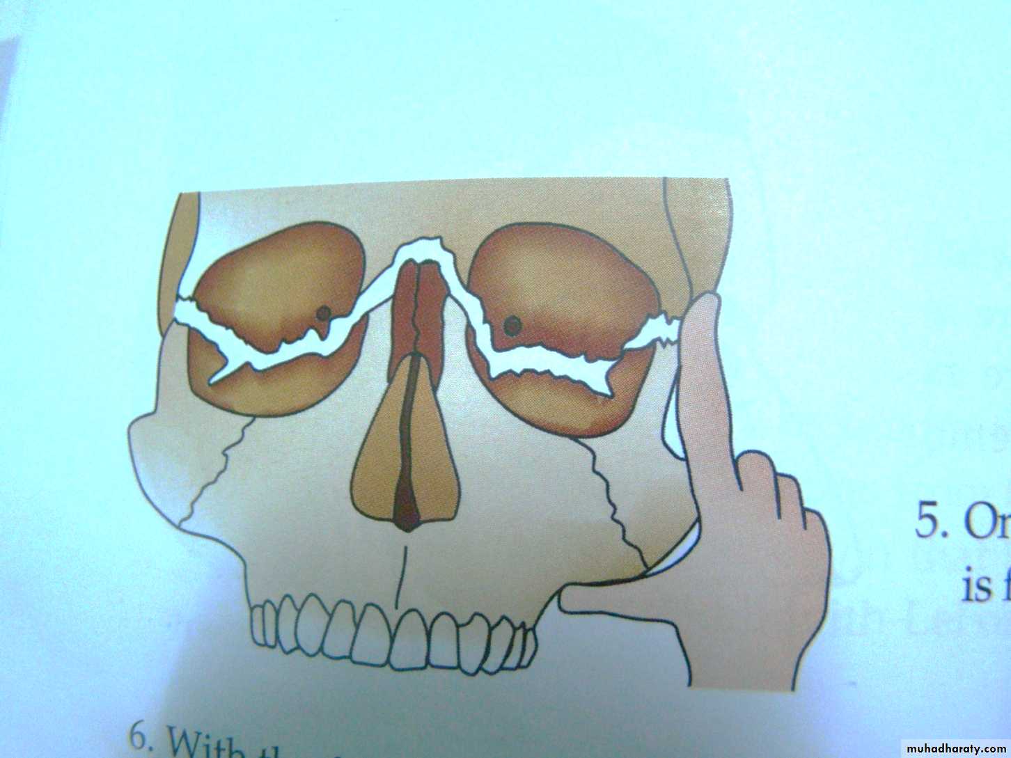
Lefort II FracturePyramidal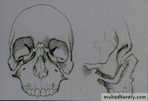
Le Fort II (also known as "pyramidal" fracture
separates the whole maxilla with part of the nasal bones and the lower part of the pterygoid platesThe fracture line runs below the frontonasal suture down on either side, crossing the frontal process of the maxillae and passes anteriorly across the lacrimal bones, immediately anterior to nasolacrimal canal. From this point the fracture line passes downward, forward and laterally crossing the inferior orbital margin.The fracture line
now extends downward and forward and laterally to traverse the lateral wall of the antrum, then the fracture line passes beneath the zygomatic buttress, traversing the pterygo maxillary fissure at a higher level and fracturing the pterygoid plate approximately midway from its base.
Difintion of Lefort II fracture
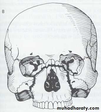
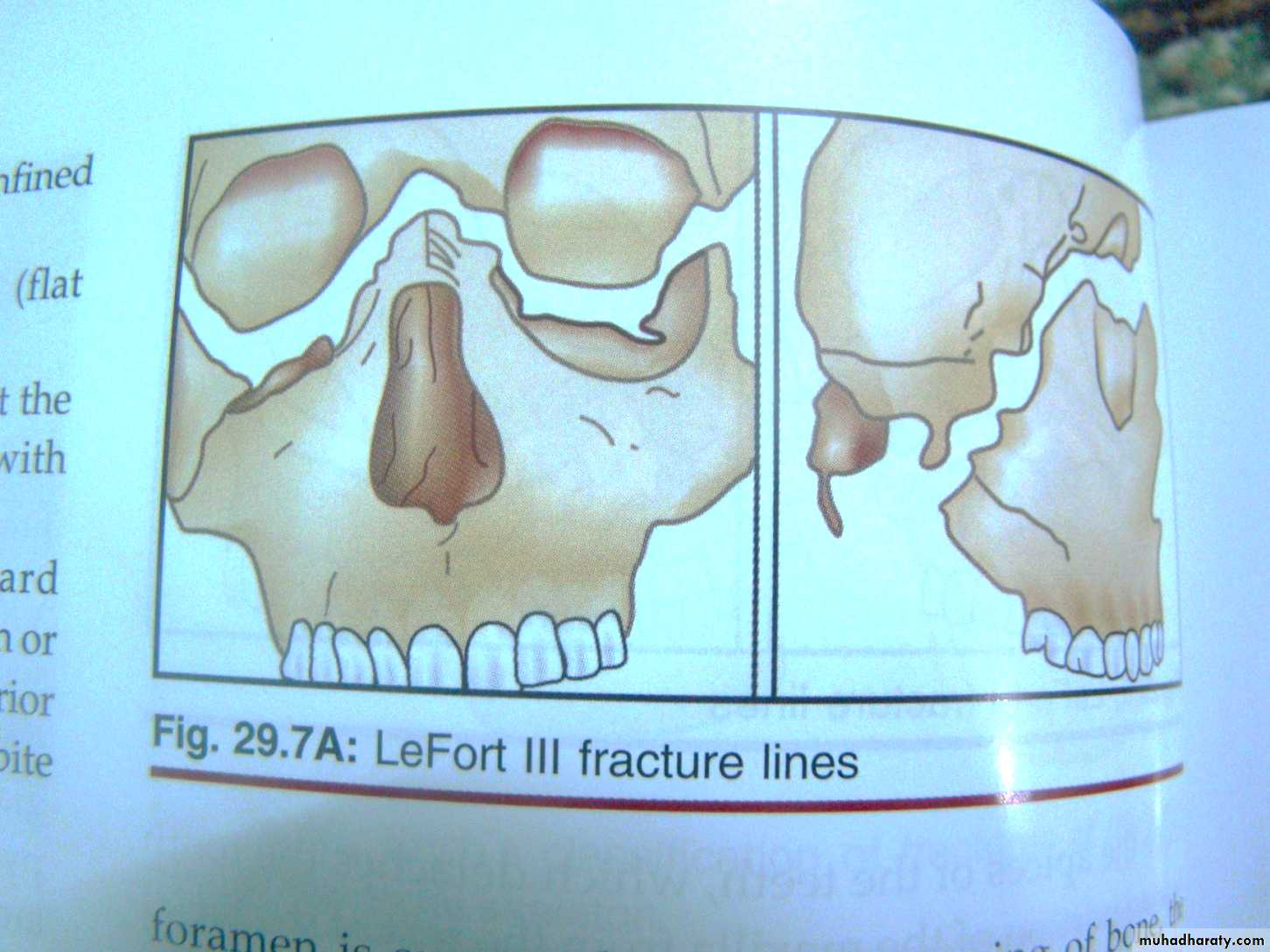
Lefort III FractureCraniofacial Dysjunction
Le Fort III separates both zygomatico-maxillary complexes plus the nasal bones, palatal bones and most of the pterygoid plates, from the rest of the cranium.
Radiographic Evaluation
• Plain Films• Lateral Skull
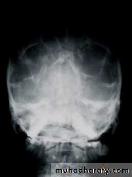
• Waters View
• Posteroanterior view of skull
• Submental vertex
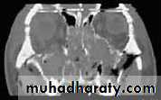
• CT Scan
• 1.5 mm cuts
• axial and coronal views
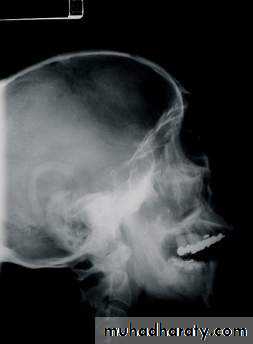
Lateral skull
Water’s View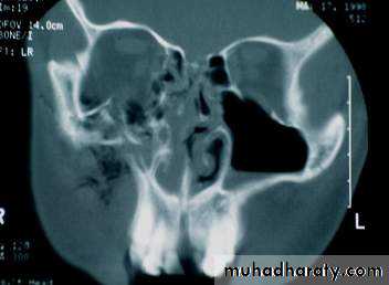
CT Scan
Maxillary FracturesLeFort I
Clinical findings:
Facial edema.
Ecchymosis in the buccal sulcus.
Disturbed occlusion of the teeth
Motion of the maxilla while the nasal bridge remains stable. - Anterior open bite due to the fractured segment having dropped down.
Percussion of upper teeth will give a typical “cracked- pot” sound.
• Radiographic findings:
• Fracture line which involves• Nasal aperture
• Inferior maxilla
• Lateral wall of maxilla
• CT of the face and head
• coronal cuts
• 3-D reconstruction
Treatment of Lefort I Fractures
• Direct exposure of all involved fractures• Reduction and anatomic realignment of the maxillary buttresses to reestablish
• Anterior projection
• Transverse width
• Occlusion
• Restoration of occlusion using IMF
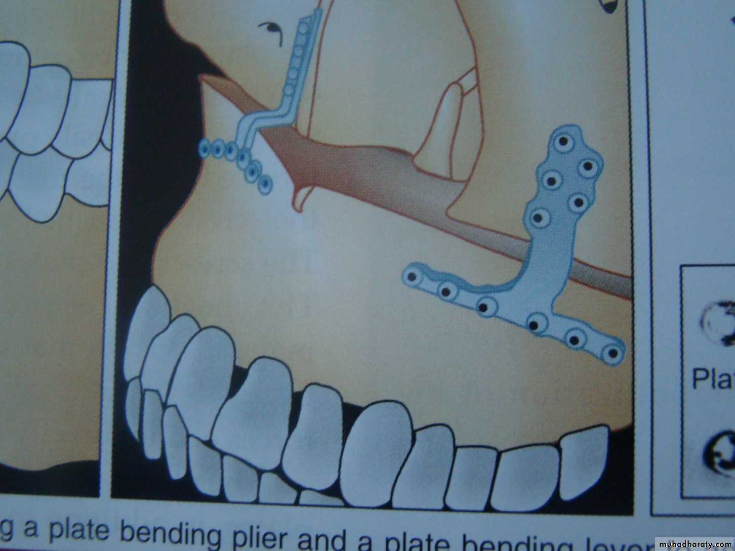
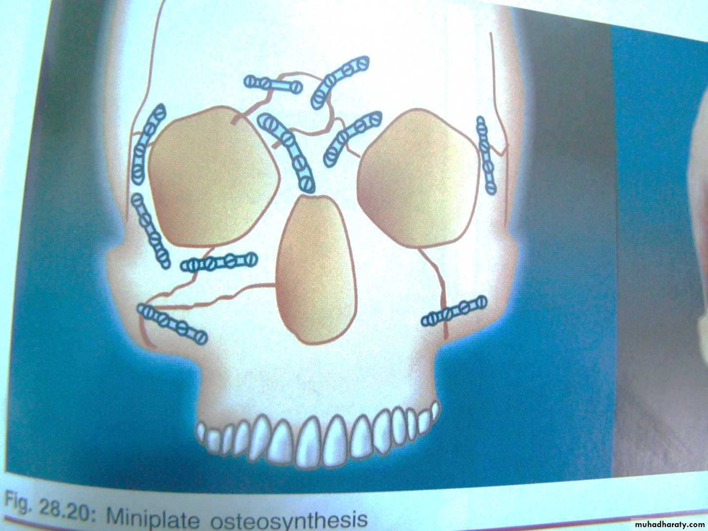
• Internal fixation using miniplate fixation
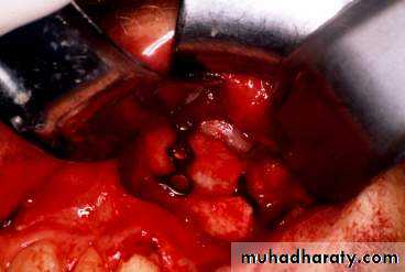
Treatment : reduction
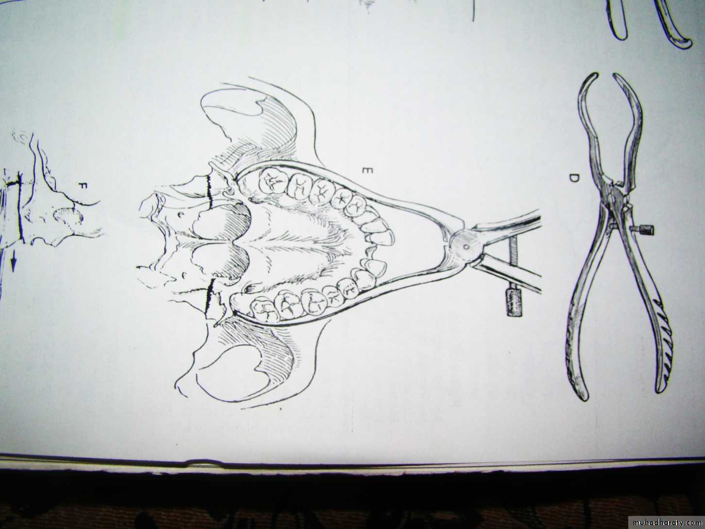
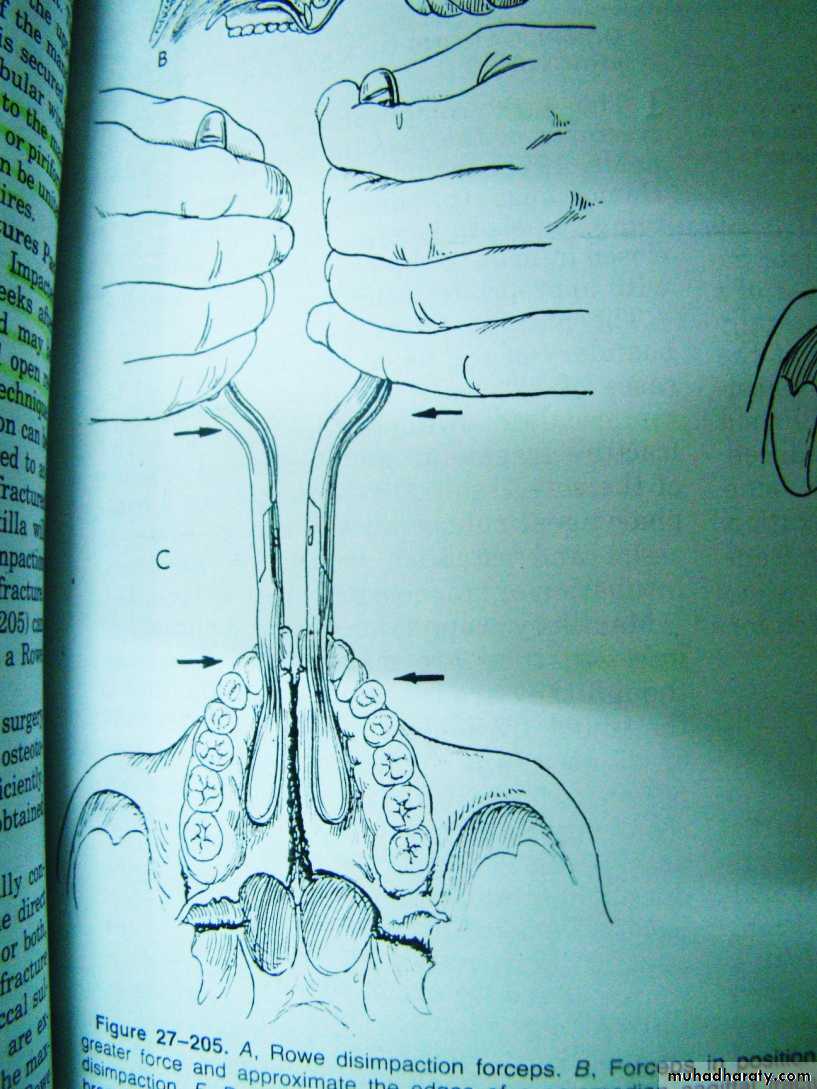
Reduction of maxillary fractures: (1) Rowe’s maxillary disimpaction forceps, (2) Hayton William’s disimpaction forcep
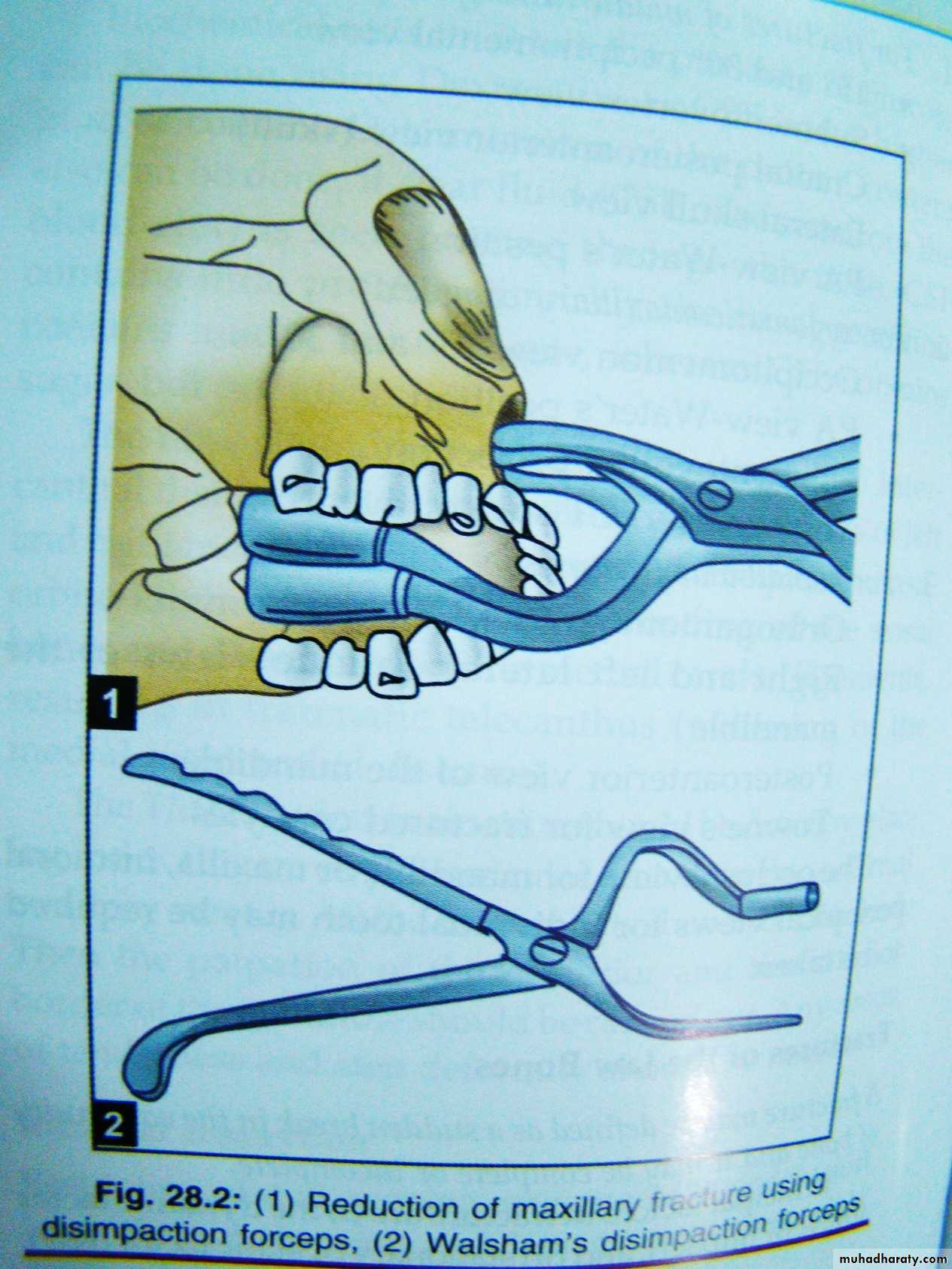
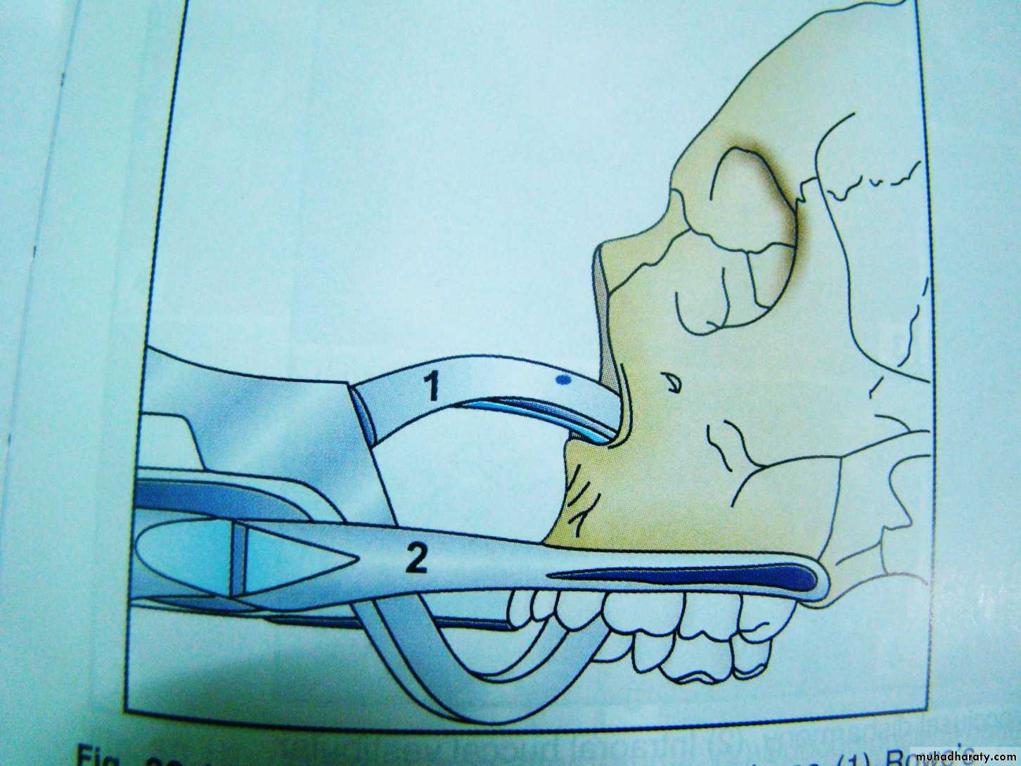
Reduction of maxilla using rowe disimpaction forceps and walsham’s disimpaction forceps
Treatment of Lefort I Fractures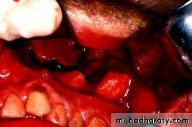
fixation of Lefort I fracture
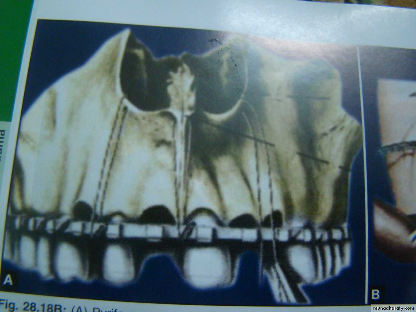
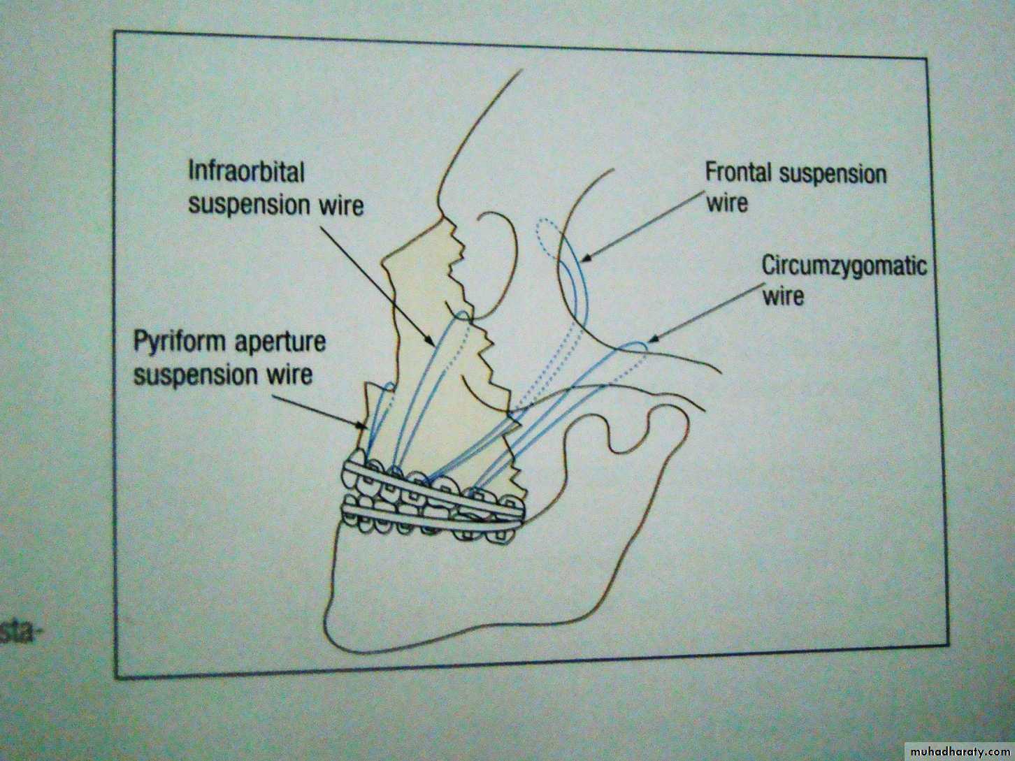
1- plating : through an intra oral approach.2- suspension.
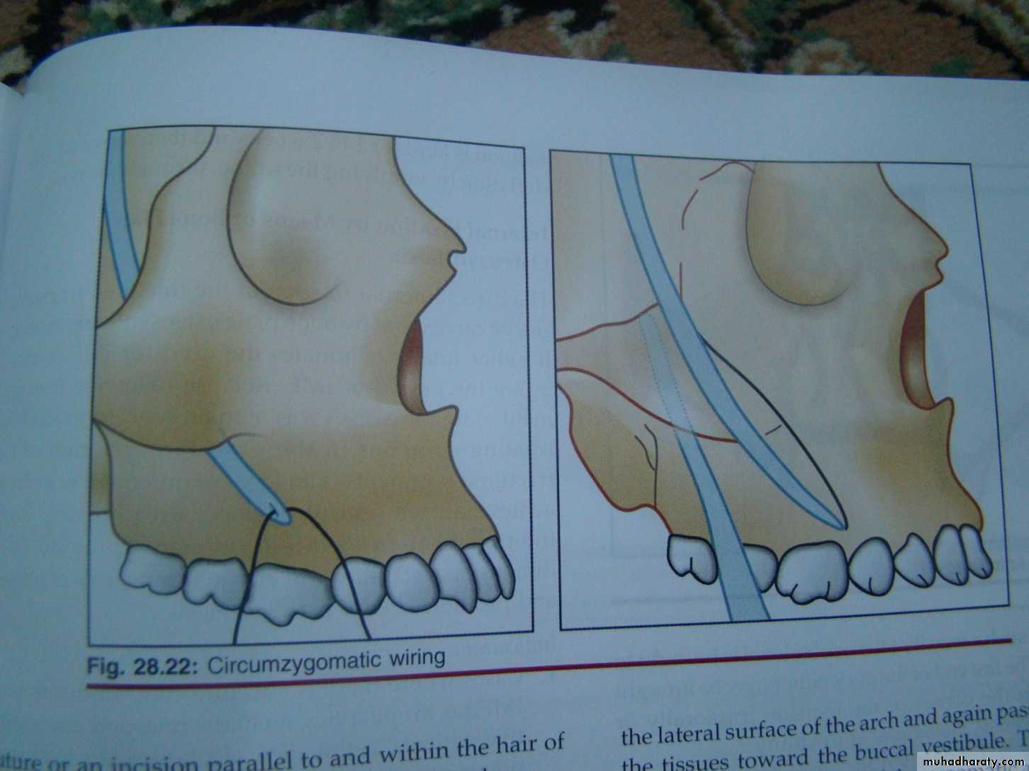
i- circum zygomatic suspension .
ii – pyriform aperture suspension.
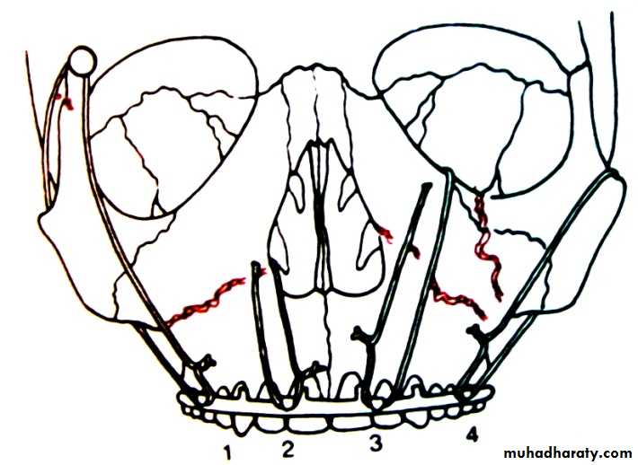
• 3- Intra osseous wiring.
Maxillary FracturesLefort II( pyramidal) or subzygomatic fracture.Definition:
Is a Pyramidal fracture that involve
Maxilla
Nasal bones
Medial aspect of the orbits( lacrimal and ethmoid bone).
Maxillary FracturesLeFort II
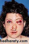
Clinical findings:1-Marked facial edema: moon face appearance.
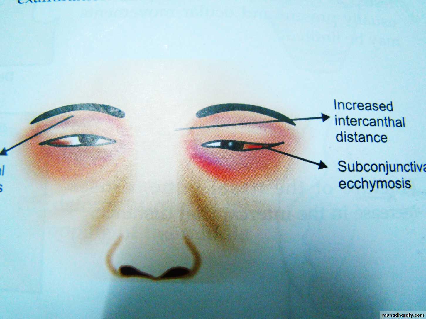
2- Nasal flattening (fracture of the nasal bone) causing Traumatic telecanthus
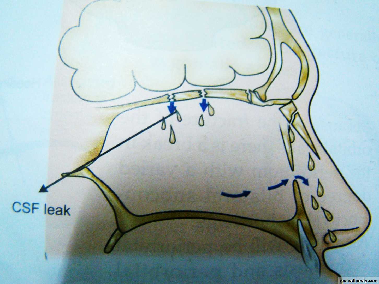
3- Epistaxis or CSF rhinorrhea due to fracture of cribriform plate of ethmoid bone.
4- Movement of the upper jaw and the nose.
5- Nasal bleeding.
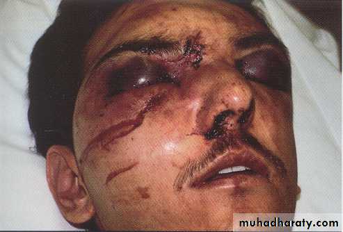
6- Bilateral circum orbital ecchymosis.
7- Subconjuctival ecchymosis8- Enophthalmos.
9-Diplopia is usually present and ocular movements may be limited.
10- Anesthesia of the infra orbital region.
Maxillary FracturesLeFort II
Radiographic imaging:Fracture involves:
Nasal bones
Medial orbit
Maxillary sinus
Frontal process of the maxilla
CT of the face and head
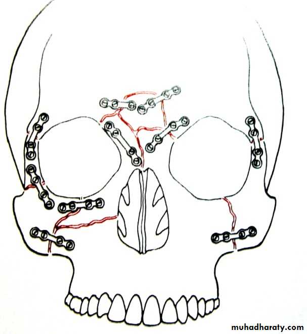
Treatment of Lefort II fracture
1- Wiring ( infraorbitally)2-Plating ( at the zygomatic buttress).
3-Orbital plate reconstruction
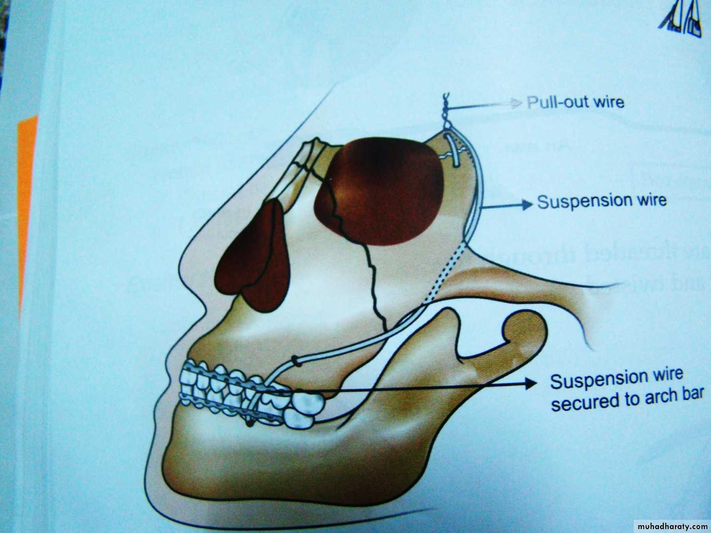
4-Internal suspension ( circumzygomatic wiring and infra orbital suspension.
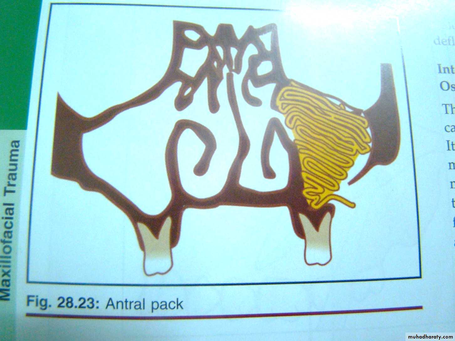
5- Antral pack through a cald-well luck surgery. pack for 2 weeks.
Maxillary FracturesLeFort III
Definition:
Fractures through:
Maxilla
Zygoma
Nasal bones
Ethmoid bones
Base of the skull
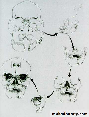
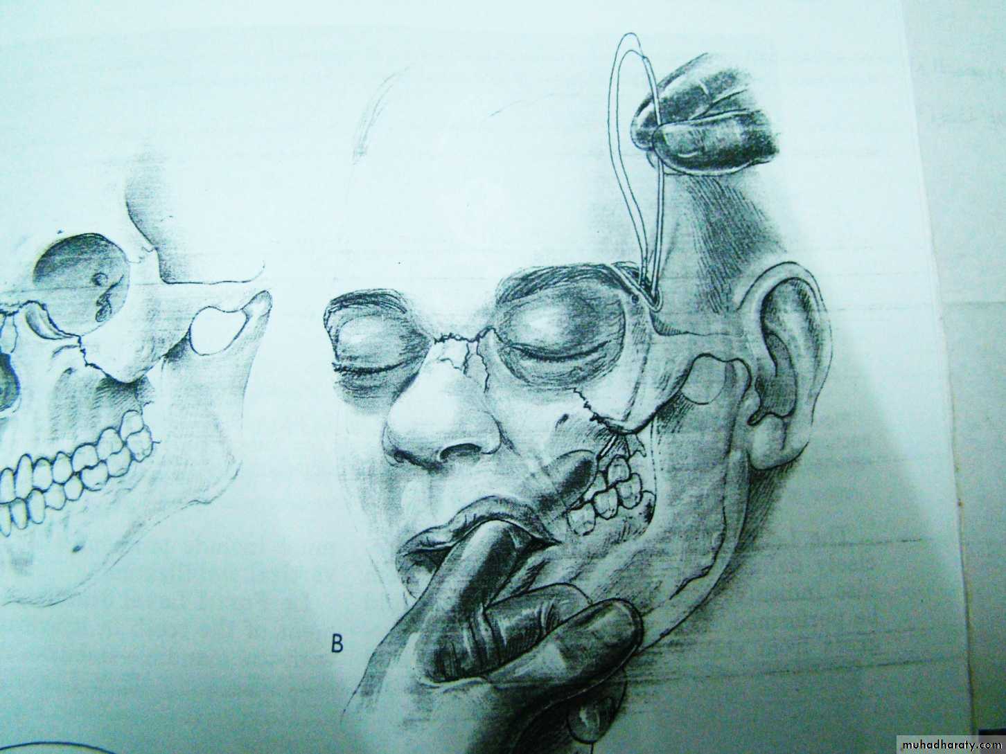
Craniofacial dysjunction may occur when the fracture extends through the zygomatico frontal suture , nasofrontal suture and across the floor of the orbits to effect complete separation of the midfacial structures from the cranium. In these fracture the maxilla may not be separated from the Zygoma or from nasal structures. The entire midfacial skeleton is completely detached from the base of the skull and suspended only by soft tissues.
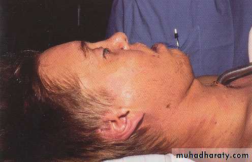
Lefort III fracture
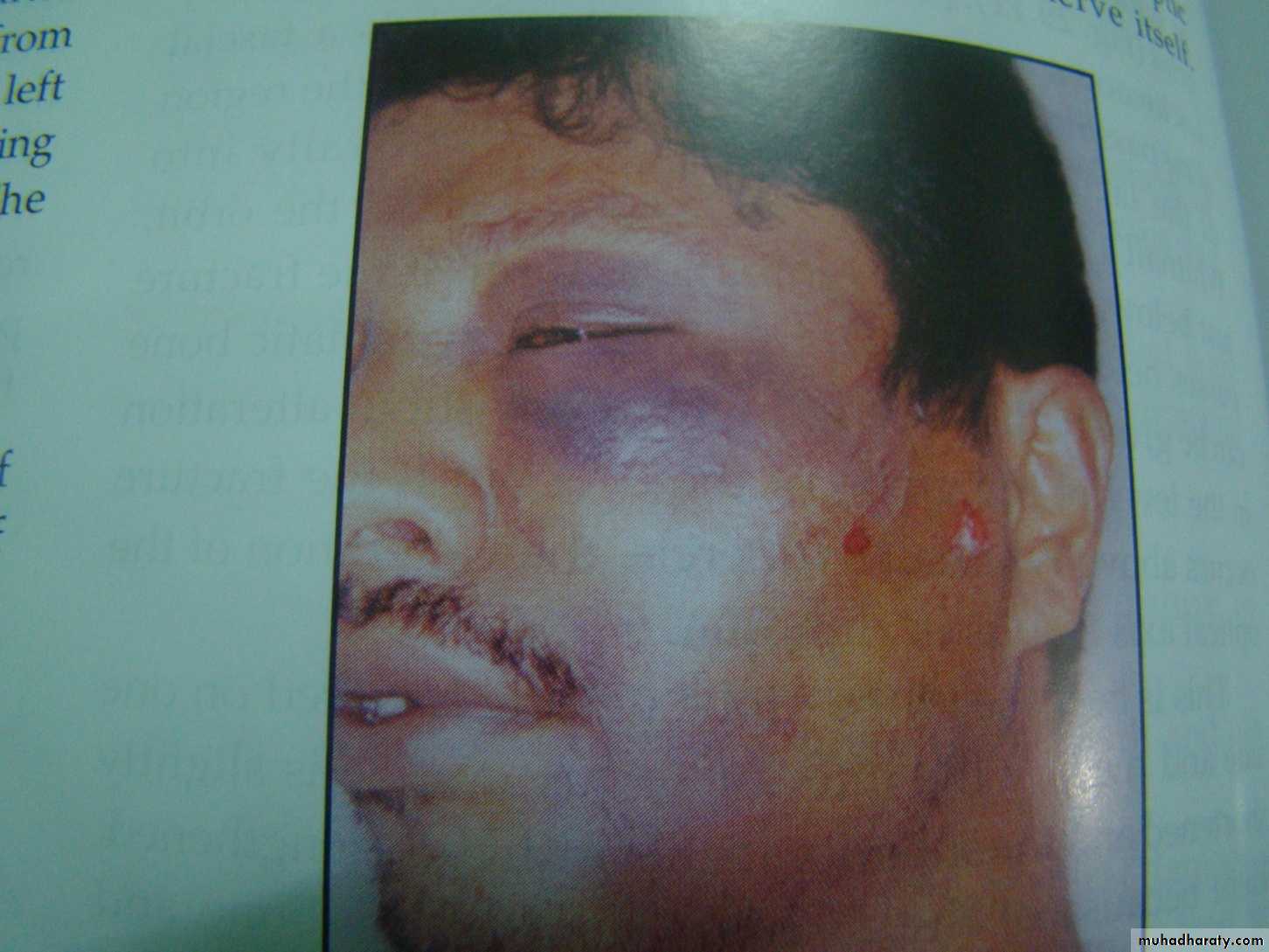
Clinical findings:
1- Dish faced deformity2- Epistaxis and CSF rhinorrhea
3- Motion of the maxilla, nasal bones and zygoma
4- Severe airway obstruction
5- Facial edema.
Maxillary FracturesLeFort III
6- Nasal flattening .
7- Telecanthus( increase the intercanthal distance).
8- Fracture ethmoid cause CSF rhinorrhea.
9- Downwards movement of the face, resulting in the lengthening of the face.
10- Hooding of one or both eyes .
11- Bilateral subconjuctival hemorrhage.
12- Circum orbital ecchymosis
13- Enophthalmos.
14- Diplopia.
Treatment of Lefort II and III
Fractures should be treated as early as the general condition of the patient allowsTeam approach to treatment
Neurosurgery
Ophthamology
ENT
Plastic surgery
Oral/Maxillofacial surgery
Treatment of Lefort II and III
Intubation must not interfere with ability to use IMF
Exposure & visualization of all fractures
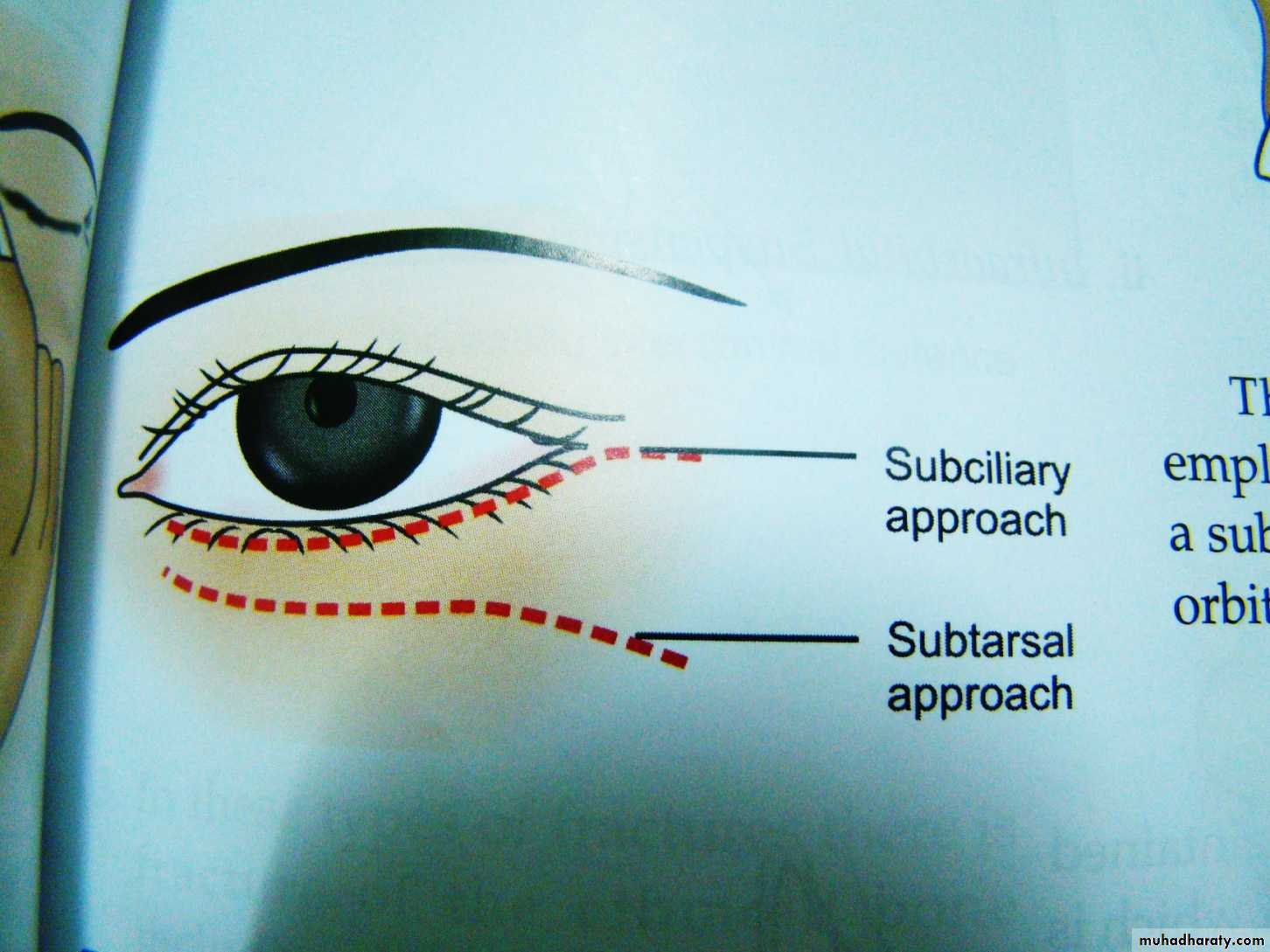
Approaches to inferior rim
Infraorbital
Subciliary
Transconjunctival
Mid lower lid
Coronal approach
Gingivobuccal incision
Teeth and occlusion are the key to reconstruction and provide the foundation upon which other facial structures are built
• Fractures
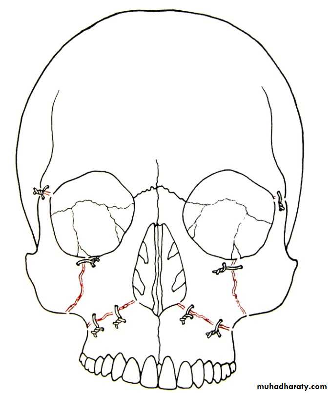
1-internal fixation: by transosseous wiring or miniplates at fracture sites like fronto zygomatic and fronto nasal.
2- frontal suspension.
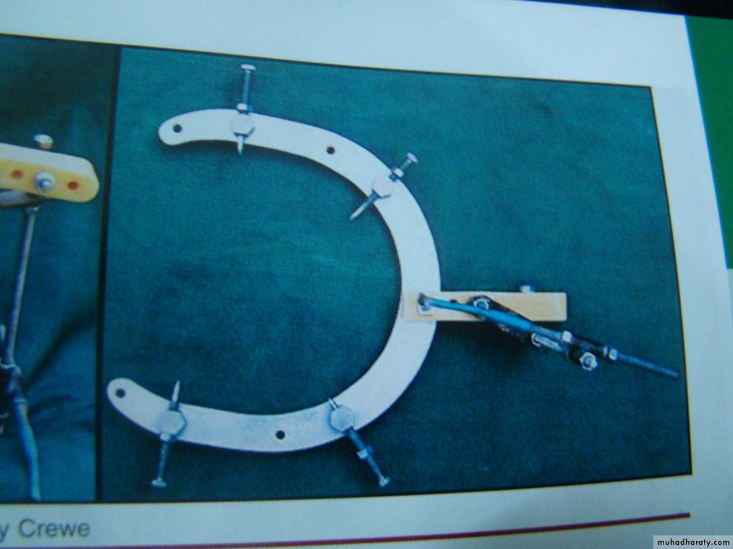
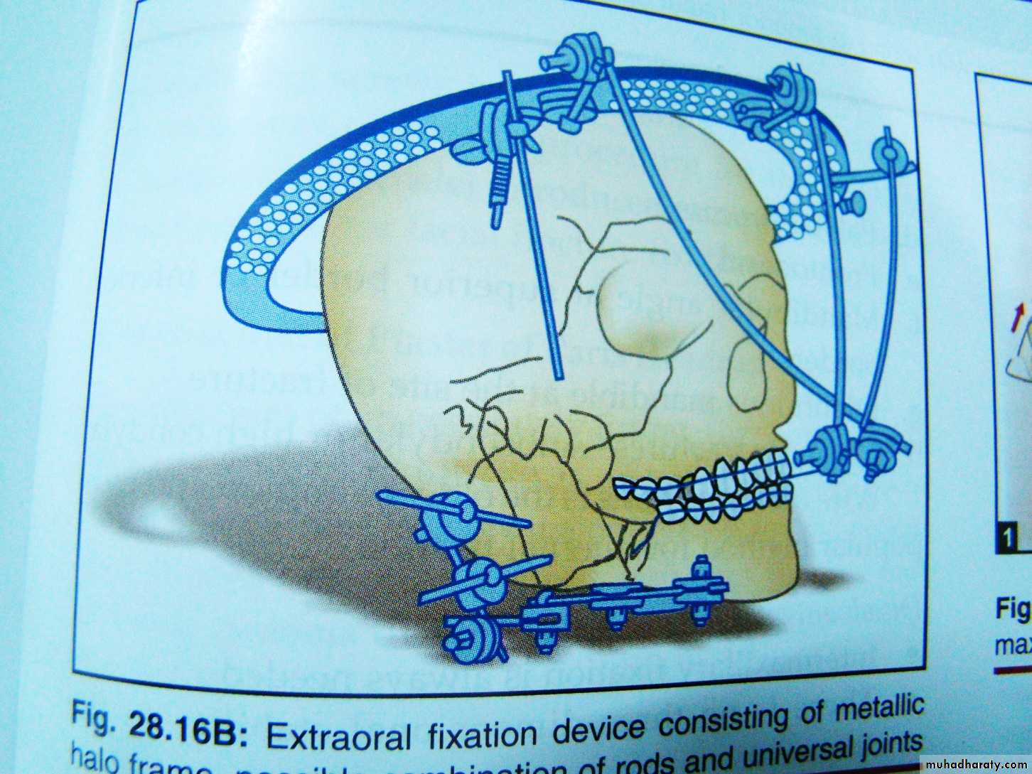
3- External fixation :
a- cranio mandibular like Box – frame, Halo frame , and plaster of Paris head cap.
b- craniomaxillary like supra orbital pins, zygomatic pins, and Halo frame .
Treatment of Lefort III
Complications of maxillary fractures:
Early complications:1- extensive hemorrhage.
2-Compromised airway.
3- infection. 4- CSF rhinorrhea.
5-bilateral blindness is a rare complication.
Late complications : 1- non union 2- malunion 3- plate exposure.4- lacrimal system obstruction 5- infra orbital anesthesia 6- devitalization of teeth 6- diplopia and enophthalmos.