Physical properties of dentine
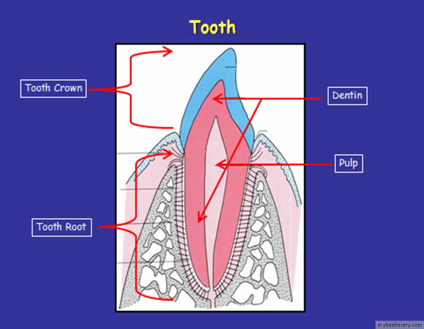
Dentin forms the bulk of the tooth. It’s covered by enamel in crown and by cementum in root.A vascular
Cellular (contains odontoblast processes )Dentin is yellowish in color it becomes darker by age.
Dentin is elastic tissue which allows the impact of mastication to occur without fracturing brittle overlying enamel. This resilience is due in part to the presence of tubules which extend from dentioenamel junction to the pulp.Physical properties of dentine
Dentin is softer than the enamel but harder than the bone and cementum.Radiographically dentin appears more radiolucent than enamel and much more radio-opaque than pulp.
Permeability :
Existence of the cytoplasmic processes across the hole length of the dentin make the dentin permeable providing a bath way for the invasion of caries , also drugs and chemicals present in a variety of dental restorative materials can also diffuse through the dentin and create a pulpal injury .
Chemical composition of dentin:-
It consists of 70% inorganic, which represented by hydroxyapatite crystals {Ca₃ (Po₄)₂, Ca (OH)}, the crystals of dentin are much smaller than those in enamel.20% organic material
10% water (by weight).Histological features of dentin
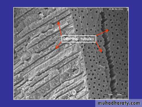
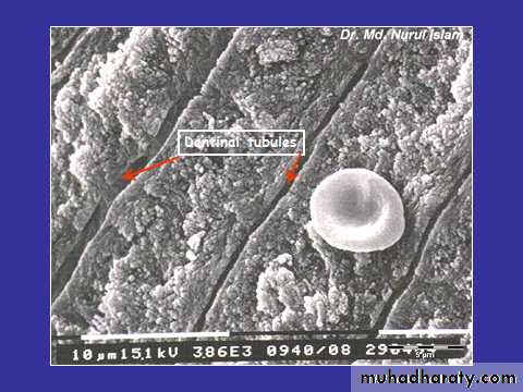
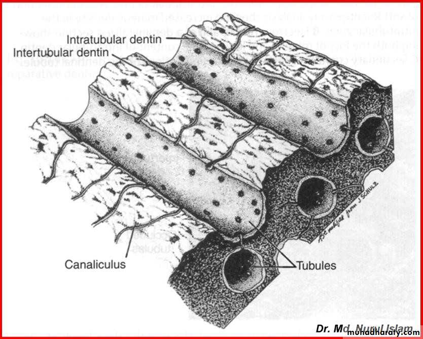
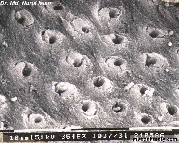
The dentin composed mainly from:-• Dentin tubules.
• Peritubular dentin.(Intra-tubular Dentin)• Intertubular dentin.
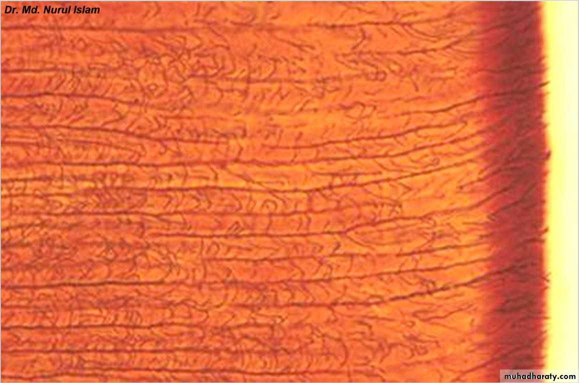
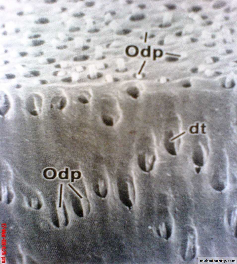
Dentinal tubule
• Long tube ( fine canals )• Running from DEJ to the pulp
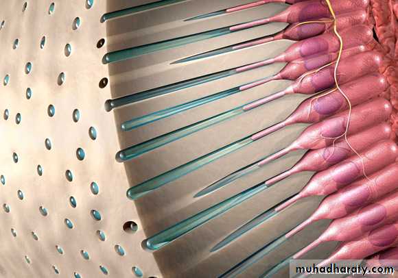
• Filled with an odontoblastic process
• As a living tissue, it contains within its tubules the process of specialized cells (odontoblasts ). The odontoblastic processes are Cytoplasmic extensions of odontoblasts , have numerous side branches that exist in the canaliculi or microtubules (lateral branches of the dentinal tubules)
Dentinal
tubules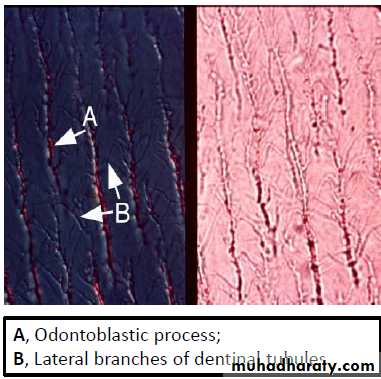
odontoblastic processes

odontoblastic processes
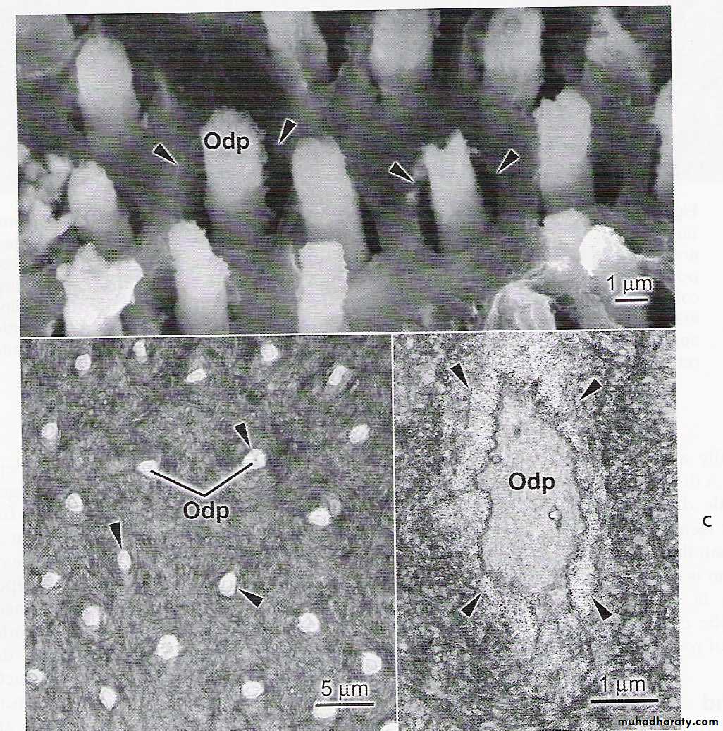
DENTINAL TUBULES AND ODONTOBLASTS

Peritubular dentin
• An area which immediately surrounds dentinal tubules• creates the wall of the dentinal tubule.
higher crystalline content
Intertubular dentin
• Bulk of the dentin material (It forms the most of the body of dentin)
• Located between the zones of Peritubular dentine
Dentinal tubules
Peritubular dentinIntertubular dentin
peritubular / intratubular dentine dentin that forms the wall of each tubulemore mineral than intertubular dentin
intertubular dentine
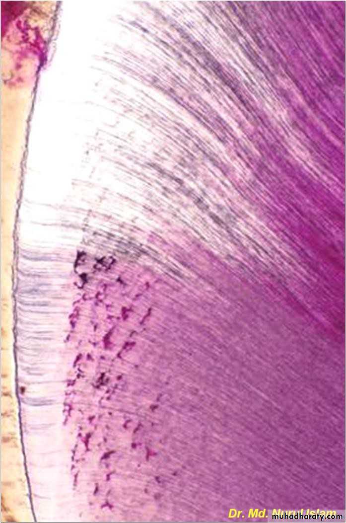
dentine between the peritubular dentinGranular layer of Tomes
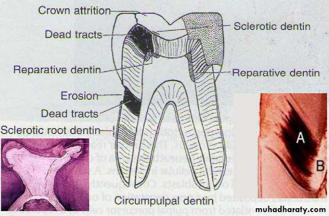
• It’s black granular zone In the roots near the cemento-dentinal junction• hypo-mineralized areas of dentin around the dentinal tubule called the Tomes granular layer
• Caused by a coalescing and looping of terminal portion of dentinal tubules
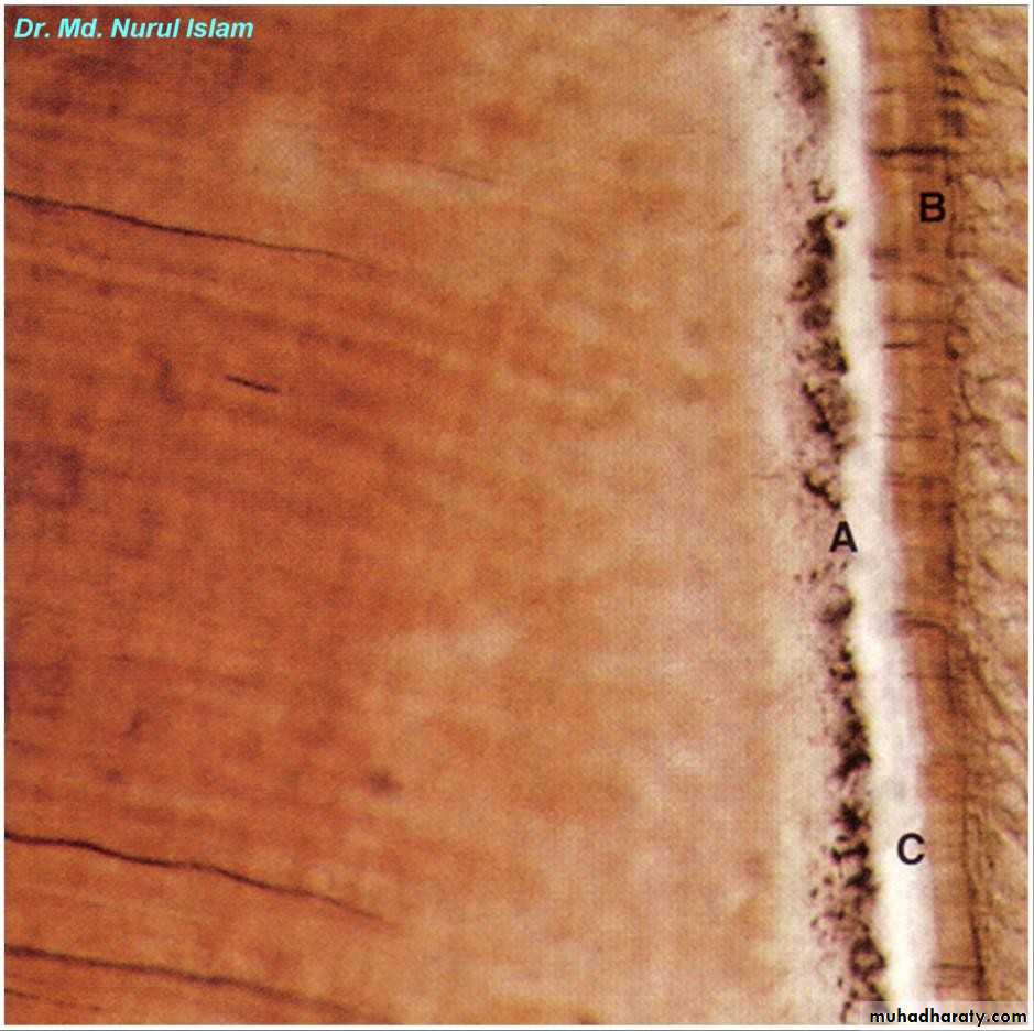
Dentin
CementumGranular layer
of Tomes
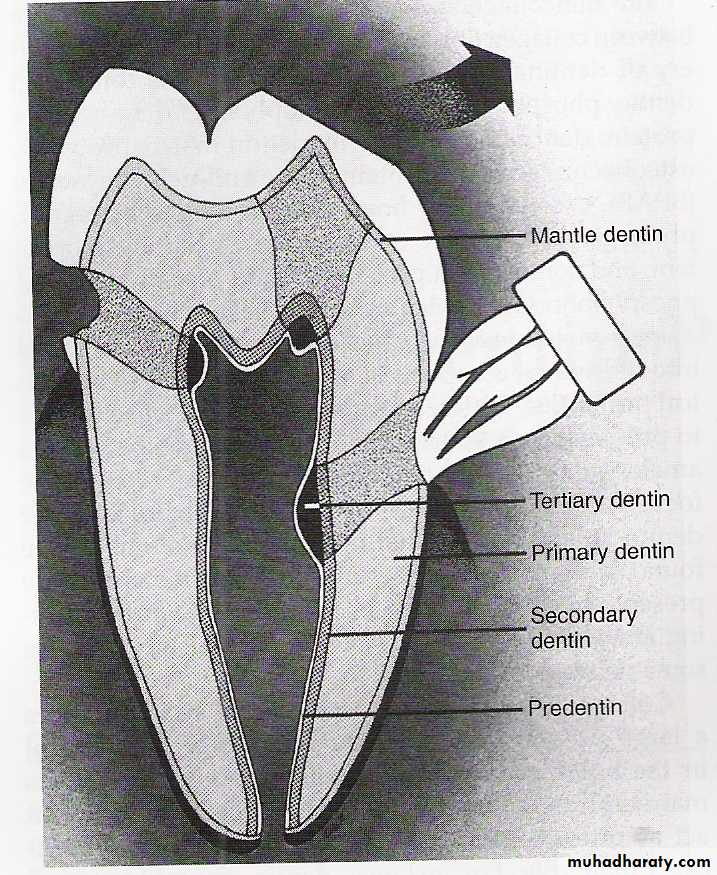
Predentin
• Predentin is the first deposited layer of un-mineralized matrix of dentin (immature uncalcified dentin consisting chiefly of fibrils )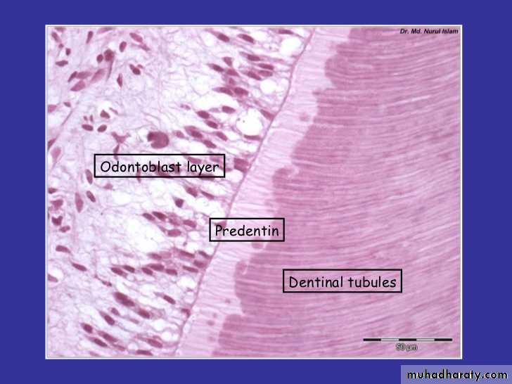
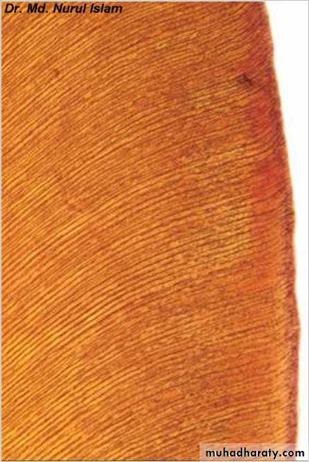
Incremental growth line
They are fine lines correspond to daily rhythmic pattern of growth.The distance between lines varies from 4:8um in crown& much less in the root . They are called VON EBNER lines.
Occasionally, some of them are accentuated because of disturbance in the matrix& mineralization. Such lines seen in ground section& known as CONTOUR LINES OF OWEN which represent hypocalcified bands.

VON EBNER lines
CONTOUR LINES OF OWEN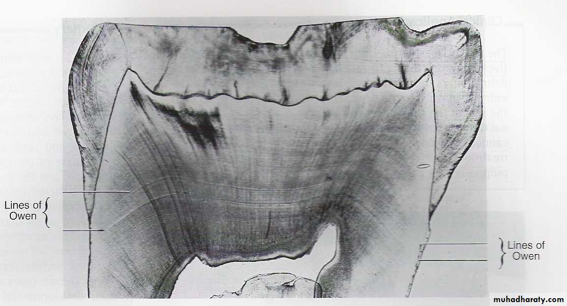
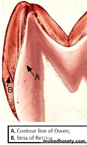
VON EBNER lines
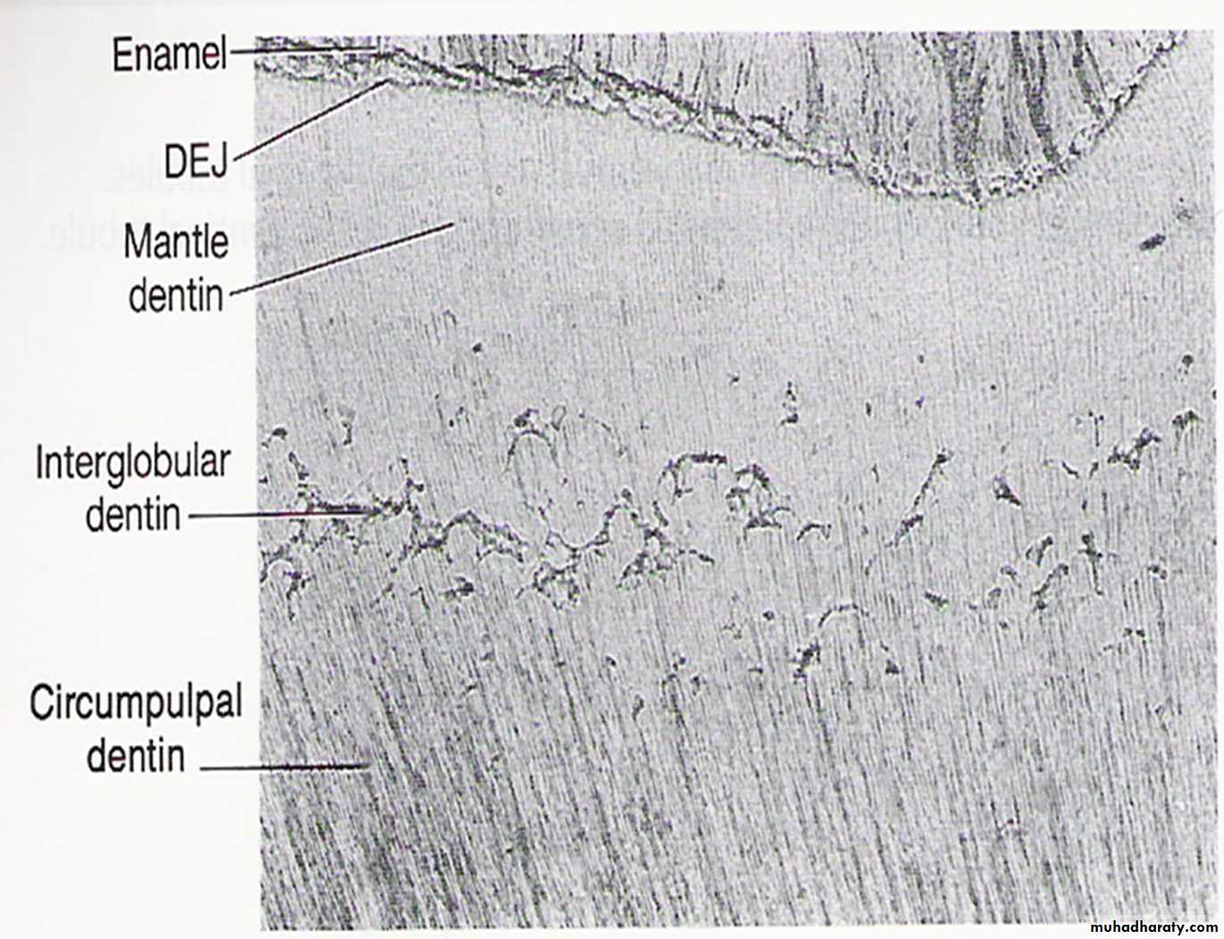
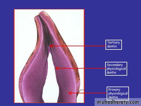
Primary dentinThe most prominent dentin in the tooth, lies between the enamel and the pulp chamber. Primary dentin include :
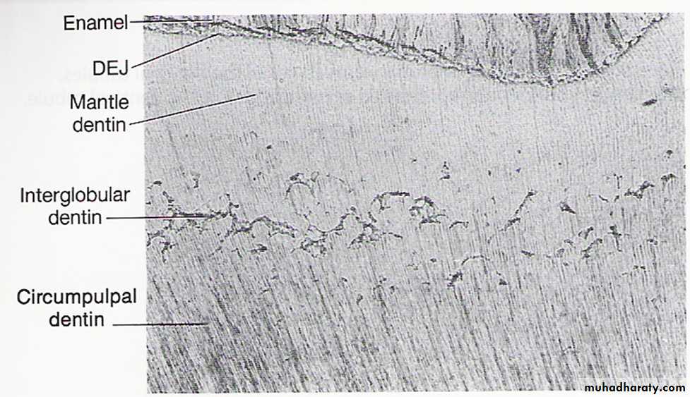
a. Mantle dentin : is the name of the first layer of dentin formed under D.E,J. in the crown . It is about 20um thick.
b. Circumpulpal dentin : forming the remaining part of primary dentin .
formed before the complete formation of the root
Slightly contain more mineral than mantle dentine
Secondary dentin
Narrow bent of dentin bordering the pulpit is separated by darkly stained line from the primary dentin.
Represent the dentin formed after root completion
Contains fewer tubules than dentine
It grows much slower than primary dentin
Considered as it protects pulp from exposure in older teeth
The growth of this type of dentin causes the decrease in the size of the pulp chamber with age
Tertiary dentin
Tertiary dentin is formed in reaction to stimulus, such as attrition ,caries or restorative dental procedureTertiary dentin can be reactionary or reparative
Reactionary dentin is that type of tertiary dentin that is deposited by the pre-existing odontoblasts
Reparative dentin is deposited by newly differentiated odontoblasts
Localized formation of dentine on the pulp dentin border
Rate of deposition depends on the degree of injury.
This dentin characterized by sparse and twisted tubules and possible cell inclusions.
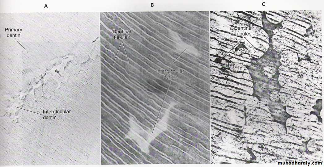
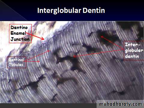
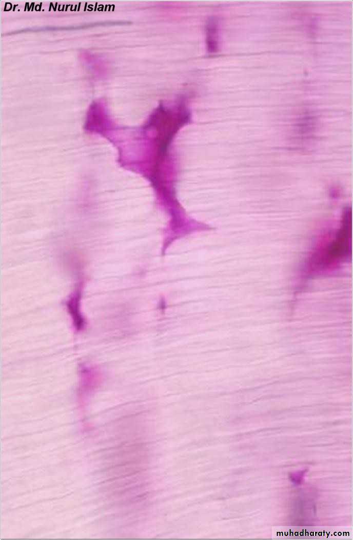
Abnormalities of dentin formationInter globular dentin
• Separating the mantle dentin and circumpulpal dentin ,It is hypo-mineralized• Seen when mineralization of dentin fails to coalesce into a homogenous mass in the globular dentin (hyper mineralized layer).
• Dentin gets entrapped
• Found next to DEJ in crown
Inter-
globulerdentin
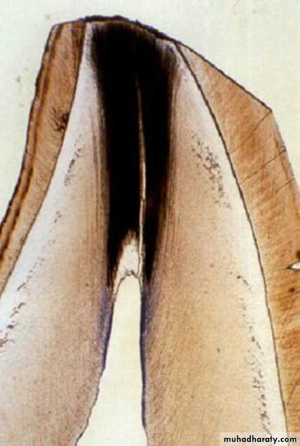
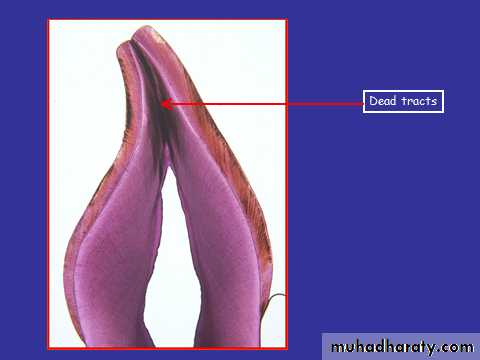
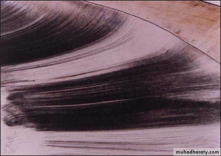
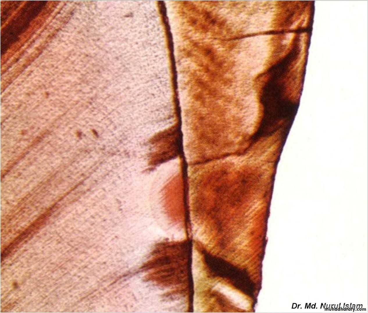
Dead tracks
• Empty dentinal tubules due to death of odontoblastic cells• Empty tubule give rise to pathway for bacteria in decay
• More rapid penetration of decay once it had reached the DEJ
• They appear black in transmitted and white in reflected light
• Their degeneration is often observed in the area of narrow pulpal horns because of crowding of odontoblasts
Sclerotic dentin (transparent dentin
• Seen in cases of caries attrition abrasion erosion or cavity preparation, sufficient stimuli are generated leading to the appearance of collagen fibers and appetite crystals in the dentinal tubules instead of the Odontoblastic processes which be degenerated and the tubules of degenerated odontoblasts are filled by collagen fibers and appetite crystals as a defensive mechanism of dentin• Seen mostly in older individuals
Sclerotic dentin
Types of Dentin
DentinPrimary physiologic
dentin
Secondary physiologic
dentin
Tertiary dentin or
reparative dentin or
reactionary dentin or
irregular secondary dentin
Mantle
dentin
Circumpulpal
dentin
Peritubular
dentin
Intertubular
dentin