THE RETICULAR FORMATION AND DIFFUSE
MODULATORY SYSTEMS(II) Neurohormonal Control of Brain Activity
Aside from direct control of brain activity by transmission of nerve signals from the lower brain areas to the cortical regions of the brain, still another physiologic mechanism is very often used to control brain activity by secreting excitatory or inhibitory neurotransmitter agents into the substance of the brain. There are four major diffuse modulatory systems:
1.The raphe nuclei and the serotonin system,
located in the pons and medulla
its neurons secrete serotonin.
it send fibers into the diencephalon, cerebral cortex, spinal cord. The serotonin secreted at the spinal cord has the ability to suppress pain. The serotonin released in the diencephalon and cerebrum plays an essential role in the control of wakefulness and sleep-wake cycles and to the control of mood and emotions.
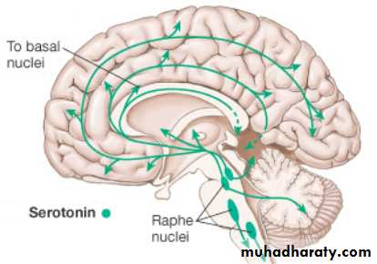
Figure; The raphe nuclei and the serotonin system.
2. The noradrenergic neuron systemoriginates in the locus coeruleus of the pons.
it functions as an “alarm center” that becomes most active when new environmental stimuli appear.
Noradrenergic neurons are widely distributed throughout the CNS and mediate an increase in the general state of arousal.
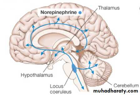
Figure; The noradrenergic neuron system
3.The substantia nigra and the dopamine system.The substantia nigra lies in the superior mesencephalon.
its neurons pass mainly to the caudate nucleus and putamen.
Other neurons pass to ventral areas of the brain, especially to the hypothalamus and the limbic system.
The dopamine is believed to act as an inhibitory transmitter in the basal ganglia, but in other areas of the brain it is possibly excitatory. Dopaminergic neurons play an important role in the brain’s reward system. The reward system is short circuited by addictive drugs, including cocaine, amphetamines, alcohol, nicotine, and opiates, which all increase dopaminergic transmission in the nucleus accumbens.
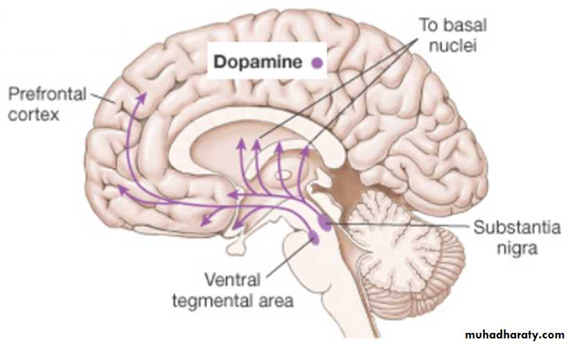
Figure; Dopaminergic neurons system.
4. The gigantocellular neurons of the reticular excitatory area and the acetylcholine system.Fibers from gigantocellular neurons (the giant cells) in the reticular excitatory area of the pons and mesencephalon divide immediately into two branches, one passing upward to the higher levels of the brain and the other passing downward into the spinal cord. The neurohormone secreted at their terminals is acetylcholine. In most places, the acetylcholine functions as an excitatory neurotransmitter. Activation of these acetylcholine neurons leads to an acutely awake and excited nervous system. Cholinergic neurons in the nucleus basalis of Meynert, a forebrain structure, are involved in the pathogenesis of Alzheimer’s disease.
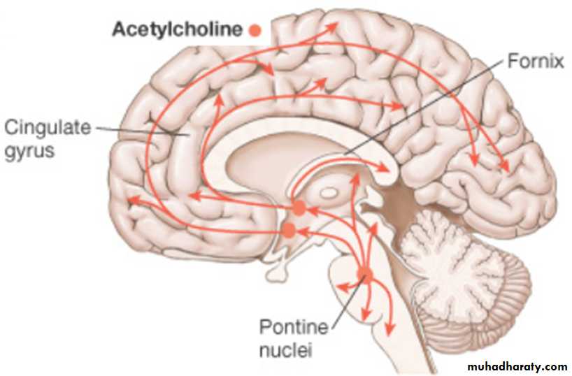
Figure; acetylcholine system..
principal causes of unconsciousness orThe neurons in the reticular formation has an important influence on the degree of wakefulness. Consciousness requires proper functioning of the reticular activating system and both cerebral hemispheres. Therefore, the principal causes of unconsciousness or coma are:
1. Lesions that damage the reticular activating system.
2. Diffuse damage to both cerebral hemispheres.
3. Global suppression of the cerebrum and/or reticular activating
system by drugs (e.g., sedative-hypnotics or alcohol) or metabolic derangements (e.g., anoxia, hypercapnia, hypoglycemia, or hepatic encephalopathy).
Mood disorders, the main two affective (mood) disorders are
1.major depression2. bipolar disorder, bipolar disorder is expressed by periods of depression alternating with bouts of mania. Drugs that enhance the diffuse serotonergic and noradrenergic modulatory systems are among the most effective treatments available for mood disorders. For example, the monoamine oxidase inhibitors (MAOIs) reduce the rate of norepinephrine and serotonin breakdown, which elevates the levels of these transmitters in the brain. More recently, serotonin-selective reuptake inhibitors (SSRIs) such as fluoxetine (e.g., Prozac) have been developed. The effectiveness of these drugs supports the monoamine hypothesis that low activity of serotonin and norepinephrine is the basis of mood disorders.
Schizophrenia. is a group of psychotic disorders characterized by a loss of the normal perceptions of reality evidenced by delusions, hallucinations, and disordered speech and behavior. Dopamine antagonists are effective when used as antipsychotic (neuroleptic) drugs due to the inhibition of dopamine signaling in the mesocorticolimbic system. The antipsychotic actions of dopamine antagonists are mediated by blocking D2-receptors. The use of nonselective dopamine antagonists inhibits the nigrostriatal dopaminergic system, resulting in adverse effects that resemble Parkinson’s disease.
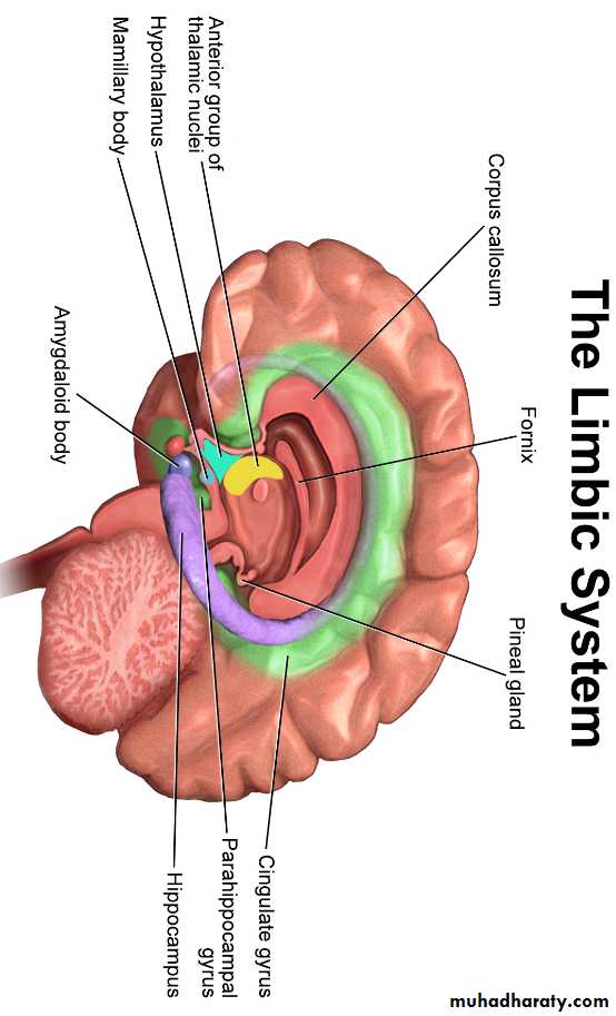
The limbic system
The concept of a "limbic system" actually encompasses two levels of structural and functional organization. The first level consists of the cortical structures on the most medial edge (the limbus) of the hemisphere:
the subcallosal area.
the cingulate gyrus and the isthmus of the cingulate gyrus.
the parahippocampal gyrus; and the uncus.
the hippocampal formation.
The second level includes a variety of subcortical nuclei and tracts
the septal nuclei and nucleus accumbens
various nuclei of the hypothalamus
the nuclei of the amygdaloid complex
parts of the dorsal thalamus,
habenular nuclei, ventral tegmental area, and periaqueductal gray.
The functions of limbic system include:,
emotion, behavior, motivation,
long-term memory ( specially in hippocampus).
Emotional component of learning( specially in amygdale).
olfaction.
Associated with pain/pleasure, rage
Emotional life is largely housed in the limbic system, and it has a great deal to do with the formation of memories.
Reward” and “Punishment” Function of the Limbic System
The limbic structures are concerned with the affective nature of sensory sensations – that is, whether the sensations are pleasant or unpleasant or also called reward or punishment or satisfaction or aversion:I. “Reward” and “Punishment” Function of the Limbic System
1.the major reward centers have been found to be located along the course of the medial forebrain bundle, especially in the lateral and ventromedial nuclei of the hypothalamus. Less potent reward centers, are found in the septum, the amygdale. Stimulation of these areas gives a sense of reward. When offered the choice of eating some delectable food, the animal often chooses the electrical stimulation.
2.Punishment Centers
The most potent areas for punishment and escape tendencies have been
found in the central gray area surrounding the aqueduct of Sylvius. Less potent punishment areas are found in the amygdala and hippocampus
Stimulation in these areas causes the animal to show all the signs of displeasure, fear, terror, pain, punishment, and even sickness. Strong stimulation of the punishment centers, causes the animal to (1) develop a defense posture, (2) extend its claws, (3) lift its tail, (4) hiss, (5) spit, (6) growl, and (7) develop piloerection, wide-open eyes, and dilated pupils. Furthermore, even the slightest provocation causes an immediate savage attack. This pattern of behavior that is called rage. Fortunately, in the normal animal, the rage phenomenon is held in check mainly by inhibitory signals from the ventromedial nuclei of the hypothalamus.
Importance of Reward or Punishment in Behavior
Almost everything that we do is related in some way to reward and punishment. If we are doing something that is rewarding, we continue to do it; if it is punishing, we cease to do it. Therefore, the reward and punishment centers undoubtedly constitute one of the most important of all the controllers of our bodily activities, our drives, our aversions, our motivations. Tranquilizers , such as chlorpromazine, usually inhibits both the reward and the punishment centers, thereby decreasing the affective reactivity of the animal.Importance of Reward or Punishment in Learning and Memory –Habituation Versus Reinforcement
If the sensory experience does not elicit a sense of either reward or punishment, repetition of the stimulus over and over leads to almost complete extinction of the cerebral cortical response, thus the animal becomes habituated to that specific sensory stimulus and thereafter ignores it.
If the stimulus does cause either reward or punishment , the cerebral cortical response becomes progressively more and more intense during repeated stimulation and the response is said to be reinforced
Functions of the Hippocampus
The hippocampus presumably has critical decision-making neuronal mechanism, determining the importance of the incoming sensory signals. It has been suggested that the hippocampus provides the drive that causes translation of short-term memory(verbal and symbolic thinking type) into long-term memory (consolidation).Thus removal of a portions of the hippocampi as treatment for epilepsy, lead to anterograde amnesia. These people can recall most previously learned memories satisfactorily. They are capable of short-term memory for seconds up to a minute or two, but their ability to establish memories lasting longer than a few minutes is either completely or almost completely abolished.
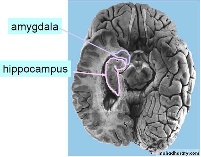
Functions of the Amygdala
The amygdala is a complex of multiple small nuclei located immediately beneath the cerebral cortex of the medial anterior pole of each temporal lobe. The amygdala receives neuronal signals from all portions of the limbic cortex, as well as from the neocortex of the temporal, parietal, and occipital lobes—especially from the auditory and visual association areas. Because of these multiple connections, the amygdala has beencalled the “window” through which the limbic system sees the place of in the world. Stimulating the Amygdala had effect on
(1)arterial pressure, heart rate,
(2) gastrointestinal motility and secretion
(3)pupillary dilation or, rarely, constriction,
(4) piloerection,
(5) secretion of various anterior pituitary hormones, especially the gonadotropins and adrenocorticotropic hormone.
(6) different types of movements associated with olfaction and eating, such as licking, chewing, and swallowing.
(7).Finally, excitation of still other portions of the amygdale can cause sexual activities that include erection, copulatory movements, ejaculation, ovulation, uterine activity, and premature labor.
Effects of Bilateral Ablation of the Amygdala—The Klüver-Bucy
Syndrome.Klüver-Bucy syndrome is a rare neurologic disorder that occurs as a result of bilateral lesions of the amygdale (e.g., herpes encephalitis). Patients exhibit hypersexuality (lack of social sexual restraint), hyperorality (strong compulsion to place objects in the mouth), visual agnosia (able to see objects, but unable to identify them), flat affect, and placidity (able to approach danger without anger or fear).