Respiratory System
Dr. Shatha Th.AhmadPhD mol.Path/UK
Aim of studying pathology of Respiratory system
1.To know the types of lesions affecting this system2. To study the gross & microscopical features of these lesions
3. To correlate the signs & symptoms
Anatomy
Respiratory tract consist of:Nose,nasopharynx,larynx,trachea,right & left bronchi.
The bronchi lead to respiratory lobule or acinus
Respiratory Lobule or Acinus
Consist ofterminal bronchiole leading to respiratory bronchioles and alveolar ducts
Alveoli arise from both respiratory bronchioles and alveolar duct
Respiratory Acinus
Upper & lower respiratory tract
The respiratory tract are roughly divided in toUpper respiratory tract : Above cricoid cartilage
Lower respiratory tract : Below cricoid cartilage
Histology
The nose, nasopharynx, bronchi are lined by pseudo stratified ciliated epithelium &contain goblet cells and neuroendocrine cells.True vocal cord are lined by squamous epithelium. Submucosa contain mucus glands.
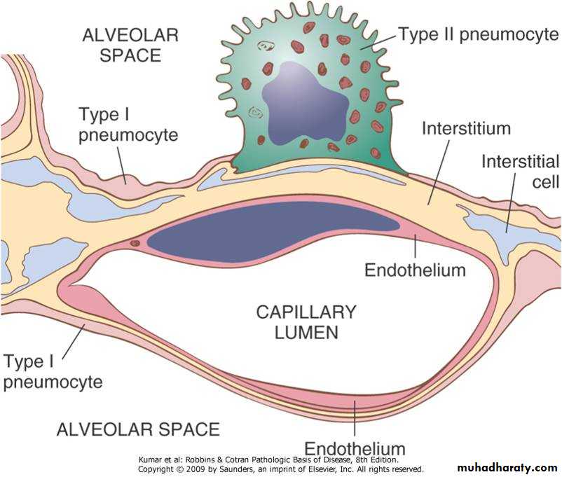
The alveoli are lined by:
Type I pneumocytes: Flattened cells
Type II pneumocytes :Rounded. It is the sours
of pulmonary surfactant & repair of type I pneumocytes
The wall of Alveolus
Infections of upper respiratory tract
Include:Viral: common cold
Bacterial : staph, streptococci etc
Fungal : Aspergillosis
Acute Rhinitis
Acute inflammation of the nasal mucosaAetiology:
• Viral e.g. common cold caused by rhinoviruses, influenza, para influenza
• Allergic: e.g. atopic rhinitis due to
• type 1 hypersensitivity reaction
• Manifestation of systemic disease: e.g. measle

Chronic Granulomatous Rhinitis
Etiology:Tuberculosis
Fungal
Leprosy
Syphilis
Wegenar granulomatosis

Tumors of Nose Nasopharynx & Sinuses
Benign:Hemangioma
squamous cell papilloma
Transitional cell papilloma & inverted papilloma:They tend to recur , difficult to eradicate & liable for malignant changes.
Angio fibroma
Malignant:
Squamous cell carcinoma
Adeno carcinoma
Nasopharyngeal carcinoma
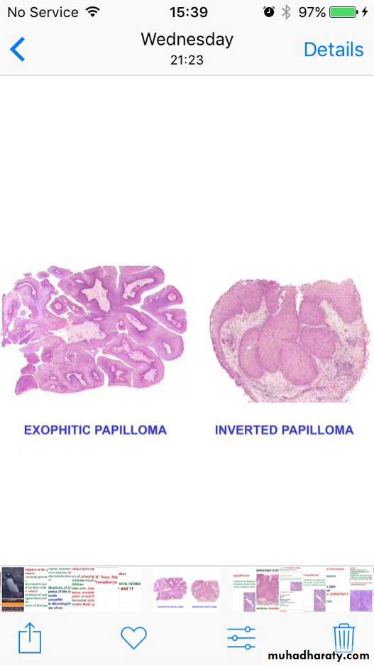

Inverted papilloma

Angiofibroma
Angiofibroma is a benign vascular tumor of the nasopharynx occur exclusively in young adult male.It is a benign tumor but grow rapidly may erodes bone and bleed profusely
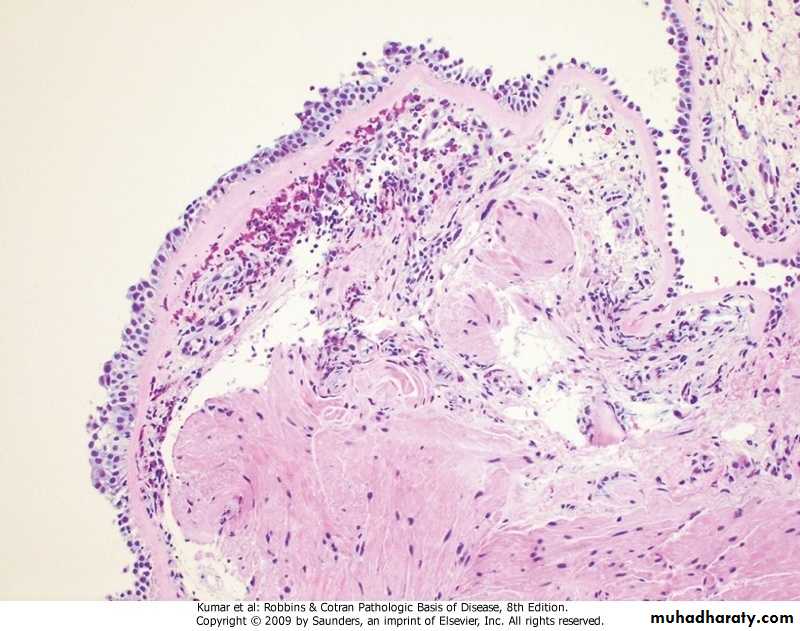
Nasal polyp
Polypoid projection from nasal mucosa , gelatinous in consistency with smooth surface. Usually bilateral (cf with neoplasm)It consist of edematous nasal mucosa contain, blood vessels, mucus glands & infiltrated by chronic inflammatory cells & eosinophils.
It result from allergic & inflammatory reaction
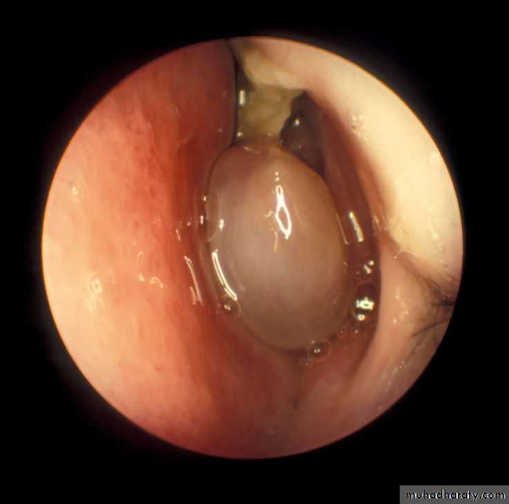
Microscopic picture of nasal polyp
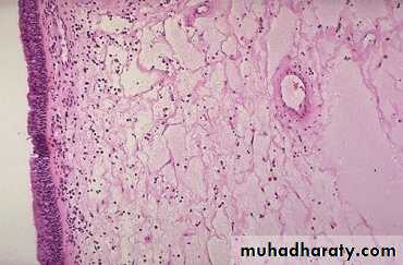
Nasal polyp
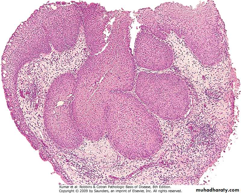
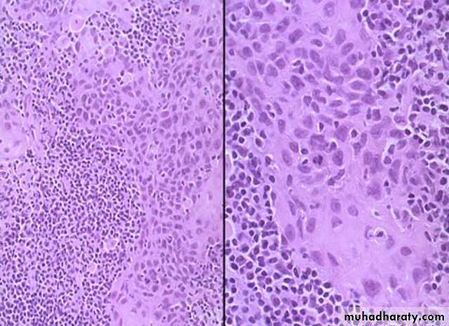
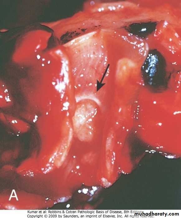
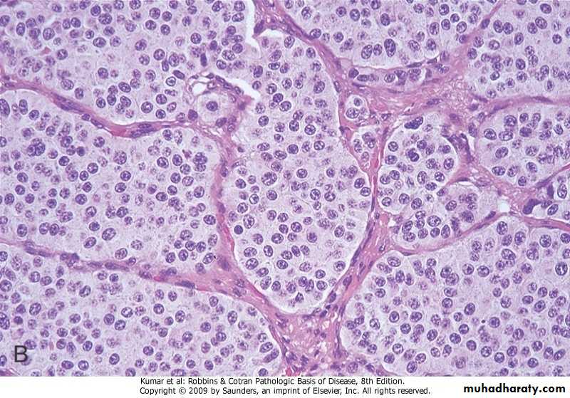
Nasopharyngeal carcinoma
Carcinoma arises from nasopharynx.Environmental and viruses (Epstein Barr virus EBV play a role in its pathogenesis
Microscopically: consist of sheets of malignant cells which may be of :
Undifferentiated
Keratinising squamous cells
With variable number of lymphocytes led to its old name of lymphoepithelioma
Sinusitis
Acute inflammation of the nasal sinuses. Usually follow extension of infection from the nose or from tooth socketsEtiology:
Viral
Bacterial
Allergic
Sinusitis (cont)
Pathology:Acute inflammation: congestion &edema lead to obstruction of sinus opening resulting in accumulation of mucus secretion followed by bacterial infection & suppuration
Complications:
Osteomyelitis
Subcutaneous abscess
Orbital cellulites
Intracranial suppuration (meningitis,brain abscess)
Larynx : Laryngitis
Nonspecific laryngitisFollow upper respiratory tract infection e.g. common cold. Air pollution predispose to laryngitis
Tuberculous laryngitis
Diphtheria ,syphilis, leprosy may involve the larynx
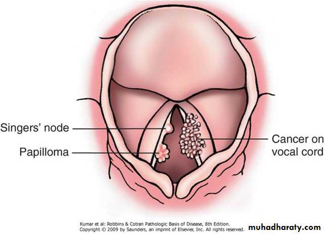
Tumors of the larynx
Benign tumors :squamous cell papilloma.
Single or Multiple
viral in origion (HPV)
Malignant:
Squamous cell carcinoma ,Verrucous ca.
(Glottic , supraglottic , subglottic).
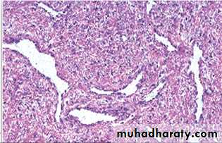
Laryngeal nodule:(singer nodule) it is not a neoplasm .It is a small nodule at the vocal cord , has smooth surface covered by normal epithelium its core consist of fibrous tissue, blood vessels ,amyloid materials.
The larynx
The LungCongenital Anomalies:
1.Agenesis
2.Tracheo-Esophageal fistula (T-E fistula)
3.Vascular abnormalities
4.Pulmonary sequestration (part without connection to air ways system)
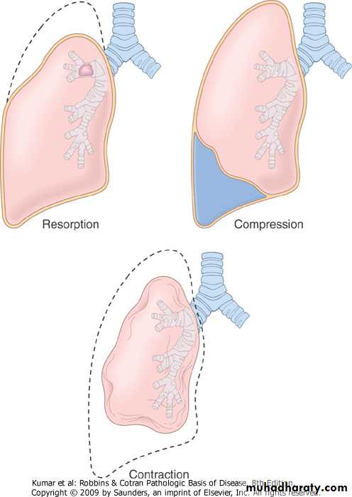
Atelectasis (collaps)
1.Neonatal collapse: Failure of lung to expand.
2.Acquired collapse: may involve a segment or the whole lung ( massive collapse).
Three types recognized:
Compression collapse e.g.Pleural effusion
Absorption collapse (Obstruction): e.g. Foreign body
Contraction collapse : e.g. Fibrosis
Acquired collapseAcquired atelectasis
Neonatal collapse
Failure of the lung to expand in newborn babyCauses:
1. Brain damage involving respiratory center
2. Congenital anomalies
3. Bronchial obstruction
Respiratory failure
Hypoxemia , arterial oxygen tension below 60 mmHg as a result of lung diseases in patient breathing air at sea level.(normal 80-100mmHg)In some patient there is retention of CO2 so the arterial tension of CO2 is over 45mm Hg (normal 35-45mmHg)
Type I respiratory failure when there is hypoxemia with no CO2 retention e.g. pneumonia, asthma
Type II respiratory failure when there is CO2 retention with hypoxemia e,g, Chronic bronchitis, Emphysema
Circulatory disorders of the lungs
Chronic passive venous congestion
Pulmonary edema
Pulmonary oedema
Causes:Heart failure (left)
Inflammatory
Toxic agents
Raised Intracranial pressure
Pulmonary embolism
Pulmonary infarction
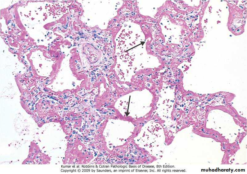
Adult Respiratory Distress Syndrome (ARDS)
A clinical syndrome caused by diffuse alveolar capillary endothelial and epithelial cell damage.Increased permeability result in exudation of fluid.
Clinically: severe respiratory distress , cyanosis & respiratory failure.
Grossly:
The lung is heavy red congested &edematous
Micro:
Diffuse alveolar wall damage( epithelial & endothelial)
Alveolar wall is lined by hyaline materials Latter on intra alveolar organization takes place
Adult Respiratory Distress Syndrome ARDS (cont)
Causes:
Sepsis
Pulmonary infections
Aspiration of gastric juice
Trauma e.g. head injury
Others
Adult Respiratory Distress Syndrome(ARDS)
Hyaline Membrane disease(cf with ARDS)
Severe respiratory distress, cyanosis and death from respiratory failureAffect infants in the first few day of life who are:
Baby delivered by caesarean section
Baby of diabetic mother
Premature baby
Hyaline Membrane Disease (cont)
Aetiology: uncertain and include:Deficiency of pulmonary surfactant
Increased permeability of pulmonary capillaries
Inhalation of amniotic fluid
Hyaline Membrane Disease (cont)
Pathology:
Collapse of the alveoli
Respiratory and terminal bronchioles are distended and lined by hyaline eosinophilic materials
Pneumonia
Definition: Inflammation of lung parenchyma .Characterized by consolidation ,Consolidation:
Replacement of the alveolar air by inflammatory exudates.
Classification of Pneumonia
1.Pathological classification2. Microbiological classification
3. Clinical classification
Pathological Classification
Depending on how the micro-organism spread in the lung:
1. Lobar Pneumonia: From alveoli to alveoli .Typically bacterial
2. Bronchopneumonia: From bronchi to alveoli
3. Interstitial Pneumonia: In the interstitial tissue of the lung . Typically viral.
Microbiological Classification
Depending on the causative micro-organism as determined by bacteriological examination e.g. Pnemococcal pneumonia ,viral pneumonia etc
Clinical Classification of pneumonia
Depends on the circumstances surrounding the infection . It helps in predicting the causative micro organism. So you can start treatment until confirmation from the lab arrive Include:1.community aquired pneumonia: strept pn. ,H.influenza. Mycoplasma, chlamydia, candida
2. Nosocomial pneumonia: Hospital acquired due to gram (- ve) bacteria , pseudomonas, penicillin resistant staph.
3.Aspiration pneumonia: Aerobic & Anaerobic bacteria
4. Pneumonia in immunocompromised patient: pneumocystis carinii, CMV,
Compare Lobar & Bronchopneumonia
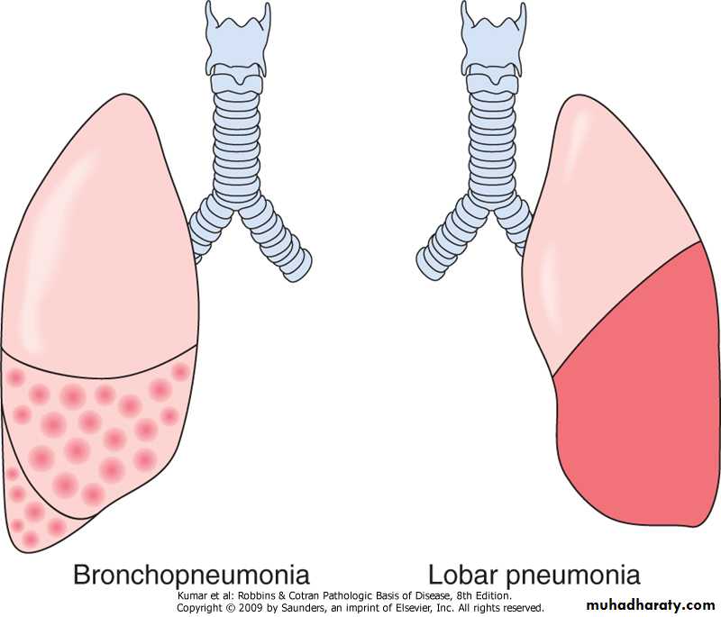
Lobar Pneumonia
Pneumonia: Classification
Lobular ( Bronchopneumonia)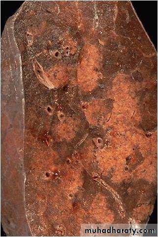
Lobar Pneumonia
Etiology: pneumococcal Pneumonia, streptococcal. PneumoniaPredisposing factors: Upper respiratory tract infection .The m.o. reach the alveoli through the bronchial tree & spread through pores of Kohn.
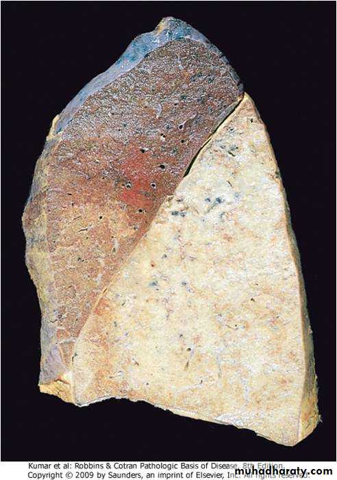
Grossly: a complete lobe is involved (consolidated)
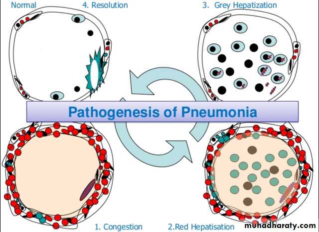
Microscopically: For descriptive purposes divided in to 4 stages
Lobar Pneumonia (cont)
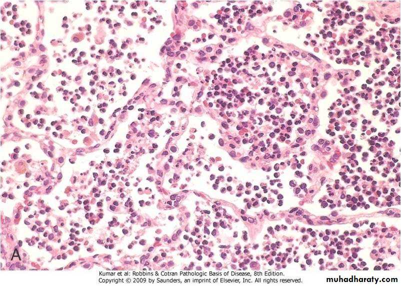
Stage 1 :Acute congestion .The affected lobe is red, firm and heavyThe alveolar capillaries are congested and the alveolar space contain fluid exudate
Stage 2: Red hepatisation , the affected lobe is firm &red similar to liver tissue
Microscopically: alveolar wall congested, alveolar lumen contain RBCs, m.o.& phagocytic cells
Lobar Pneumonia (cont)
Stage 3: Gray hepatisation,Gross: the affected lobe is firm & gray
Micro: the congestion in alveolar capillaries subside,The alveolar lumen contain large number of polymorphs, fibrin & macrophages
Lobar Pneumonia: Gray Hepatization
Lobar Pneumonia (cont)
Stage 4: Resolution,Gross: The affected lobe return to its normal appearance
Micro: the inflammatory exudate is liquefied & removed by expectoration ,& by lymphatic
The alveoli return normal with out residual defect
Clinical picture of Pneumonia
Fever , shivering with cough and rusty sputumChest pain from involvement of pleura (pleurisy) .
Bronchial breathing &sometime pleural rub.
Chest x-ray show consolidated lob.
Complications of Lobar Pneumonia
• 1. Organization• 2. Pleurisy & pleural effusion
• 3. Empyema
• 4. Lung abscess
• 5. Septicemia
• 6. Cardiac complications
Bronchopneumonia
Patchy consolidation centered around inflamed bronchi & bronchiole. Multifocal & may be bilateral.Predisposing factors:
1. Both extreme of age
2, Debilitating disease
3. Pre existing respiratory diseases e.g. chronic bronchitis , emphysema, measles , influenza
Pathology of Bronchopneumonia
Gross: Lesions are multiple & may be bilateral, affect basal segments of lower lobes.
Micro: Acute inflammation of bronchi ,extend to involve surrounding alveoli which become consolidated. May involve the pleura
Clinically: Fever cough sputum dyspnea
Complication of Bronchopneumonia
1. Organization & lung fibrosis.Resolution is unusual in bronchopneumonia
2.Damage of bronchial wall predisposing to bronchiectasis
3. Lung abscess
4, Empyema
Obstructive Pulmonary Disease
Diffuse pulmonary disease having increased resistance to air flow due to partial or complete obstruction at any level ,not fully reversible e,g. Chronic bronchitis, emphysema, asthma bronchiectasis.Cf. Restrictive pulmonary diseases which is failure of the lung to expand .
It is due to:
Chest wall lesions .
Infiltrative lesions of lung tissue
Chronic Obstructive Pulmonary Disease (COPD)
A clinical term include:
1.Emphysema
2. Chronic bronchitis
3. Asthma
4. Bronchiectasis
Emphysema
Permanent increase in the size of air spaces distal to the terminal bronchioles due to dilatation or destruction of the their wall with little or no fibrosis ,there will be:A. lack of elastic recoil
B. goblet cell hyperplasia & mucus plugC. inflammatory edema
D, muscle hypertrophy
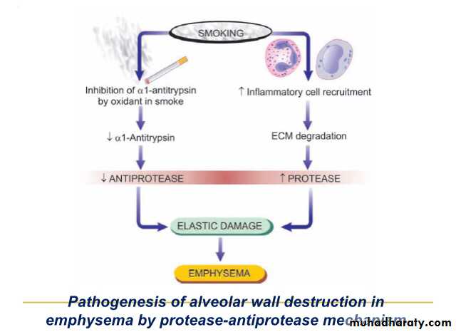
Pathogenesis of Emphysema
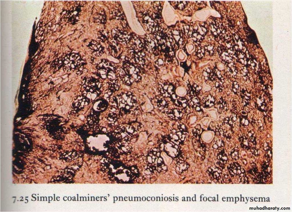
1. Air born factors e.g smoke, coal dust :Accumulation of dust in & around the wall of respiratory bronchiole lead to destruction of their wall and dilatation under the pressure of inspired air leading to focal dust emphysema.
Smoke: produce free radicles, inactivate antitrypsin, increase neutrophil elastase
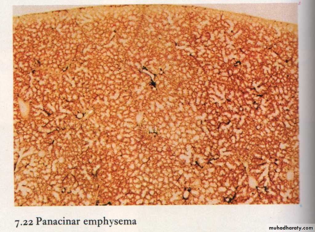
2. Hereditary factors: e.g. deficiency of α- antitrypsin leading to destruction of lung tissue and lead to panacinar emphysema
Pathogenesis of Emphysema (cont)
α-antitrypsin present normally in the serum prevents digestion of lung tissue by proteolytic enzyme released from WBC and alveolar macrophages.
Absence of this enzyme allow digestion of lung tissue leading to panacinar emphysema
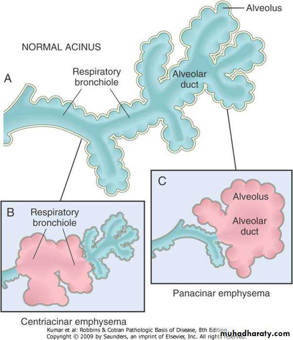
Classification of Emphysema
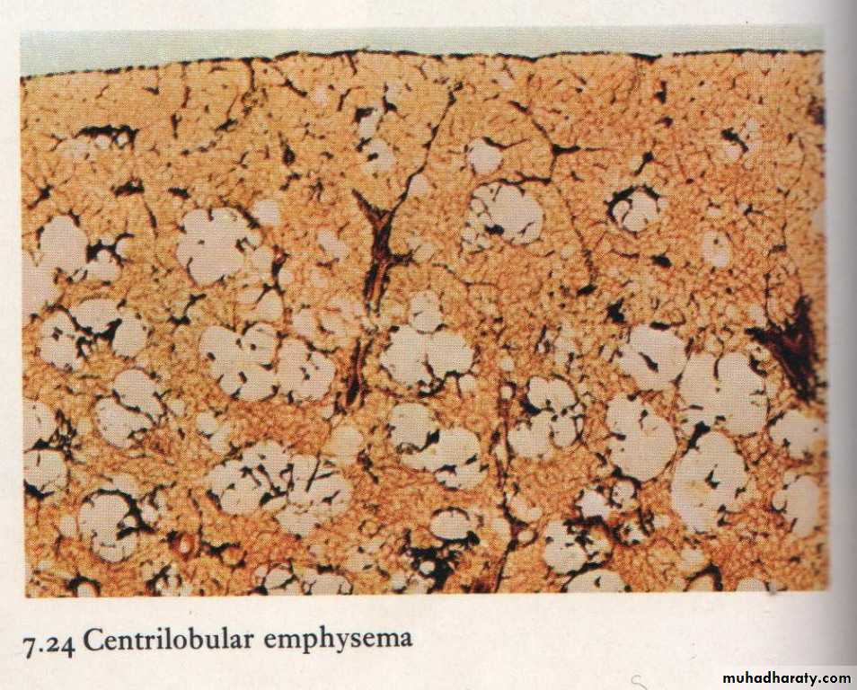
Depending on the microanatomy of the respiratory acinus :1. Bronchiolar emphysema :Include
a. Focal dust emphysema
b. Centrilobular emphysema (most common 95% of the cases)
2. Alveolar emphysema: Include
a. Alveolar duct emphysema
b. Panacinar emphysema
Respiratory Acinus
Pathology of Emphysema
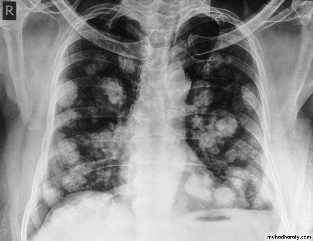
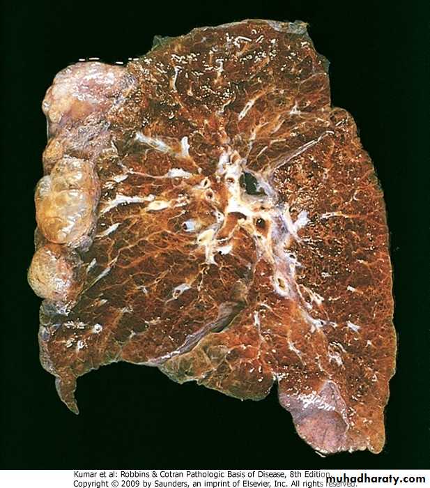
Gross: The lung is voluminous , pits on pressure due to lack of elasticity. A dilated air space may become cystic and project on the surface forming what is called emphysematous bullies
Clinically the patient have barrel shape chest
Chest x-ray show the diaphragm is lowered and the anterior surface of the heart is covered
Emphysema Pathology (cont)
In bronchiolar Emphysema:The proximal part of the respiratory acinus is involved by dilatation (i.e. the respiratory bronchiole)In Alveolar Emphysema : The alveoli &alveolar duct are involved to begin with , later the entire acinus is involved ( panacinar).
Gross appearance of emphysema
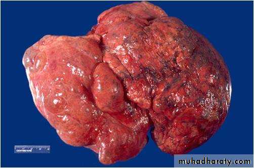
Emphysematous Bullae
Coal dust Emphysema
Clinically
About 1/3 of lung tissue is destroyed before symptom appears,Dyspnea , cough & sometime wheezes weight loss, Barrel chest.
Respiratory failure, core pulmonale due to pulmonary hypertension
On examination
X-ray finding
Respiratory function test show low FEV1
Interstitial Emphysema
Presence of air in the interstitial tissue of the lung due to:1. Laceration of lung tissue by trauma
2. Rupture of alveolar wall by severe cough
Compensatory Emphysema
Over distention of air spaces due to collapse or resection of lung tissue.
Chronic bronchitis
Definition: Chronic inflammation of the bronchial tree with cough and productive sputum for a period of at least 3 months in two successive yearsMore common in male
Causes respiratory disability
Chronic bronchitis: Aetiology
Chronic irritation of the bronchial epithelium by cigarette smoke & air pollutantBacterial infection
Chronic bronchitis: Pathogenesis
Chronic irritation lead to hypertrophy & hyperplasia of the mucus glands & goblet cells in the bronchial wall leading to excessive mucus production.In typical chronic bronchitis inflammation is not important.
Reid Index: The ratio of thickness of mucus gland layer to thickness of bronchial wall (normally 0.4)
Chronic bronchitis: Complications
Bronchopneumonia: excessive secretion predispose for infection
Emphysema
Respiratory failure :due to obstruction of bronchi by mucus ---- low ventilation lead to type II respiratory failure
Right sided heart failure
Bronchial Asthma
Chronic inflammatory condition of the air passages characterized by recurrent attacks of:dyspnea
Wheezing
Cough
Feeling of tightness in the chest
Severe & prolonged attack is called status asthmaticus
Bronchial Asthma ( cont )
Aetiology :Hereditary factors
Allergy : The allergen may be inhaled as pollen, ingested as protein or injected as drugs
Psychological factors
Pathogenesis of Bronchial Asthma
These symptom are due to narrowing of the bronchial lumen ( bronchospasm) due to the muscular spasm and plugging of the lumen by thick mucusAccording to β-adrenergic theory asthma is due to inherited or acquired defeciency of adenyl cyclase which is the β-receptor for catecholamine as a result of this deficiency there is activation of α-receptors that induce bronchospasm
Various inflammatory stimuli and variety of cells ( esinophils , mast cells macrophages ) are involved in the pathogenesis of asthma
β
Bronchial Asthma ( cont)
Types of asthma: asthma has many predisposing factors and variety of clinical presentation that make classification so difficult .One of these classification is :Extrinsic asthma
Intrinsic asthma
Types of Bronchial Asthma (cont)
Extrinsic asthma (mmune-mediated,atopic);Usually start in chilhood , due to atopic hypersensitivity to allergen mediated by IgE
IgE is fixed to mast cells, so inhalation of the allergen lead to Ag Ab reaction & release of broncho constrictor substances from mast cells leading to broncho spasm.
Triggering allergen include pollen , drugs etc
Family history and skin test are usually positive.
Types of Bronchial Asthma (cont)
Intrinsic Asthma(non immune-mediated,non-atopic);Usually develop in adulthood without history of atopic hypersensitivity (due to nonimmune causes e.g. viral infection ).
Hyperirritability of the bronchial tree is the underlying cause.
Family history and skin tests are usually negative
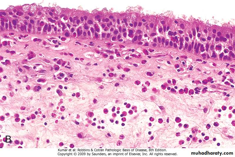
Bronchial Asthma : Pathology
Gross: The lung are overinflatedMicroscopical:
1.The lumen of the bronchi and bronchiole contain thick mucus plug (containing whorls of epithelium called curschmann spirals),charcot leyden crystals and eosinophils
2. The basement membrane shows characteristic hyaline thickening
3. The submucosa shows congestion . edema and infiltration by esinophil & mast cells
4. The bronchial muscle are hypertrophied
Bronchial mucosa in Asthma
Bronchial Asthma Complications
1. Emphysema2. Respiratory infections
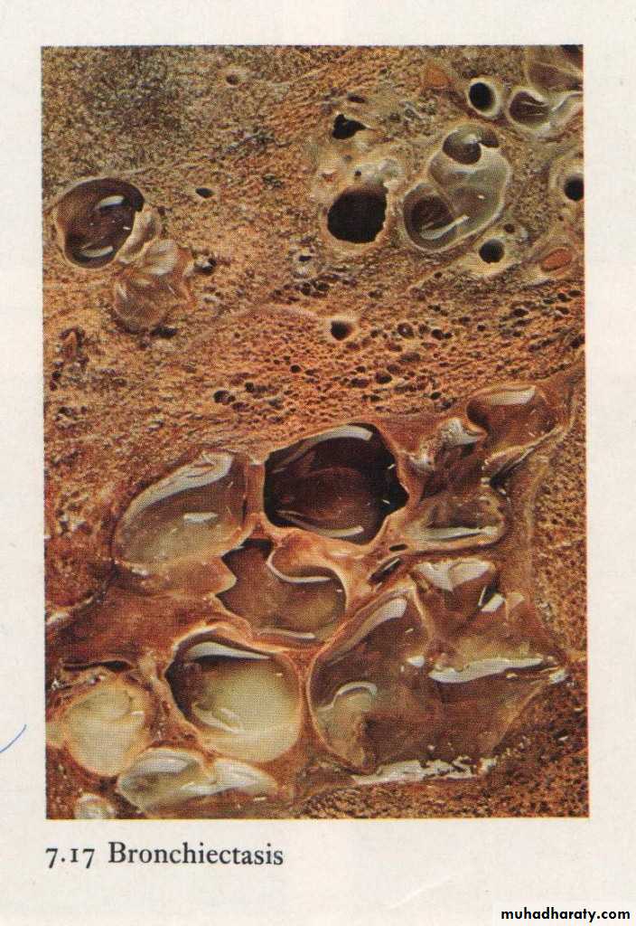
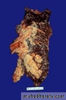
Bronchiectasis
Abnormal and permanent dilatation of the bronchi .It is common and affects all age group characterized by cough with large amount of foul odor sputum. Hemoptysis
Etiology of Bronchiectasis
• 1. Infections : e.g. T.B, Whooping cough• 2. Bronchial Obstraction: Foreign body,tumour
• 3. Fibrocystic disease of the pancrease
• 4. Congenital abnormality of the cilia (Kartageners syndrome)
Sequence of events in Bronchiectasis
Obstraction → Resorption of air → accumulat ion of secretions & stasis → infection which damage the wall of the bronchi→ Dilatation by intraluminal pressure or traction by the negative pressure of the pleuraBronchiectasis :Pathology
Gross:It may affects any part of the lung but the basal segments are commonly involved
The dilatation may be Cylindrical (involve all the circuference ) or Sacular (involve part of the circumferences),
Bronchogram demonstrate the dilatation nicely
Grossly: Bronchiectasis
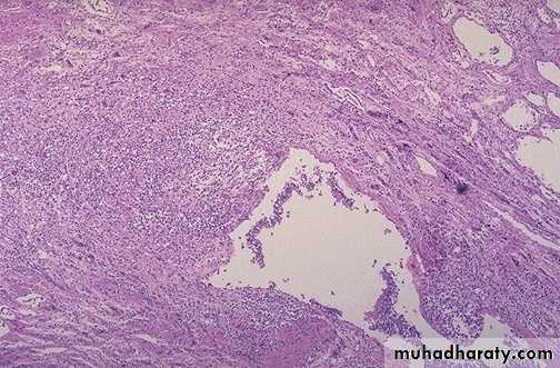
Bronchiectasis :Pathology
Microscopically: The affected bronchi show:Dilatation of the lumen which contain pus
Ulceration of the mucosa and squamous metaplsia
Destruction of muscle and elastic fibers
Microscopical: Bronchiectasis
Bronchiectasis :Complications
1.Pneumonia and lung abscess2.Pleurisy and empyema
3.Pyaemia with metastatic abscess
4. Destruction of lung tissue leading to right sided heart failure
5. In long standing cases Amyloidosis
Restrictive Lung Diseases
Reduced expansion of lung parenchyma so total lung capacity is reduced (cf obstructive lung diseases FV1 is reduced) Include:Chest wall disease e.g. polio, obesity, pleural diseases.
Chronic interstitial and infiltrative diseases e.g. pneumoconiosis, interstitial fibrosis,sarcoidosis,immunological diseases
Pneumoconiosis
Group of lung diseases caused by inhalation of dust.The type of disease depends on the type of dust. Some dust are inert causes little or no damage, other cause severe destruction & fibrosis of the lung. Some induce immunological reaction. Some predispose to T.B or malignancy.
Factors that determine the severity of lung disease in pneumoconiosis
1. Physical state of the dust: 1-5 M reach alveoli. Larger one removed by bronchi2. Chemical composition
3. Concentration of dust
4. Duration of exposure
5. possible presence of other particles
Classification of pneumoconiosis
1.pneumoconiosis due to inhalation of inorganic dust e.g. Anthracosis, coal worker pneumoconiosis, silicosis, Asbestosis, berylliosis
2. Pneumoconiosis due to inhalation of organic dust e.g. Byssinosis ,Extrinsic allergic alveolitis
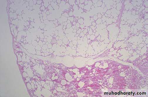
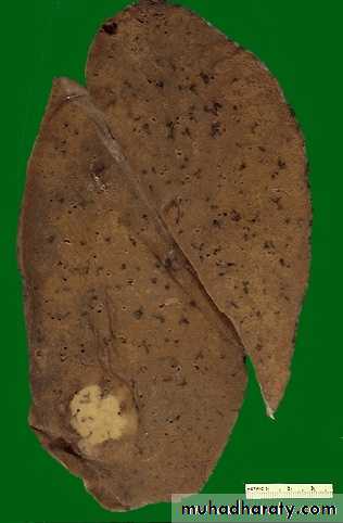
Anthracosis
Black discoloration of the lung due to inhalation of carbon .Carbon particles are inert so there will be no significant effect on the lung, Lung function is normal
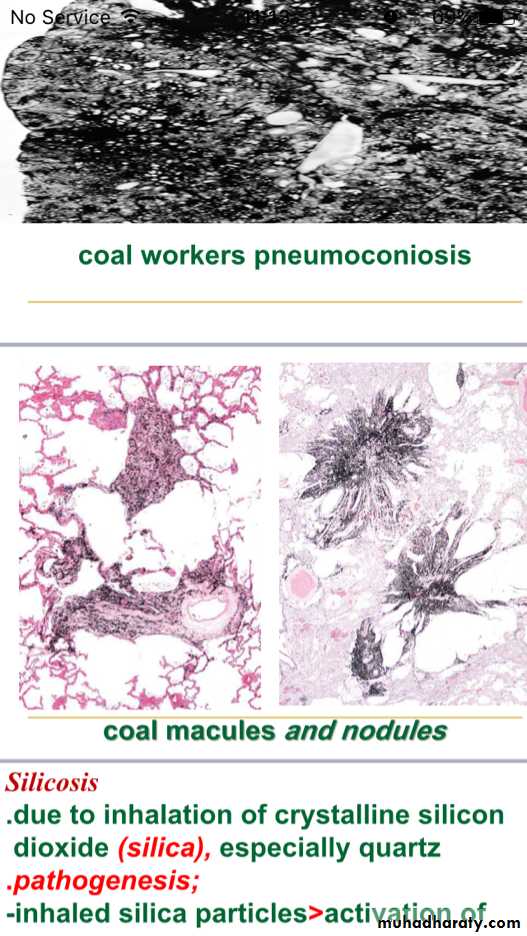
Coal worker pneumoconiosis
Lung fibrosis due to inhalation of coal dust in coal workerIt cause fibrosis of the lung and focal duct emphysema .Two type recognized:
1.Simple coal worker pneumoconiosis. Characterized by small nodules of fibrosis 2-5mm
2. Progressive massive fibrosis. Characterized by large nodules more than 10 mm.. Associated silica particles may play a role in this type
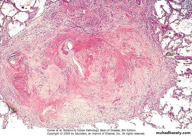
Silicosis
Lung fibrosis due to inhalation of silica containing particles e.g miner of gold and ironSilica produce collagenous fibrous tissue in concentric laminated layers (silicotic nodule) due to formation of silicic acid or due to immunological mechanism
Silica particle may be carried to the regional lymph node leading to enlargement and fibrosis of the lymph node
Effect of silicosis
1. Fibrosis of the lung → pulmonary hypertension → Right sided heart failure
2. Tuberculosis coexist in 80% of the cases due to depression of cell mediated immunity Silica inhibit alveolar macrophage to destroy phagocytosed T.B. bacilli
Silicotic nodules
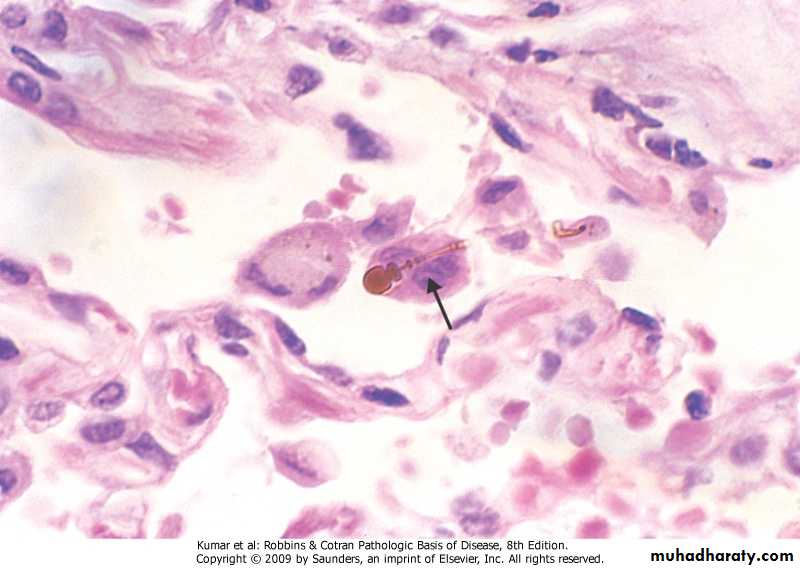
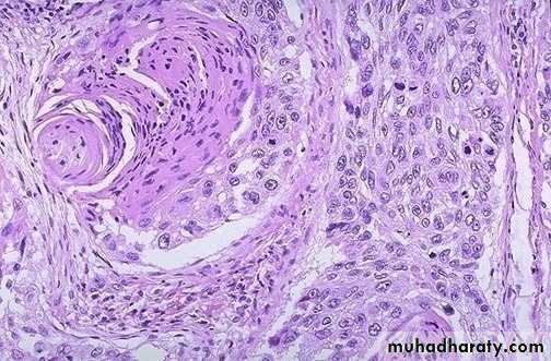
Asbestosis
Lung fibrosis due to inhalation of asbestos fibersSmall fibers are ingested by macrophage which release fiberogenic substances
Long fibers cannot be ingested but surrounded by iron rich protienaceous material producing asbestos bodies (golden brown bodies )
Pathology of asbestosis
Gross:Localized pleural thickening (plaque)
Diffuse pleural thickening
Pleural effusion
Diffues lung fibrosis (Asbestosis)
Brochogenic carcinoma & Mesothelioma
Micro : Dense fibrosis around asbestose bodies
Asbestos body
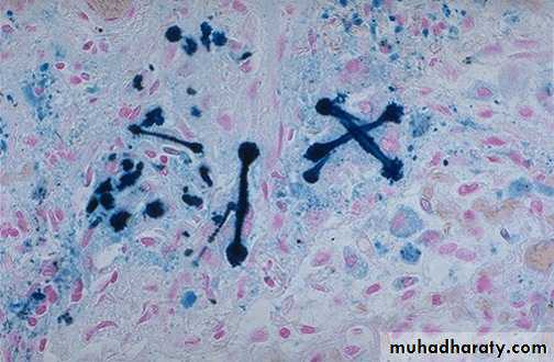
Asbestos body, iron stain
Effects of asbestosis
1. Lung fibrosis and right sided heart failure2. Increased incidence of bronchial carcinoma and mesothelioma (pleural and pereitoneal)
Pulmonary siderosis
Lung fibrosis due to inhalation of ironOccur in haematite minors
Predispose to malignancy
Pneumoconiosis due to organic dust
Byssinosis: Inhalation of cotton dust lead to:chronic bronchitis , asthma , emphysema
Extrinsic allergic alveolitis:
Inhalation of organic materials like fungi, bird dropping and mould which act as antigen and react with circulating antibody ( Arthus reaction) cause lung damage
The Pleura
Acute pleurisy:
Acute inflammation of pleura. It may due to :
Secondary to lung infection,
Sub diaphragmatic lesion
Perforating chest wall injury
Clinically: chest pain, fever,
Pleural effusion: collection of fluid in the pleural cavity.(hydrothorax) It may lead to collapse of the lung and interfere with respiration
Pleural effusion (cont)
Pleural effusion may be:Transudate pleural effusion :is due to :
Heart failure, hypoproteinaemia , or Meigs
syndrome(ovarian fibroma associated with right sided pleural effusion).
Pleural effusion(cont)
Exudative pleural effusion:1. Infections e.g. T.B. Pneumonia .
2. Systemic diseases e.g. Uraemia
3. Lymphatic obstruction by tumor cells
4. Malignant effusion
Empyrma:collection of pus in the pleural cavity
haemothrax:collection of blood in the pleural cavity.
pneumothorax:air collection in the pleural cavity
hydropneumothorax:this is seen when there is fisula between the lung and the pleural cavity
Tension Pneumothorax
A valve like connection between pleural cavity and the lung lesion which allow air to enter the pleura during inspiration and prevents its escape during expiration this Lead to a rise in the intrapleural pressure leading to collapse of the lung & shift of mediastinum to the other side. It is serious condition cause severe respiratory distressTreatment by chest tube
Tumors of pleura
1. Primary tumors:Malignant Mesothelioma arises from mesothelial cells.
Macroscopically: Form whitish mass compressing the lung. Asbestosis is a predisposing factor
Microscopically: it may have carcinomatous or sarcomatous appearace,
2. Secondary tumours:
More common, commonly from breast , bronchus. Usually present as haemorrhagic pleural effusion
Lung tumours
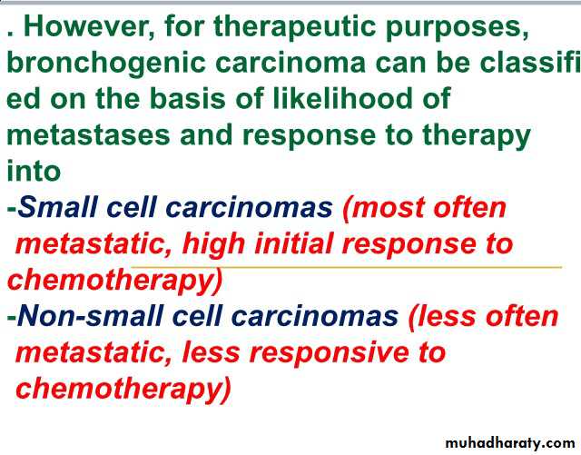
Primary tumours :Carcinoma 90-95%
Carcinoid 5%
Sarcoma and others 2-5 %
Secondary tumours:
The lung is a common sites for secodaries from many organs e.g. breast,GIT,ovaries ,bone etc.
Bronchogenic carcinoma
A common cancer, It is number one cancer in men.
Age incidence 50-70 Y
Present with cough sputum haemoptysis .In late stages with secondaries
abscess, bronchpneumonia, not responding to treatment
Chest x-ray show shadow in the lung
Etiology of bronchogenic carcinoma
Genetic abnormality that transform benign epithelium to neoplastic tissueRisk factors:
1. Smoking: Smoker have 10 time greater risk than nonsmoker. Benzpyrene is the carcinogenic substance
2, Radioactive substances e.g. Radium
3. Atmospheric pollution e.g.industrial fumes
4. occupational hazards e.g. asbestose
Etiology of lung cancer @@
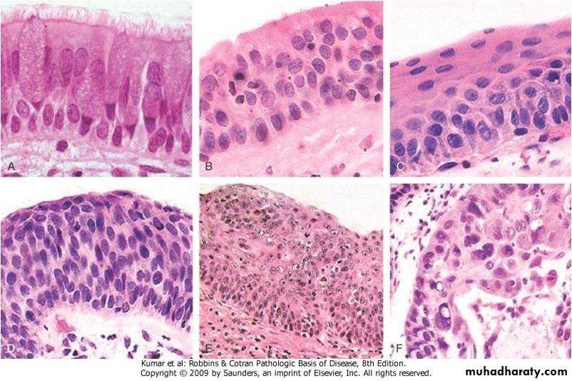
Most lung cancer arises by stepwise accumullation of genetic abnormalities that transform benign bronchial epith to neoplastic oneCont @@
1.Tobacco smoking: there is statistical and clinical obserevation establishing a positive relationship between lung cancer and smoking
Statistical evidence:87% of lung cancer affect active smokers & this depends on:
Daily smoke
Tendency to inhale
Duration of smoking
Cont @@@
Clinical evidence:obtained from observation of histological changes in bronchial epithelium in smokers . There is sequential changes leading to squamous cell carcinoma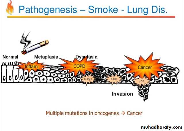
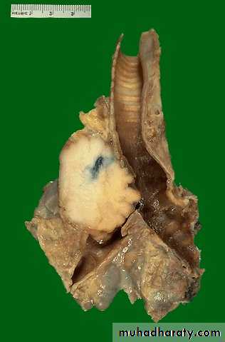
Pathology of bronchogenic carcinoma
Site:1.Central 55% arises from main bronchus
2. Perpheral 40 % arises from small bronchi and bronchiole
Diffuse 5%
Pathology of bronchogenic carcinoma (cont)
Histopathological classification:1. Squamous cell carcinoma ; arises from squamous metaplastic epithelium---dysplasia---carcinoma in situ---invasive carcinoma.
It is usually poorly differentiated
Bronchogenic carcinoma
Sequence of changes in bronchial epithelium
Squamous cell carcinoma of bronchus
Pathology of bronchogenic carcinoma (cont)
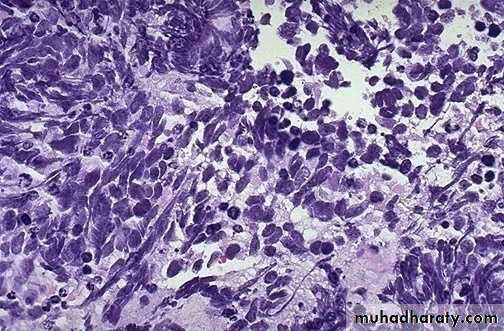
2,Oat cell carcinoma : Arises from neuroendocrine cells in the bronchial mucosa . Consist of small hyperchromatic cells similar to aot seeds. Arranges in sheetsBy E/M the cells contain neurosecretory granules
Oat cell carcinoma -Bronchus
Oat cell carcinoma -Bronchus
Pathology of bronchogenic carcinoma (cont)
3 ,Adenocarcinoma : Arises from mucus gland in the bronchial mucosa, Consist of malignant glands with mucus secretion4.Broncho alveolar carcinoma: A form of adenocarcinoma arises from terminal bronchoalveolar region It grows on preexisting structure (alveolar wall) without its destruction
Pathology of bronchogenic carcinoma (cont)
5,Large cell carcinoma: Undifferentiated carcinoma consist of large hyperchromatic cells with some giant malignant cells.It is probably of squamous or adeno carcinoma that is so undifferentiated to know the histogenesisAdenocarcinoma -Bronchus
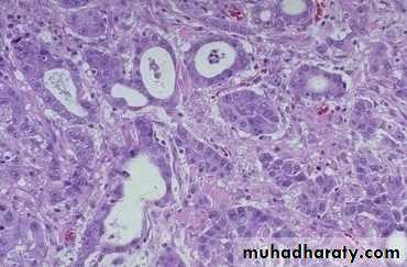
Adenocarcinoma -Bronchus
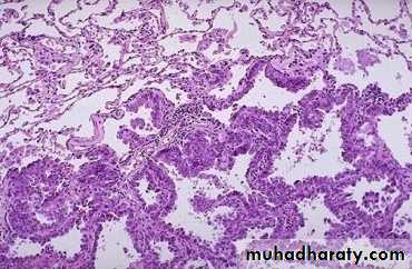
Bronchoalveolar carcinoma
Large Cell Carcinoma of Bronchus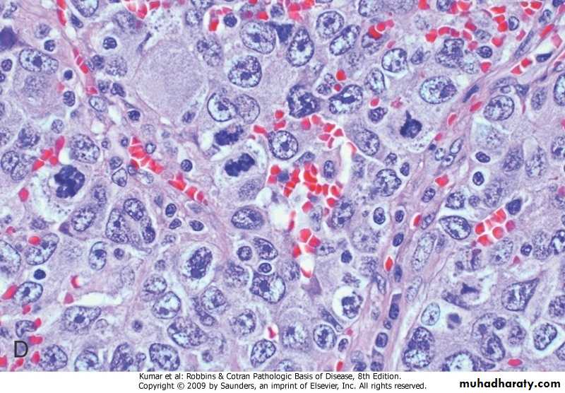
Spread of bronchogenic carcinoma
1.Direct spread: to pleura, pericardium,Esophagus, left recurrent laryngeal nerve. Tumor at the apex of the lung may involve brachial plexus causing pain and muscle atrophy
Involving the cervical sympathetic chain leading to Horner syndrome ( contracted pupil , ptoses & ipsilateral facial anhydrosis)
Spread of bronchogenic carcinoma (cont)
2. Lymphatic spread: To the hilar trachiobronchial , mediastinal supraclavicular lymph node leading to enlargment of the lymph node ( lymphadeno pathy)Spread of bronchogenic carcinoma (cont)
3.Blood spread: to the liver .bone. Adrenal brain etc
Paramalignant syndrome in bronchogenic carcinoma
Effects ocure in patients with bronchogenic carcinoma which is neither due to the primary tumour nor to the secondary.But probably due to substances secreted by the tumorIt may be:
1.Endocrine syndrome2,Neurological syndromeق
Paramalignant syndrome in bronchogenic carcinoma (cont)
1.Endocrine syndrome :a. Cushing syndrome: oat cell carcinoma secrete ACTH
b. Secretion of ADH by oat cell lead to water retention and brain edema
c. Hypercalcaemia due to secretion of parathyroid like hormone by squamus cell carcinoma
Paramalignant syndrome in bronchogenic carcinoma (cont)
2,Neurological syndrome:a. peripheral neuropathy,
b. encephalopathy
c. myopathy
3. Dermatomyocytis
4. Pulmonary osteoarthropathy with clubbing of fingers
Diagnosis of bronchogenic carcinoma
History, Clinical examination
Sputum cytology
Bronchoscopic biopsy
Percutaneous fine needle aspiration
Open biopsy
Scalene lymph node aspiration & biopsy
Prognosis of Bronchogenic carcinoma
It is poor. The overall 5 year survival rate is 16%.Carcinoid tumor
Low grade malignant tumorAffect younger age group than carcinoma
Both sexes are affected equally
It form a nodule may be central or peripheral
It can metastasise
May produce vasoactive amines leading to carcinoid syndrome
Histologically consist of uniform cells
Bronchial carcinoid
Bronchial carcinoid
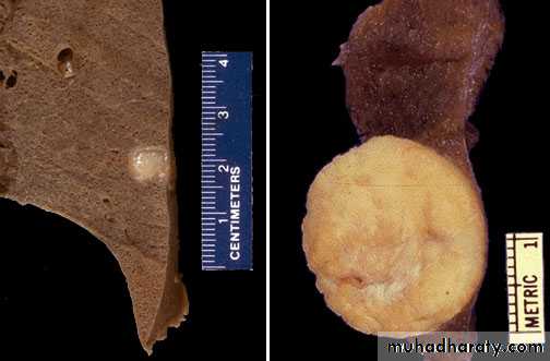
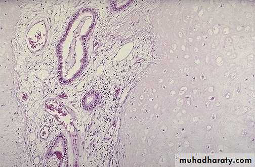
Lung Hamartoma
Lung hamartoma form mass few cm in diameter , discovered by routine chest x-ray(coin lesion).Consist of mixture of lung tissue (cartillage. Glands.fibrous tissue epithelial tissue) in disorganized pattern
Secondary tumors in the lung
The lung is a common site of secodary's due to the high blood supply.Secondaries reach the lung by blood from GIT, Female G.T., bone breast giving rise to cannon ball metastasis etc
By lymphatics from breast